4EBP1 Antibody - BSA Free
Novus Biologicals | Catalog # NB200-157
![Knockout Validated: 4EBP1 Antibody [NB200-157]](https://resources.rndsystems.com/images/products/4EBP1-Antibody-Knockout-Validated-NB200-157-img0011.jpg "Western Blot: 4EBP1 Antibody [NB200-157]")
Key Product Details
Validated by
Knockout/Knockdown
Species Reactivity
Validated:
Human
Cited:
Human
Applications
Knockout Validated, Immunohistochemistry, Immunohistochemistry-Paraffin, Western Blot
Label
Unconjugated
Antibody Source
Polyclonal Rabbit IgG
Format
BSA Free
Loading...
Product Specifications
Immunogen
The immunogen recognized by this antibody maps to a region between residue 75 and the C-terminus (residue 118) of human eukaryotic translation initiation factor 4E binding protein 1 using the numbering given in entry NP_004086.1 (GeneID 1978).
Localization
Cytoplasmic
Clonality
Polyclonal
Host
Rabbit
Isotype
IgG
Scientific Data Images for 4EBP1 Antibody - BSA Free
Western Blot: 4EBP1 Antibody [NB200-157]
Western Blot: 4EBP1 Antibody [NB200-157] - Western blot shows lysates of HeLa human cervical epithelial carcinoma parental cell line and 4EBP1 knockout (KO) HeLa cell line. PVDF membrane was probed with 0.5 ug/ml of Rabbit Anti-Human 4EBP1 Polyclonal Antibody (Catalog # NB200-157) followed by HRP-conjugated Anti-Rabbit IgG Secondary Antibody (Catalog #HAF008). Specific band was detected for 4EBP1 at approximately 17-20 kDa (as indicated) in the parental HeLa cell line, but is not detectable in the knockout HeLa cell line. This experiment was conducted under reducing conditions.![Western Blot: 4EBP1 Antibody [NB200-157]](https://resources.rndsystems.com/images/products/4EBP1-Antibody-Western-Blot-NB200-157-img0009.jpg "Western Blot: 4EBP1 Antibody [NB200-157]")
Western Blot: 4EBP1 Antibody [NB200-157]
Western Blot: 4EBP1 Antibody [NB200-157] - Whole cell lysate (100 ug) from RINm5F insulinoma exposed to high or low concentration of leucine. Antibody used at 0.25 ug/ml.![Immunohistochemistry: 4EBP1 Antibody [NB200-157]](https://resources.rndsystems.com/images/products/4EBP1-Antibody-Immunohistochemistry-NB200-157-img0010.jpg "Immunohistochemistry: 4EBP1 Antibody [NB200-157]")
Immunohistochemistry: 4EBP1 Antibody [NB200-157]
Immunohistochemistry: 4EBP1 Antibody [NB200-157] - Sample: FFPE section of human breast carcinoma. Antibody: Affinity purified rabbit anti-4EBP1 used at a dilution of 1:20,000 ( 0.05ug/ml). Detection: DAB staining using Immunohistochemistry Accessory Kit.
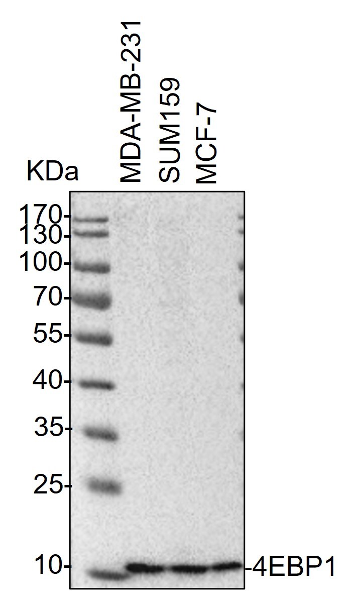
Western Blot: Rabbit Polyclonal 4EBP1 Antibody [NB200-157] -
Western Blot: Rabbit Polyclonal 4EBP1 Antibody [NB200-157] - Whole cell lysates from MDA-MB-231, SUM159 and MCF-7 cells were loaded with 50 ug/lane. 10% SDS-PAGE. 4EBP1 Antibody (NB200-157) was used for primary antibody: 1:2000, 4C, overnight. Image from a verified customer review.Applications for 4EBP1 Antibody - BSA Free
Application
Recommended Usage
Immunohistochemistry
1:5000-1:20000
Immunohistochemistry-Paraffin
1:5000-1:20000
Western Blot
1:1000-1:6000
Application Notes
For IHC: Epitope retrieval with citrate buffer pH 6.0 is recommended.
Reviewed Applications
Read 1 review rated 5 using NB200-157 in the following applications:
Formulation, Preparation, and Storage
Purification
Immunogen affinity purified
Formulation
Tris-Citrate/Phosphate (pH 7.0 - 8.0)
Format
BSA Free
Preservative
0.09% Sodium Azide
Concentration
1.0 mg/ml
Shipping
The product is shipped with polar packs. Upon receipt, store it immediately at the temperature recommended below.
Stability & Storage
Store at 4C. Do not freeze.
Background: 4EBP1
Long Name
Eukaryotic Translation Initiation Factor 4E Binding Protein 1
Alternate Names
EIF4EBP1, PHAS-I
Gene Symbol
EIF4EBP1
UniProt
Additional 4EBP1 Products
Product Documents for 4EBP1 Antibody - BSA Free
Certificate of Analysis
To download a Certificate of Analysis, please enter a lot or batch number in the search box below.
Product Specific Notices for 4EBP1 Antibody - BSA Free
This product is for research use only and is not approved for use in humans or in clinical diagnosis. Primary Antibodies are guaranteed for 1 year from date of receipt.
Related Research Areas
Citations for 4EBP1 Antibody - BSA Free
 Powered by Bioz
Customer Reviews for 4EBP1 Antibody - BSA Free (1)
5 out of 5
1 Customer Rating
Have you used 4EBP1 Antibody - BSA Free?
Submit a review and receive an Amazon gift card!
$25/€18/£15/$25CAN/¥2500 Yen for a review with an image
$10/€7/£6/$10CAN/¥1110 Yen for a review without an image
Submit a review
Customer Images
Showing
1
-
1 of
1 review
Showing All
Filter By:
-
Application: Western BlotVerified Customer | Posted 12/20/2023Western Blot: whole cell lysates from MDA-MB-231, SUM159 and MCF-7 cells were loaded with 50 ug/lane. 10% SDS-PAGE. 4EBP1 Antibody (NB200-157) was used for primary antibody: 1:2000, 4℃, overnight.

There are no reviews that match your criteria.
Protocols
Find general support by application which include: protocols, troubleshooting, illustrated assays, videos and webinars.
- Antigen Retrieval Protocol (PIER)
- Antigen Retrieval for Frozen Sections Protocol
- Appropriate Fixation of IHC/ICC Samples
- Cellular Response to Hypoxia Protocols
- Chromogenic IHC Staining of Formalin-Fixed Paraffin-Embedded (FFPE) Tissue Protocol
- Chromogenic Immunohistochemistry Staining of Frozen Tissue
- ClariTSA™ Fluorophore Kits
- Detection & Visualization of Antibody Binding
- Fluorescent IHC Staining of Frozen Tissue Protocol
- Graphic Protocol for Heat-induced Epitope Retrieval
- Graphic Protocol for the Preparation and Fluorescent IHC Staining of Frozen Tissue Sections
- Graphic Protocol for the Preparation and Fluorescent IHC Staining of Paraffin-embedded Tissue Sections
- Graphic Protocol for the Preparation of Gelatin-coated Slides for Histological Tissue Sections
- IHC Sample Preparation (Frozen sections vs Paraffin)
- Immunofluorescent IHC Staining of Formalin-Fixed Paraffin-Embedded (FFPE) Tissue Protocol
- Immunohistochemistry (IHC) and Immunocytochemistry (ICC) Protocols
- Immunohistochemistry Frozen Troubleshooting
- Immunohistochemistry Paraffin Troubleshooting
- Preparing Samples for IHC/ICC Experiments
- Preventing Non-Specific Staining (Non-Specific Binding)
- Primary Antibody Selection & Optimization
- Protocol for Heat-Induced Epitope Retrieval (HIER)
- Protocol for Making a 4% Formaldehyde Solution in PBS
- Protocol for VisUCyte™ HRP Polymer Detection Reagent
- Protocol for the Preparation & Fixation of Cells on Coverslips
- Protocol for the Preparation and Chromogenic IHC Staining of Frozen Tissue Sections
- Protocol for the Preparation and Chromogenic IHC Staining of Frozen Tissue Sections - Graphic
- Protocol for the Preparation and Chromogenic IHC Staining of Paraffin-embedded Tissue Sections
- Protocol for the Preparation and Chromogenic IHC Staining of Paraffin-embedded Tissue Sections - Graphic
- Protocol for the Preparation and Fluorescent IHC Staining of Frozen Tissue Sections
- Protocol for the Preparation and Fluorescent IHC Staining of Paraffin-embedded Tissue Sections
- Protocol for the Preparation of Gelatin-coated Slides for Histological Tissue Sections
- R&D Systems Quality Control Western Blot Protocol
- TUNEL and Active Caspase-3 Detection by IHC/ICC Protocol
- The Importance of IHC/ICC Controls
- Troubleshooting Guide: Immunohistochemistry
- Troubleshooting Guide: Western Blot Figures
- Western Blot Conditions
- Western Blot Protocol
- Western Blot Protocol for Cell Lysates
- Western Blot Troubleshooting
- Western Blot Troubleshooting Guide
- View all Protocols, Troubleshooting, Illustrated assays and Webinars
Loading...
Associated Pathways


