ABCA4 Antibody (3F4) - Azide Free
Novus Biologicals | Catalog # NBP1-30032
![Immunohistochemistry-Frozen: ABCA4 Antibody (3F4) [NBP1-30032]](https://resources.rndsystems.com/images/products/ABCA4-Antibody-3F4-Immunohistochemistry-Frozen-NBP1-30032-img0006.jpg "Immunohistochemistry-Frozen: ABCA4 Antibody (3F4) [NBP1-30032]")
Loading...
Key Product Details
Validated by
Knockout/Knockdown
Species Reactivity
Validated:
Human, Mouse, Bovine, Canine, Feline, Xenopus
Cited:
Bovine, Canine, Frog - Xenopus (African Clawed Frog)
Applications
Validated:
Knockout Validated, Immunohistochemistry, Immunohistochemistry-Frozen, Western Blot, Immunocytochemistry/ Immunofluorescence
Cited:
Immunohistochemistry-Frozen, Western Blot, Immunocytochemistry/ Immunofluorescence
Label
Unconjugated
Antibody Source
Monoclonal Mouse IgG1 Clone # 3F4
Format
Azide Free
Loading...
Product Specifications
Immunogen
Partially purified bovine 220-kDa disc rim protein. Accession # F1MWM0
Epitope
PLHPRTAGASR (PMID: 9092582)
Reactivity Notes
Please note that this antibody is reactive to Mouse and derived from the same host, Mouse. Additional Mouse on Mouse blocking steps may be required for IHC and ICC experiments. Please contact Technical Support for more information. Canine reactivity reported in scientific literature (PMID: 30889179). Feline reactivity reported from a verified customer review.
Clonality
Monoclonal
Host
Mouse
Isotype
IgG1
Theoretical MW
220 kDa.
Disclaimer note: The observed molecular weight of the protein may vary from the listed predicted molecular weight due to post translational modifications, post translation cleavages, relative charges, and other experimental factors.
Disclaimer note: The observed molecular weight of the protein may vary from the listed predicted molecular weight due to post translational modifications, post translation cleavages, relative charges, and other experimental factors.
Scientific Data Images for ABCA4 Antibody (3F4) - Azide Free
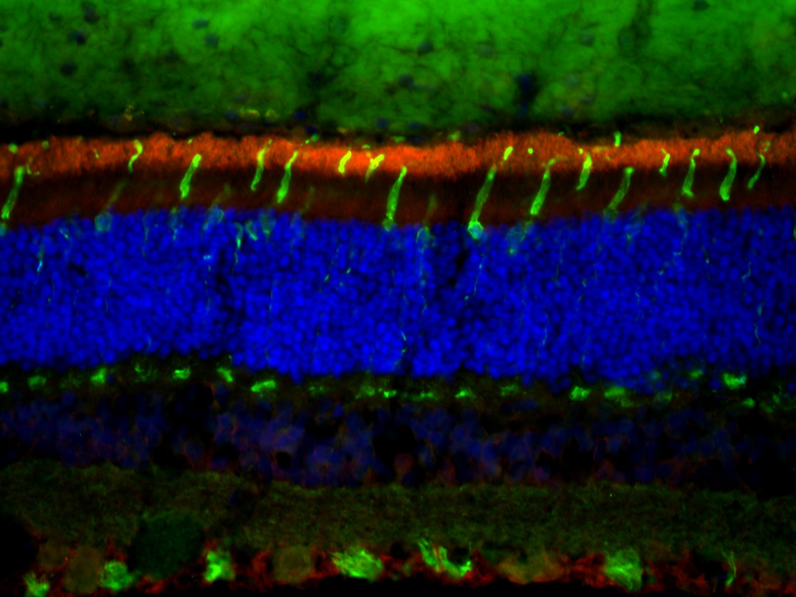
Immunohistochemistry-Frozen: ABCA4 Antibody (3F4) [NBP1-30032]
Immunohistochemistry-Frozen: ABCA4 Antibody (3F4) [NBP1-30032] - IHC-Fr of normal feline retina with antibodies ABCA4 3F4 (red) and HCAR (green). ABCA4 antibody (1:1000) labels outer segments of photoreceptors and HCAR antibody labels cones. IHC-Fr image submitted by a verified customer review.![Immunohistochemistry: ABCA4 Antibody (3F4) [NBP1-30032]](https://resources.rndsystems.com/images/products/ABCA4-Antibody-3F4-Immunohistochemistry-NBP1-30032-img0001.jpg "Immunohistochemistry: ABCA4 Antibody (3F4) [NBP1-30032]")
Immunohistochemistry: ABCA4 Antibody (3F4) [NBP1-30032]
Immunohistochemistry: ABCA4 Antibody (3F4) [NBP1-30032] - staining of adult mouse retina showing specific immunolabeling of the ABCA4 protein. Photo courtesy of Mary Raven, University of California, Santa Barbara, CA.![Immunohistochemistry: ABCA4 Antibody (3F4) [NBP1-30032]](https://resources.rndsystems.com/images/products/ABCA4-Antibody-3F4-Immunohistochemistry-NBP1-30032-img0003.jpg "Immunohistochemistry: ABCA4 Antibody (3F4) [NBP1-30032]")
Immunohistochemistry: ABCA4 Antibody (3F4) [NBP1-30032]
ABCA4-Antibody-3F4-Immunohistochemistry-NBP1-30032-img0003.jpg![Immunohistochemistry: ABCA4 Antibody (3F4) [NBP1-30032]](https://resources.rndsystems.com/images/products/ABCA4-Antibody-3F4-Immunohistochemistry-NBP1-30032-img0004.jpg "Immunohistochemistry: ABCA4 Antibody (3F4) [NBP1-30032]")
Immunohistochemistry: ABCA4 Antibody (3F4) [NBP1-30032]
ABCA4-Antibody-3F4-Immunohistochemistry-NBP1-30032-img0004.jpg![Knockout Validated: ABCA4 Antibody (3F4) [NBP1-30032]](https://resources.rndsystems.com/images/products/ABCA4-Antibody-3F4-Western-Blot-NBP1-30032-img0002.jpg "Western Blot: ABCA4 Antibody (3F4) [NBP1-30032]")
![Knockout Validated: ABCA4 Antibody (3F4) [NBP1-30032]](https://resources.rndsystems.com/images/products/ABCA4-Antibody-3F4-Immunohistochemistry-NBP1-30032-img0005.jpg "Immunohistochemistry: ABCA4 Antibody (3F4) [NBP1-30032]")
 [NBP1-30032] -")
Western Blot: ABCA4 Antibody (3F4) [NBP1-30032] -
Western Blot: ABCA4 Antibody (3F4) [NBP1-30032] - Characterization of ABCA4 mRNA expression & western blot analyses of ABCA4 protein levels in the canine retina.(A) Relative ABCA4 mRNA expression levels by quantitative RT-PCR in three different regions in three dogs with different genotypes (ABCA4+/+, ABCA4+/-, & ABCA4-/-), normalized to GAPDH expression. (B) Western blot analyses of ABCA4 (above), GAPDH (middle), & RHO (below) protein levels in retinal tissue of dogs with the three different genotypes. Image collected & cropped by CiteAb from the following publication (https://pubmed.ncbi.nlm.nih.gov/30889179), licensed under a CC-BY license. Not internally tested by Novus Biologicals. [NBP1-30032] -")
Immunocytochemistry/ Immunofluorescence: ABCA4 Antibody (3F4) [NBP1-30032] -
Immunocytochemistry/ Immunofluorescence: ABCA4 Antibody (3F4) [NBP1-30032] - Fluorescence histochemistry of ABCA4, cone photoreceptors, & autofluorescence in the canine retina.(A-C) Fluorescence micrographs showing ABCA4 expression (red), FITC-conjugated peanut agglutinin (PNA, green), & DAPI nuclear staining (blue) in wild-type (ABCA4+/+), heterozygous (ABCA4+/-), & affected (ABCA4-/-) retinas. PNA labels cone photoreceptors. Autofluorescence, indicative of lipofuscin accumulation, was seen in the ABCA4-/- RPE. (D) Bar graph with the average number of DAPI-stained nuclei within a given region of the ONL & the INL. (E-G) Fluorescence micrographs of RPE without immunohistochemistry show autofluorescence. (H) Bar graph with background-corrected mean autofluorescence-intensity in the RPE. Note the reduction of ABCA4-immunoreactivity & PNA binding, higher autofluorescence, & fewer nuclei in the ONL in the ABCA4-/- compared to ABCA4+/+ or ABCA4+/- retinas. All scale bars = 50 μm; RPE = retinal pigment epithelium; ONL = outer nuclear layer; INL = inner nuclear layer; Because there was only one individual per genotype, the statistics are valid for the technical replicates. ANOVA with Tukey’s post hoc test, n = 6; **P < 0.01; ***P < 0.001; mean ± S.D. Image collected & cropped by CiteAb from the following publication (https://pubmed.ncbi.nlm.nih.gov/30889179), licensed under a CC-BY license. Not internally tested by Novus Biologicals. [NBP1-30032] -")
Immunocytochemistry/ Immunofluorescence: ABCA4 Antibody (3F4) [NBP1-30032] -
Immunocytochemistry/ Immunofluorescence: ABCA4 Antibody (3F4) [NBP1-30032] - Fluorescence histochemistry of ABCA4, cone photoreceptors, & autofluorescence in the canine retina.(A-C) Fluorescence micrographs showing ABCA4 expression (red), FITC-conjugated peanut agglutinin (PNA, green), & DAPI nuclear staining (blue) in wild-type (ABCA4+/+), heterozygous (ABCA4+/-), & affected (ABCA4-/-) retinas. PNA labels cone photoreceptors. Autofluorescence, indicative of lipofuscin accumulation, was seen in the ABCA4-/- RPE. (D) Bar graph with the average number of DAPI-stained nuclei within a given region of the ONL & the INL. (E-G) Fluorescence micrographs of RPE without immunohistochemistry show autofluorescence. (H) Bar graph with background-corrected mean autofluorescence-intensity in the RPE. Note the reduction of ABCA4-immunoreactivity & PNA binding, higher autofluorescence, & fewer nuclei in the ONL in the ABCA4-/- compared to ABCA4+/+ or ABCA4+/- retinas. All scale bars = 50 μm; RPE = retinal pigment epithelium; ONL = outer nuclear layer; INL = inner nuclear layer; Because there was only one individual per genotype, the statistics are valid for the technical replicates. ANOVA with Tukey’s post hoc test, n = 6; **P < 0.01; ***P < 0.001; mean ± S.D. Image collected & cropped by CiteAb from the following publication (https://pubmed.ncbi.nlm.nih.gov/30889179), licensed under a CC-BY license. Not internally tested by Novus Biologicals.Applications for ABCA4 Antibody (3F4) - Azide Free
Application
Recommended Usage
Immunohistochemistry
1:100
Immunohistochemistry-Frozen
1:10 - 1:500
Western Blot
1:1000
Application Notes
Although not confirmed, this product may be useful in Immunohistochemistry-Paraffin. Immunohistochemistry-Frozen was reported in scientific literature. Use in Immunocytochemistry/immunofluorescence reported in scientific literature (PMID: 30889179).
Reviewed Applications
Read 1 review rated 5 using NBP1-30032 in the following applications:
Formulation, Preparation, and Storage
Purification
Protein G purified
Formulation
10 mM HEPES (pH 7.5), 0.15 M NaCl, 0.1 mg/mL BSA, 50% Glycerol
Format
Azide Free
Preservative
No Preservative
Concentration
Please see the vial label for concentration. If unlisted please contact technical services.
Shipping
The product is shipped with polar packs. Upon receipt, store it immediately at the temperature recommended below.
Stability & Storage
Store at -20C. Avoid freeze-thaw cycles.
Background: ABCA4
Alternate Names
ABC10, ABCRRMP, ARMD2DKFZp781N1972, ATP binding cassette transporter, ATP-binding cassette sub-family A member 4, ATP-binding cassette transporter, retinal-specific, ATP-binding cassette, sub-family A (ABC1), member 4, ATP-binding transporter, retina-specific, CORD3, EC 3.6.3, FFMFLJ17534, photoreceptor rim protein, retinal-specific ATP-binding cassette transporter, retina-specific ABC transporter, RIM ABC transporter, RIM protein, RmP, RP19, Stargardt disease protein, STGD, STGD1
Gene Symbol
ABCA4
Additional ABCA4 Products
Product Documents for ABCA4 Antibody (3F4) - Azide Free
Certificate of Analysis
To download a Certificate of Analysis, please enter a lot or batch number in the search box below.
Product Specific Notices for ABCA4 Antibody (3F4) - Azide Free
This product is for research use only and is not approved for use in humans or in clinical diagnosis. Primary Antibodies are guaranteed for 1 year from date of receipt.
Citations for ABCA4 Antibody (3F4) - Azide Free
 Powered by Bioz
Customer Reviews for ABCA4 Antibody (3F4) - Azide Free (1)
5 out of 5
1 Customer Rating
Have you used ABCA4 Antibody (3F4) - Azide Free?
Submit a review and receive an Amazon gift card!
$25/€18/£15/$25CAN/¥2500 Yen for a review with an image
$10/€7/£6/$10CAN/¥1110 Yen for a review without an image
Submit a review
Customer Images
Showing
1
-
1 of
1 review
Showing All
Filter By:
-
Application: Immunohistochemistry-FrozenSample Tested: cat retinaSpecies: FelineVerified Customer | Posted 09/25/2020IHC of normal cat retina with ABCA4 3F4 (red) and HCAR (green). ABCA4 (1:1000) labels outer segments of photoreceptors and HCAR labels cones.Cat retina was fixed in PFA, embedded in OCT and frozen in liquid nitrogen. OCT block was sectioned (10um) and subjected to IHC.

There are no reviews that match your criteria.
Protocols
Find general support by application which include: protocols, troubleshooting, illustrated assays, videos and webinars.
- Antigen Retrieval Protocol (PIER)
- Antigen Retrieval for Frozen Sections Protocol
- Appropriate Fixation of IHC/ICC Samples
- Cellular Response to Hypoxia Protocols
- Chromogenic IHC Staining of Formalin-Fixed Paraffin-Embedded (FFPE) Tissue Protocol
- Chromogenic Immunohistochemistry Staining of Frozen Tissue
- ClariTSA™ Fluorophore Kits
- Detection & Visualization of Antibody Binding
- Fluorescent IHC Staining of Frozen Tissue Protocol
- Graphic Protocol for Heat-induced Epitope Retrieval
- Graphic Protocol for the Preparation and Fluorescent IHC Staining of Frozen Tissue Sections
- Graphic Protocol for the Preparation and Fluorescent IHC Staining of Paraffin-embedded Tissue Sections
- Graphic Protocol for the Preparation of Gelatin-coated Slides for Histological Tissue Sections
- ICC Cell Smear Protocol for Suspension Cells
- ICC Immunocytochemistry Protocol Videos
- ICC for Adherent Cells
- IHC Sample Preparation (Frozen sections vs Paraffin)
- Immunocytochemistry (ICC) Protocol
- Immunocytochemistry Troubleshooting
- Immunofluorescence of Organoids Embedded in Cultrex Basement Membrane Extract
- Immunofluorescent IHC Staining of Formalin-Fixed Paraffin-Embedded (FFPE) Tissue Protocol
- Immunohistochemistry (IHC) and Immunocytochemistry (ICC) Protocols
- Immunohistochemistry Frozen Troubleshooting
- Immunohistochemistry Paraffin Troubleshooting
- Preparing Samples for IHC/ICC Experiments
- Preventing Non-Specific Staining (Non-Specific Binding)
- Primary Antibody Selection & Optimization
- Protocol for Heat-Induced Epitope Retrieval (HIER)
- Protocol for Making a 4% Formaldehyde Solution in PBS
- Protocol for VisUCyte™ HRP Polymer Detection Reagent
- Protocol for the Fluorescent ICC Staining of Cell Smears - Graphic
- Protocol for the Fluorescent ICC Staining of Cultured Cells on Coverslips - Graphic
- Protocol for the Preparation & Fixation of Cells on Coverslips
- Protocol for the Preparation and Chromogenic IHC Staining of Frozen Tissue Sections
- Protocol for the Preparation and Chromogenic IHC Staining of Frozen Tissue Sections - Graphic
- Protocol for the Preparation and Chromogenic IHC Staining of Paraffin-embedded Tissue Sections
- Protocol for the Preparation and Chromogenic IHC Staining of Paraffin-embedded Tissue Sections - Graphic
- Protocol for the Preparation and Fluorescent ICC Staining of Cells on Coverslips
- Protocol for the Preparation and Fluorescent ICC Staining of Non-adherent Cells
- Protocol for the Preparation and Fluorescent ICC Staining of Stem Cells on Coverslips
- Protocol for the Preparation and Fluorescent IHC Staining of Frozen Tissue Sections
- Protocol for the Preparation and Fluorescent IHC Staining of Paraffin-embedded Tissue Sections
- Protocol for the Preparation of Gelatin-coated Slides for Histological Tissue Sections
- Protocol for the Preparation of a Cell Smear for Non-adherent Cell ICC - Graphic
- R&D Systems Quality Control Western Blot Protocol
- TUNEL and Active Caspase-3 Detection by IHC/ICC Protocol
- The Importance of IHC/ICC Controls
- Troubleshooting Guide: Immunohistochemistry
- Troubleshooting Guide: Western Blot Figures
- Western Blot Conditions
- Western Blot Protocol
- Western Blot Protocol for Cell Lysates
- Western Blot Troubleshooting
- Western Blot Troubleshooting Guide
- View all Protocols, Troubleshooting, Illustrated assays and Webinars
Loading...