Actin Gamma 1 Antibody (NH3) - BSA Free
Novus Biologicals | Catalog # NB100-64792
![Immunocytochemistry/ Immunofluorescence: Actin Gamma 1 Antibody (NH3) [NB100-64792]](https://resources.rndsystems.com/images/products/F-Actin-Antibody-NH3-Immunocytochemistry-Immunofluorescence-NB100-64792-img0001.jpg "Immunocytochemistry/ Immunofluorescence: Actin Gamma 1 Antibody (NH3) [NB100-64792]")
Loading...
Key Product Details
Species Reactivity
Validated:
Human
Cited:
Human, Mouse
Applications
Validated:
Immunohistochemistry, Immunohistochemistry-Paraffin, Immunohistochemistry-Frozen, Western Blot, ELISA, Flow Cytometry, Immunocytochemistry/ Immunofluorescence
Cited:
Immunohistochemistry, Western Blot, Immunocytochemistry/ Immunofluorescence, IF/IHC
Label
Unconjugated
Antibody Source
Monoclonal Mouse IgM Clone # NH3
Format
BSA Free
Loading...
Product Specifications
Immunogen
Human monocytes and U937 cell line
Reactivity Notes
Predicted cross-reactivities: Rat, Rabbit, Mouse
Please note that this antibody is reactive to Mouse and derived from the same host, Mouse. Additional Mouse on Mouse blocking steps may be required for IHC and ICC experiments. Please contact Technical Support for more information.
Please note that this antibody is reactive to Mouse and derived from the same host, Mouse. Additional Mouse on Mouse blocking steps may be required for IHC and ICC experiments. Please contact Technical Support for more information.
Specificity
Recognizes human Filamentous actin (F-actin). The binds to the N-terminal region of actin, but not to the extreme N-terminal 40 amino acids. In tissue sections the stains the cytoplasm of macrophages strongly, and gives granular, localized nuclear staining of all cell types.
Clonality
Monoclonal
Host
Mouse
Isotype
IgM
Scientific Data Images for Actin Gamma 1 Antibody (NH3) - BSA Free
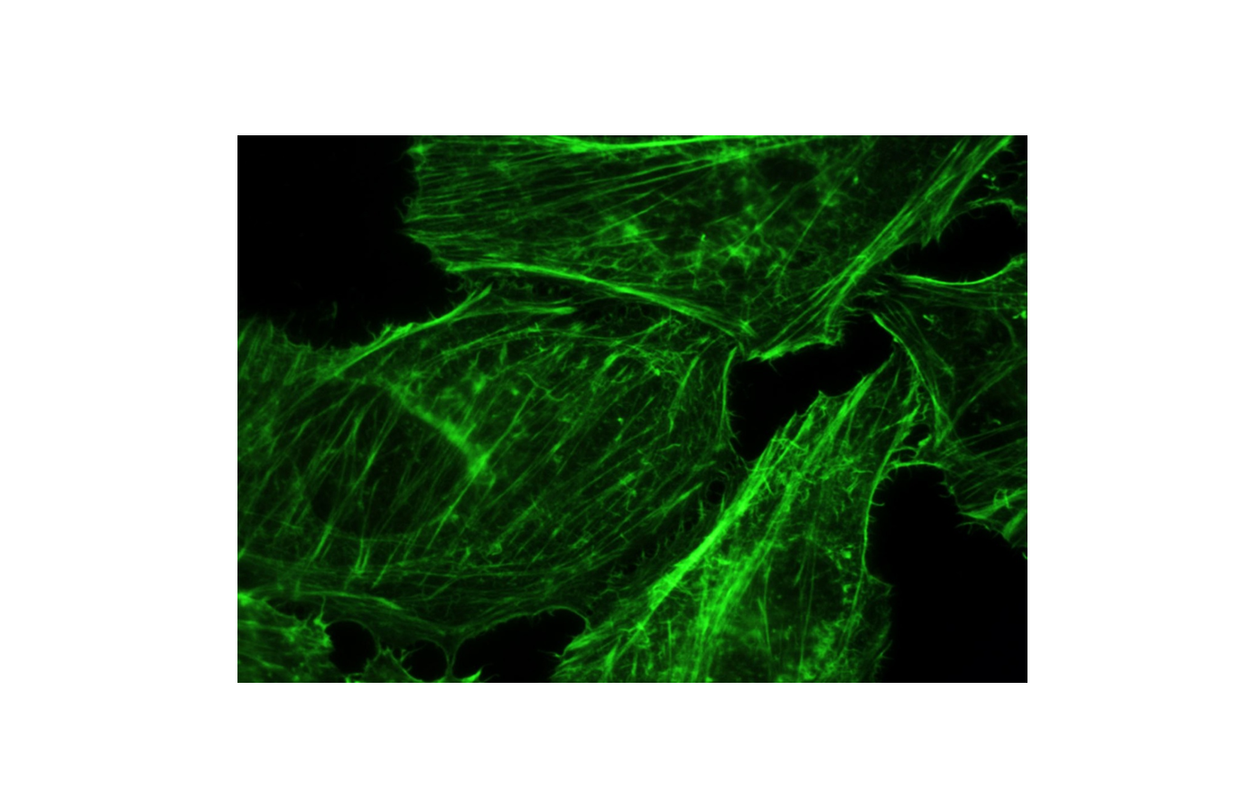
Immunocytochemistry/ Immunofluorescence: Actin Gamma 1 Antibody (NH3) [NB100-64792]
Immunocytochemistry/Immunofluorescence: Actin Gamma 1 Antibody (NH3) [NB100-64792] - The distribution of stress fiber was observed by F-actin staining. Image from verified customer review.![Immunohistochemistry-Paraffin: Actin Gamma 1 Antibody (NH3) [NB100-64792]](https://resources.rndsystems.com/images/products/Actin-Gamma-1-Antibody-NH3-Immunohistochemistry-Paraffin-NB100-64792-img0002.jpg "Immunohistochemistry-Paraffin: Actin Gamma 1 Antibody (NH3) [NB100-64792]")
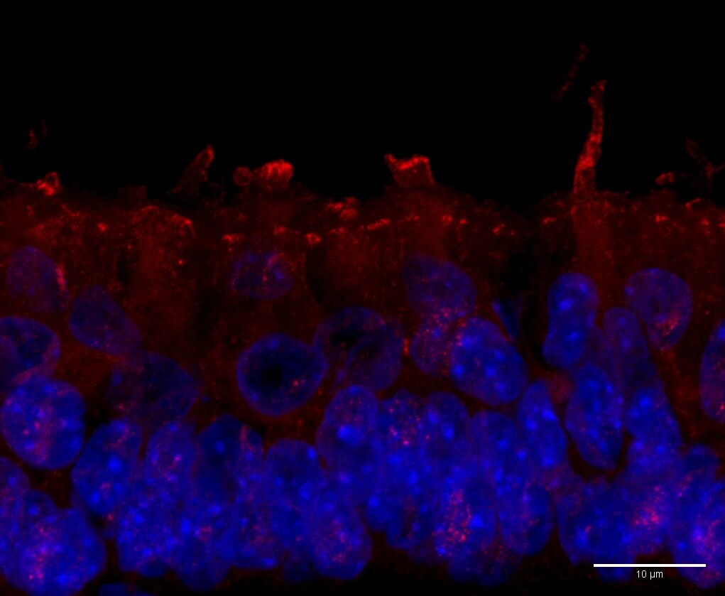
Immunohistochemistry-Paraffin: Actin Gamma 1 Antibody (NH3) [NB100-64792]
Immunohistochemistry-Paraffin: Actin Gamma 1 Antibody (NH3) [NB100-64792] - Human inner ear tissue section stained with Actin Gamma 1 antibody at 1:100 overnight at 4C and detected with Alexa Fluor 488 Donkey anti-mouse at 1:200. Nuclei stained with DAPI. IHC-P image submitted by a verified customer review. - BSA Free [NB100-64792] -")
Western Blot: Actin Gamma 1 Antibody (NH3) - BSA Free [NB100-64792] -
Effect of TSAIII on cell migration, invasion, and F-actin expression of human osteosarcoma cells. (A) Human 143-B and HOS osteosarcoma cells were treated with various concentrations of timosaponin AIII (TSAIII; 0, 2, 4, and 6 μM) for 24 h, and cell migration and invasion abilities were measured. (B) Cytoskeletal F-actin expression in human 143-B and HOS osteosarcoma cells exposed to TSAIII (0, 2, 4, 6 μM) was measured through immunoblotting. Glyceraldehyde-3-phosphate dehydrogenase (GAPDH) was used as the internal control. (C) Distribution of cytoskeletal F-actin in 143-B and HOS cells was further confirmed using immunofluorescence analysis. * p < 0.05; ** p < 0.01 versus control. Scale bar: 50 μm. Image collected and cropped by CiteAb from the following open publication (https://pubmed.ncbi.nlm.nih.gov/33799345), licensed under a CC-BY license. Not internally tested by Novus Biologicals. - BSA Free [NB100-64792] -")
Western Blot: Actin Gamma 1 Antibody (NH3) - BSA Free [NB100-64792] -
Synergistically inhibitory effect of TSAIII and PF on migration and invasion of human osteosarcoma cells. (A) Human 143-B and HOS osteosarcoma cells were treated with various concentrations of TSAIII (0 and 4 μM) and/or PF (0 and 2 μM) and then harvested to detect the expression and activation of cytoskeletal-related proteins through immunoblotting. Glyceraldehyde-3-phosphate dehydrogenase (GAPDH) was used as the internal control. (B) Using immunofluorescence analysis, the expression of cytoskeletal F-actin was observed in human 143-B and HOS osteosarcoma cells. (C) The migration and invasion abilities of human 143-B and HOS osteosarcoma cells were measured after treatment with TSAIII in the presence or absence of PF for 24 h. ** p < 0.01 versus control; # p < 0.05 versus treatment with PF alone (mean +/- standard error, n = 3). PF denotes PF-573228 (focal adhesion kinase inhibitor). Scale bar: 50 μm. Image collected and cropped by CiteAb from the following open publication (https://pubmed.ncbi.nlm.nih.gov/33799345), licensed under a CC-BY license. Not internally tested by Novus Biologicals. - BSA Free [NB100-64792] -")
Western Blot: Actin Gamma 1 Antibody (NH3) - BSA Free [NB100-64792] -
Synergistically inhibitory effect of TSAIII and Cyclo on migration and invasion of human osteosarcoma cells. (A) Human 143-B and HOS osteosarcoma cells were treated with various concentrations of TSAIII (0 and 4 μM) and/or Cyclo (0 and 50 μM) and then harvested to detect the expression and activation of cytoskeletal-related proteins through immunoblotting. Glyceraldehyde-3-phosphate dehydrogenase (GAPDH) was used as the internal control. (B) A change in the expression of cytoskeletal F-actin was observed in human 143-B and HOS osteosarcoma cells using immunofluorescence analysis. (C) The migration and invasion capacities of human 143-B and HOS osteosarcoma cells were measured after treatment with TSAIII in the presence or absence of Cyclo for 18 h (migration) or 24 h (invasion). ** p < 0.01 versus control; # p < 0.05 versus treatment with Cyclo alone. Cyclo denotes Cyclo(RGDyK) (Intergin inhibitor). Scale bar: 50 μm. Image collected and cropped by CiteAb from the following open publication (https://pubmed.ncbi.nlm.nih.gov/33799345), licensed under a CC-BY license. Not internally tested by Novus Biologicals. - BSA Free [NB100-64792] -")
Immunocytochemistry/ Immunofluorescence: Actin Gamma 1 Antibody (NH3) - BSA Free [NB100-64792] -
Effect of TSAIII on cell migration, invasion, and F-actin expression of human osteosarcoma cells. (A) Human 143-B and HOS osteosarcoma cells were treated with various concentrations of timosaponin AIII (TSAIII; 0, 2, 4, and 6 μM) for 24 h, and cell migration and invasion abilities were measured. (B) Cytoskeletal F-actin expression in human 143-B and HOS osteosarcoma cells exposed to TSAIII (0, 2, 4, 6 μM) was measured through immunoblotting. Glyceraldehyde-3-phosphate dehydrogenase (GAPDH) was used as the internal control. (C) Distribution of cytoskeletal F-actin in 143-B and HOS cells was further confirmed using immunofluorescence analysis. * p < 0.05; ** p < 0.01 versus control. Scale bar: 50 μm. Image collected and cropped by CiteAb from the following open publication (https://pubmed.ncbi.nlm.nih.gov/33799345), licensed under a CC-BY license. Not internally tested by Novus Biologicals. - BSA Free [NB100-64792] -")
Immunohistochemistry: Actin Gamma 1 Antibody (NH3) - BSA Free [NB100-64792] -
In vivo metastasis of TSAIII in human osteosarcoma cells. For the animal assay of lung metastasis, human osteosarcoma cells were harvested and injected into the tail veins of five-week-old immunodeficient mice (C.B17/IcrPrkdcscid/CrlNarl). The mice were then fed TSAIII (5 and 10 mg/kg) through oral gavage. After two months, the mice were euthanised and (A) the histopathology of the lungs in metastatic tumour-bearing animals was analysed. The lungs were fixed in neutral-buffered formalin and stained with haematoxylin and eosin. The F-actin expression were detected with immunohistochemistry assay. (B) The nodule numbers were then counted in the mice. ** p < 0.01 versus control (n = 5). Scale bar: 100 μm. Image collected and cropped by CiteAb from the following open publication (https://pubmed.ncbi.nlm.nih.gov/33799345), licensed under a CC-BY license. Not internally tested by Novus Biologicals.Applications for Actin Gamma 1 Antibody (NH3) - BSA Free
Application
Recommended Usage
ELISA
1:10
Flow Cytometry
1:10
Immunocytochemistry/ Immunofluorescence
1:10 - 1:500
Immunohistochemistry
1:10 - 1:500
Immunohistochemistry-Frozen
1:10 - 1:500
Western Blot
1:100 - 1:500
Application Notes
Actin Gamma 1 antibody validated for ICC/IF and IHC-P from verified customer reviews.
Reviewed Applications
Read 2 reviews rated 4 using NB100-64792 in the following applications:
Flow Cytometry Panel Builder
Bio-Techne Knows Flow Cytometry
Save time and reduce costly mistakes by quickly finding compatible reagents using the Panel Builder Tool.
Advanced Features
- Spectra Viewer - Custom analysis of spectra from multiple fluorochromes
- Spillover Popups - Visualize the spectra of individual fluorochromes
- Antigen Density Selector - Match fluorochrome brightness with antigen density

Formulation, Preparation, and Storage
Purification
IgM purified
Formulation
PBS
Format
BSA Free
Preservative
0.09% Sodium Azide
Concentration
1.0 mg/ml
Shipping
The product is shipped with polar packs. Upon receipt, store it immediately at the temperature recommended below.
Stability & Storage
Store at 4C short term. Aliquot and store at -20C long term. Avoid freeze-thaw cycles.
Background: Actin Gamma 1
Alternate Names
ACT, ACTB, ACTG, actin, cytoplasmic 2, actin, gamma 1, cytoskeletal gamma-actin, deafness, autosomal dominant 20; deafness, autosomal dominant 26, DFNA20, DFNA26, Gamma-actin
Entrez Gene IDs
71 (Human)
Gene Symbol
ACTG1
UniProt
Additional Actin Gamma 1 Products
Product Documents for Actin Gamma 1 Antibody (NH3) - BSA Free
Certificate of Analysis
To download a Certificate of Analysis, please enter a lot or batch number in the search box below.
Product Specific Notices for Actin Gamma 1 Antibody (NH3) - BSA Free
This product is for research use only and is not approved for use in humans or in clinical diagnosis. Primary Antibodies are guaranteed for 1 year from date of receipt.
Citations for Actin Gamma 1 Antibody (NH3) - BSA Free
 Powered by Bioz
Customer Reviews for Actin Gamma 1 Antibody (NH3) - BSA Free (2)
4 out of 5
2 Customer Ratings
Have you used Actin Gamma 1 Antibody (NH3) - BSA Free?
Submit a review and receive an Amazon gift card!
$25/€18/£15/$25CAN/¥2500 Yen for a review with an image
$10/€7/£6/$10CAN/¥1110 Yen for a review without an image
Submit a review
Customer Images
Showing
1
-
2 of
2 reviews
Showing All
Filter By:
-
Application: Immunohistochemistry-ParaffinSample Tested: inner earSpecies: HumanVerified Customer | Posted 10/30/2019Human inner ear tissue was incubated with anti-actin antibody at dilution 1:100 overnight at 4C and detected with Alexa Fluor 488 Donkey anti-mouse at dilution 1:200. Nuclei stained with DAPI.

-
Application: ImmunofluorescenceSample Tested:Species: HumanVerified Customer | Posted 07/05/2013The distribution of stress fiber was observed by F-actin staining (Green, Novus)
There are no reviews that match your criteria.
Protocols
Find general support by application which include: protocols, troubleshooting, illustrated assays, videos and webinars.
- 7-Amino Actinomycin D (7-AAD) Cell Viability Flow Cytometry Protocol
- Antigen Retrieval Protocol (PIER)
- Antigen Retrieval for Frozen Sections Protocol
- Appropriate Fixation of IHC/ICC Samples
- Cellular Response to Hypoxia Protocols
- Chromogenic IHC Staining of Formalin-Fixed Paraffin-Embedded (FFPE) Tissue Protocol
- Chromogenic Immunohistochemistry Staining of Frozen Tissue
- ClariTSA™ Fluorophore Kits
- Detection & Visualization of Antibody Binding
- ELISA Sample Preparation & Collection Guide
- ELISA Troubleshooting Guide
- Extracellular Membrane Flow Cytometry Protocol
- Flow Cytometry Protocol for Cell Surface Markers
- Flow Cytometry Protocol for Staining Membrane Associated Proteins
- Flow Cytometry Staining Protocols
- Flow Cytometry Troubleshooting Guide
- Fluorescent IHC Staining of Frozen Tissue Protocol
- Graphic Protocol for Heat-induced Epitope Retrieval
- Graphic Protocol for the Preparation and Fluorescent IHC Staining of Frozen Tissue Sections
- Graphic Protocol for the Preparation and Fluorescent IHC Staining of Paraffin-embedded Tissue Sections
- Graphic Protocol for the Preparation of Gelatin-coated Slides for Histological Tissue Sections
- How to Run an R&D Systems DuoSet ELISA
- How to Run an R&D Systems Quantikine ELISA
- How to Run an R&D Systems Quantikine™ QuicKit™ ELISA
- ICC Cell Smear Protocol for Suspension Cells
- ICC Immunocytochemistry Protocol Videos
- ICC for Adherent Cells
- IHC Sample Preparation (Frozen sections vs Paraffin)
- Immunocytochemistry (ICC) Protocol
- Immunocytochemistry Troubleshooting
- Immunofluorescence of Organoids Embedded in Cultrex Basement Membrane Extract
- Immunofluorescent IHC Staining of Formalin-Fixed Paraffin-Embedded (FFPE) Tissue Protocol
- Immunohistochemistry (IHC) and Immunocytochemistry (ICC) Protocols
- Immunohistochemistry Frozen Troubleshooting
- Immunohistochemistry Paraffin Troubleshooting
- Intracellular Flow Cytometry Protocol Using Alcohol (Methanol)
- Intracellular Flow Cytometry Protocol Using Detergents
- Intracellular Nuclear Staining Flow Cytometry Protocol Using Detergents
- Intracellular Staining Flow Cytometry Protocol Using Alcohol Permeabilization
- Intracellular Staining Flow Cytometry Protocol Using Detergents to Permeabilize Cells
- Preparing Samples for IHC/ICC Experiments
- Preventing Non-Specific Staining (Non-Specific Binding)
- Primary Antibody Selection & Optimization
- Propidium Iodide Cell Viability Flow Cytometry Protocol
- Protocol for Heat-Induced Epitope Retrieval (HIER)
- Protocol for Liperfluo
- Protocol for Making a 4% Formaldehyde Solution in PBS
- Protocol for VisUCyte™ HRP Polymer Detection Reagent
- Protocol for the Characterization of Human Th22 Cells
- Protocol for the Characterization of Human Th9 Cells
- Protocol for the Fluorescent ICC Staining of Cell Smears - Graphic
- Protocol for the Fluorescent ICC Staining of Cultured Cells on Coverslips - Graphic
- Protocol for the Preparation & Fixation of Cells on Coverslips
- Protocol for the Preparation and Chromogenic IHC Staining of Frozen Tissue Sections
- Protocol for the Preparation and Chromogenic IHC Staining of Frozen Tissue Sections - Graphic
- Protocol for the Preparation and Chromogenic IHC Staining of Paraffin-embedded Tissue Sections
- Protocol for the Preparation and Chromogenic IHC Staining of Paraffin-embedded Tissue Sections - Graphic
- Protocol for the Preparation and Fluorescent ICC Staining of Cells on Coverslips
- Protocol for the Preparation and Fluorescent ICC Staining of Non-adherent Cells
- Protocol for the Preparation and Fluorescent ICC Staining of Stem Cells on Coverslips
- Protocol for the Preparation and Fluorescent IHC Staining of Frozen Tissue Sections
- Protocol for the Preparation and Fluorescent IHC Staining of Paraffin-embedded Tissue Sections
- Protocol for the Preparation of Gelatin-coated Slides for Histological Tissue Sections
- Protocol for the Preparation of a Cell Smear for Non-adherent Cell ICC - Graphic
- Protocol: Annexin V and PI Staining by Flow Cytometry
- Protocol: Annexin V and PI Staining for Apoptosis by Flow Cytometry
- Quantikine HS ELISA Kit Assay Principle, Alkaline Phosphatase
- Quantikine HS ELISA Kit Principle, Streptavidin-HRP Polymer
- R&D Systems Quality Control Western Blot Protocol
- Sandwich ELISA (Colorimetric) – Biotin/Streptavidin Detection Protocol
- Sandwich ELISA (Colorimetric) – Direct Detection Protocol
- TUNEL and Active Caspase-3 Detection by IHC/ICC Protocol
- The Importance of IHC/ICC Controls
- Troubleshooting Guide: ELISA
- Troubleshooting Guide: Fluorokine Flow Cytometry Kits
- Troubleshooting Guide: Immunohistochemistry
- Troubleshooting Guide: Western Blot Figures
- Western Blot Conditions
- Western Blot Protocol
- Western Blot Protocol for Cell Lysates
- Western Blot Troubleshooting
- Western Blot Troubleshooting Guide
- View all Protocols, Troubleshooting, Illustrated assays and Webinars
Loading...