AIF-1/Iba1 Antibody (JM36-62)
Novus Biologicals | Catalog # NBP2-75397
Recombinant Monoclonal Antibody
![Western Blot: AIF-1/Iba1 Antibody (JM36-62) [NBP2-75397]](https://resources.rndsystems.com/images/products/AIF-1-Iba1-Antibody-JM36-62-Western-Blot-NBP2-75397-img0008.jpg "Western Blot: AIF-1/Iba1 Antibody (JM36-62) [NBP2-75397]")
Loading...
Key Product Details
Validated by
Biological Validation
Species Reactivity
Human, Mouse, Rat
Applications
Immunohistochemistry, Immunohistochemistry-Paraffin, Immunohistochemistry-Frozen, Western Blot, Flow Cytometry, Immunocytochemistry/ Immunofluorescence, Immunoprecipitation
Label
Unconjugated
Antibody Source
Recombinant Monoclonal Rabbit IgG Clone # JM36-62 expressed in HEK293
Loading...
Product Specifications
Immunogen
Synthetic peptide within N-terminal human AIF-1/Iba1. (SwissProt: P55008 Human; SwissProt: O70200 Mouse; SwissProt: P55009 Rat)
Localization
Cell membrane, Cell projection, Cytoplasm, Cytoskeleton, Membrane.
Clonality
Monoclonal
Host
Rabbit
Isotype
IgG
Theoretical MW
17 kDa.
Disclaimer note: The observed molecular weight of the protein may vary from the listed predicted molecular weight due to post translational modifications, post translation cleavages, relative charges, and other experimental factors.
Disclaimer note: The observed molecular weight of the protein may vary from the listed predicted molecular weight due to post translational modifications, post translation cleavages, relative charges, and other experimental factors.
Scientific Data Images for AIF-1/Iba1 Antibody (JM36-62)
Western Blot: AIF-1/Iba1 Antibody (JM36-62) [NBP2-75397]
Western Blot: AIF-1/Iba1 Antibody (JM36-62) [NBP2-75397] - Analysis of Iba1 on THP-1 cell lysates. Proteins were transferred to a PVDF membrane and blocked with 5% BSA in PBS for 1 hour at room temperature. The primary antibody (1:500) was used in 5% BSA at room temperature for 2 hours. Goat Anti-Rabbit IgG - HRP Secondary Antibody at 1:5000 dilution was used for 1 hour at room temperature.![Immunocytochemistry/ Immunofluorescence: AIF-1/Iba1 Antibody (JM36-62) [NBP2-75397]](https://resources.rndsystems.com/images/products/AIF-1-Iba1-Antibody-JM36-62-Immunocytochemistry-Immunofluorescence-NBP2-75397-img0002.jpg "Immunocytochemistry/ Immunofluorescence: AIF-1/Iba1 Antibody (JM36-62) [NBP2-75397]")
Immunocytochemistry/ Immunofluorescence: AIF-1/Iba1 Antibody (JM36-62) [NBP2-75397]
Immunocytochemistry/Immunofluorescence: AIF-1/Iba1 Antibody (JM36-62) [NBP2-75397] - Staining AIF-1/Iba1 in SH-SY5Y cells using (green). The nuclear counter stain is DAPI (blue). Cells were fixed in paraformaldehyde, permeabilised with 0.25% Triton X-100, PBS.![Immunohistochemistry-Paraffin: AIF-1/Iba1 Antibody (JM36-62) [NBP2-75397]](https://resources.rndsystems.com/images/products/AIF-1-Iba1-Antibody-JM36-62-Immunohistochemistry-Paraffin-NBP2-75397-img0015.jpg "Immunohistochemistry-Paraffin: AIF-1/Iba1 Antibody (JM36-62) [NBP2-75397]")
Immunohistochemistry-Paraffin: AIF-1/Iba1 Antibody (JM36-62) [NBP2-75397]
Immunohistochemistry-Paraffin: AIF-1/Iba1 Antibody (JM36-62) [NBP2-75397] - Immunofluorescence analysis of paraffin-embedded rat brain tissue labeling Iba1 with Rabbit anti-AIF-1/Iba1 antibody washed with PBS, and then probed with the primary antibody (green) at 1/100 dilution overnight at 4, washed with PBS. Goat Anti-Rabbit IgG H&L (iFluor(TM) 488) was used as the secondary antibody at 1/1,000 dilution. Nuclei were counterstained with DAPI (blue).![Flow Cytometry: AIF-1/Iba1 Antibody (JM36-62) [NBP2-75397]](https://resources.rndsystems.com/images/products/AIF-1-Iba1-Antibody-JM36-62-Flow-Cytometry-NBP2-75397-img0001.jpg "Flow Cytometry: AIF-1/Iba1 Antibody (JM36-62) [NBP2-75397]")
Flow Cytometry: AIF-1/Iba1 Antibody (JM36-62) [NBP2-75397]
Flow Cytometry: AIF-1/Iba1 Antibody (JM36-62) [NBP2-75397] - Analysis of THP-1 cells withAIF-1:Iba1 Antibody (JM36-62) at 1:100 dilution (red) compared with an unlabelled control (cells without incubation with primary antibody; black).![Immunohistochemistry-Paraffin: AIF-1/Iba1 Antibody (JM36-62) [NBP2-75397]](https://resources.rndsystems.com/images/products/AIF-1-Iba1-Antibody-JM36-62-Immunohistochemistry-Paraffin-NBP2-75397-img0003.jpg "Immunohistochemistry-Paraffin: AIF-1/Iba1 Antibody (JM36-62) [NBP2-75397]")
Immunohistochemistry-Paraffin: AIF-1/Iba1 Antibody (JM36-62) [NBP2-75397]
Immunohistochemistry-Paraffin: AIF-1/Iba1 Antibody (JM36-62) [NBP2-75397] - Analysis of paraffin-embedded human lung cancer tissue using AIF-1/Iba1 Antibody (JM36-62). Counterstained with hematoxylin.![Immunohistochemistry-Paraffin: AIF-1/Iba1 Antibody (JM36-62) [NBP2-75397]](https://resources.rndsystems.com/images/products/AIF-1-Iba1-Antibody-JM36-62-Immunohistochemistry-Paraffin-NBP2-75397-img0004.jpg "Immunohistochemistry-Paraffin: AIF-1/Iba1 Antibody (JM36-62) [NBP2-75397]")
Immunohistochemistry-Paraffin: AIF-1/Iba1 Antibody (JM36-62) [NBP2-75397]
Immunohistochemistry-Paraffin: AIF-1/Iba1 Antibody (JM36-62) [NBP2-75397] - Analysis of paraffin-embedded human spleen tissue using AIF-1/Iba1 Antibody (JM36-62). Counterstained with hematoxylin.![Immunohistochemistry-Paraffin: AIF-1/Iba1 Antibody (JM36-62) [NBP2-75397]](https://resources.rndsystems.com/images/products/AIF-1-Iba1-Antibody-JM36-62-Immunohistochemistry-Paraffin-NBP2-75397-img0005.jpg "Immunohistochemistry-Paraffin: AIF-1/Iba1 Antibody (JM36-62) [NBP2-75397]")
Immunohistochemistry-Paraffin: AIF-1/Iba1 Antibody (JM36-62) [NBP2-75397]
Immunohistochemistry-Paraffin: AIF-1/Iba1 Antibody (JM36-62) [NBP2-75397] - Analysis of paraffin-embedded mouse brain tissue using AIF-1/Iba1 Antibody (JM36-62). Counterstained with hematoxylin.![Immunohistochemistry-Paraffin: AIF-1/Iba1 Antibody (JM36-62) [NBP2-75397]](https://resources.rndsystems.com/images/products/AIF-1-Iba1-Antibody-JM36-62-Immunohistochemistry-Paraffin-NBP2-75397-img0006.jpg "Immunohistochemistry-Paraffin: AIF-1/Iba1 Antibody (JM36-62) [NBP2-75397]")
Immunohistochemistry-Paraffin: AIF-1/Iba1 Antibody (JM36-62) [NBP2-75397]
Immunohistochemistry-Paraffin: AIF-1/Iba1 Antibody (JM36-62) [NBP2-75397] - Analysis of paraffin-embedded mouse spleen tissue using AIF-1/Iba1 Antibody (JM36-62). Counterstained with hematoxylin.![Immunohistochemistry-Paraffin: AIF-1/Iba1 Antibody (JM36-62) [NBP2-75397]](https://resources.rndsystems.com/images/products/AIF-1-Iba1-Antibody-JM36-62-Immunohistochemistry-Paraffin-NBP2-75397-img0007.jpg "Immunohistochemistry-Paraffin: AIF-1/Iba1 Antibody (JM36-62) [NBP2-75397]")
Immunohistochemistry-Paraffin: AIF-1/Iba1 Antibody (JM36-62) [NBP2-75397]
Immunohistochemistry-Paraffin: AIF-1/Iba1 Antibody (JM36-62) [NBP2-75397] - Analysis of paraffin-embedded rat lung tissue using AIF-1/Iba1 Antibody (JM36-62). Counterstained with hematoxylin.![Immunohistochemistry-Paraffin: AIF-1/Iba1 Antibody (JM36-62) [NBP2-75397]](https://resources.rndsystems.com/images/products/AIF-1-Iba1-Antibody-JM36-62-Immunohistochemistry-Paraffin-NBP2-75397-img0009.jpg "Immunohistochemistry-Paraffin: AIF-1/Iba1 Antibody (JM36-62) [NBP2-75397]")
Immunohistochemistry-Paraffin: AIF-1/Iba1 Antibody (JM36-62) [NBP2-75397]
Immunohistochemistry-Paraffin: AIF-1/Iba1 Antibody (JM36-62) [NBP2-75397] - Immunofluorescence staining of paraffin- embedded human spleen tissue using anti-AIF-1/Iba1rabbit polyclonal antibody. The section was pre-treated using heat mediated antigen retrieval with Tris-EDTA buffer (pH 9.0) for 20 minutes.(sodium citrate buffer (pH6) for 20 mins.) The tissues were blocked in 10% negative goat serum for 1 hour at room temperature, washed with PBS, and then probed with at 1/50 dilution for 10 hours at 4C and detected using Alexa Fluor(R) 488 conjugate-Goat anti-Rabbit IgG (H+L) Secondary Antibody at a dilution of 1:500 for 1 hour at room temperature.![Immunohistochemistry-Paraffin: AIF-1/Iba1 Antibody (JM36-62) [NBP2-75397]](https://resources.rndsystems.com/images/products/AIF-1-Iba1-Antibody-JM36-62-Immunohistochemistry-Paraffin-NBP2-75397-img0010.jpg "Immunohistochemistry-Paraffin: AIF-1/Iba1 Antibody (JM36-62) [NBP2-75397]")
Immunohistochemistry-Paraffin: AIF-1/Iba1 Antibody (JM36-62) [NBP2-75397]
Immunohistochemistry-Paraffin: AIF-1/Iba1 Antibody (JM36-62) [NBP2-75397] - Immunofluorescence staining of paraffin- embedded mouse brain tissue using anti-AIF-1/Iba1 antibody.The section was pre-treated using heat mediated antigen retrieval with Tris-EDTA buffer (pH 9.0) for 20 minutes. The tissues were blocked in 10% negative goat serum for 1 hour at room temperature, washed with PBS, and then probed with at 1/50 dilution for 10 hours at 4 and detected using Alexa Fluor(R) 488 conjugate-Goat anti-Rabbit IgG (H+L) Secondary Antibody at a dilution of 1:500 for 1 hour at room temperature.![Immunohistochemistry-Paraffin: AIF-1/Iba1 Antibody (JM36-62) [NBP2-75397]](https://resources.rndsystems.com/images/products/AIF-1-Iba1-Antibody-JM36-62-Immunohistochemistry-Paraffin-NBP2-75397-img0011.jpg "Immunohistochemistry-Paraffin: AIF-1/Iba1 Antibody (JM36-62) [NBP2-75397]")
Immunohistochemistry-Paraffin: AIF-1/Iba1 Antibody (JM36-62) [NBP2-75397]
Immunohistochemistry-Paraffin: AIF-1/Iba1 Antibody (JM36-62) [NBP2-75397] - Analysis of paraffin-embedded mouse hippocampus tissue with Rabbit anti-AIF-1/Iba1 antibody. The section was pre-treated using heat mediated antigen retrieval with Tris-EDTA buffer (pH 9.0) for 20 minutes. The tissues were blocked in 1% BSA for 20 minutes at room temperature, washed with ddH2O and PBS, and then probed with the primary antibody for 1 hour at room temperature. The detection was performed using an HRP conjugated compact polymer system. DAB was used as the chromogen. Tissues were counterstained with hematoxylin and mounted with DPX.![Immunohistochemistry-Paraffin: AIF-1/Iba1 Antibody (JM36-62) [NBP2-75397]](https://resources.rndsystems.com/images/products/AIF-1-Iba1-Antibody-JM36-62-Immunohistochemistry-Paraffin-NBP2-75397-img0012.jpg "Immunohistochemistry-Paraffin: AIF-1/Iba1 Antibody (JM36-62) [NBP2-75397]")
Immunohistochemistry-Paraffin: AIF-1/Iba1 Antibody (JM36-62) [NBP2-75397]
Immunohistochemistry-Paraffin: AIF-1/Iba1 Antibody (JM36-62) [NBP2-75397] - Analysis of paraffin-embedded human kidney tissue with Rabbit anti-AIF-1/Iba1 antibody. The section was pre-treated using heat mediated antigen retrieval with Tris-EDTA buffer (pH 9.0) for 20 minutes. The tissues were blocked in 1% BSA for 20 minutes at room temperature, washed with ddH2O and PBS, and then probed with the primary antibody for 1 hour at room temperature. The detection was performed using an HRP conjugated compact polymer system. DAB was used as the chromogen. Tissues were counterstained with hematoxylin and mounted with DPX.![Immunohistochemistry-Paraffin: AIF-1/Iba1 Antibody (JM36-62) [NBP2-75397]](https://resources.rndsystems.com/images/products/AIF-1-Iba1-Antibody-JM36-62-Immunohistochemistry-Paraffin-NBP2-75397-img0013.jpg "Immunohistochemistry-Paraffin: AIF-1/Iba1 Antibody (JM36-62) [NBP2-75397]")
Immunohistochemistry-Paraffin: AIF-1/Iba1 Antibody (JM36-62) [NBP2-75397]
Immunohistochemistry-Paraffin: AIF-1/Iba1 Antibody (JM36-62) [NBP2-75397] - Analysis of paraffin-embedded human spleen tissue with Rabbit anti-AIF-1/Iba1 antibody. The section was pre-treated using heat mediated antigen retrieval with Tris-EDTA buffer (pH 9.0) for 20 minutes. The tissues were blocked in 1% BSA for 20 minutes at room temperature, washed with ddH2O and PBS, and then probed with the primary antibody for 1 hour at room temperature. The detection was performed using an HRP conjugated compact polymer system. DAB was used as the chromogen. Tissues were counterstained with hematoxylin and mounted with DPX.![Immunohistochemistry-Paraffin: AIF-1/Iba1 Antibody (JM36-62) [NBP2-75397]](https://resources.rndsystems.com/images/products/AIF-1-Iba1-Antibody-JM36-62-Immunohistochemistry-Paraffin-NBP2-75397-img0014.jpg "Immunohistochemistry-Paraffin: AIF-1/Iba1 Antibody (JM36-62) [NBP2-75397]")
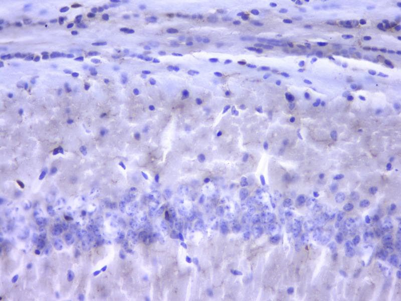
Immunohistochemistry-Paraffin: AIF-1/Iba1 Antibody (JM36-62) [NBP2-75397]
Immunohistochemistry-Paraffin: AIF-1/Iba1 Antibody (JM36-62) [NBP2-75397] - Analysis of paraffin-embedded mouse brain tissue with Rabbit anti-Iba1 antibody. The section was pre-treated using heat mediated antigen retrieval with Tris-EDTA buffer (pH 9.0) for 20 minutes. The tissues were blocked in 1% BSA for 20 minutes at room temperature, washed with ddH2O and PBS, and then probed with the primary antibody for 1 hour at room temperature. The detection was performed using an HRP conjugated compact polymer system. DAB was used as the chromogen. Tissues were counterstained with hematoxylin and mounted with DPX.Applications for AIF-1/Iba1 Antibody (JM36-62)
Application
Recommended Usage
Flow Cytometry
1:50-1:200
Immunocytochemistry/ Immunofluorescence
1:50-1:100
Immunohistochemistry
1:10 - 1:500
Immunohistochemistry-Frozen
1:50
Immunohistochemistry-Paraffin
1:100-1:500
Immunoprecipitation
1:10 - 1:500
Western Blot
1:500-1:1000
Application Notes
IP: Use at an assay dependent concentration.
Reviewed Applications
Read 1 review rated 1 using NBP2-75397 in the following applications:
Flow Cytometry Panel Builder
Bio-Techne Knows Flow Cytometry
Save time and reduce costly mistakes by quickly finding compatible reagents using the Panel Builder Tool.
Advanced Features
- Spectra Viewer - Custom analysis of spectra from multiple fluorochromes
- Spillover Popups - Visualize the spectra of individual fluorochromes
- Antigen Density Selector - Match fluorochrome brightness with antigen density

Formulation, Preparation, and Storage
Purification
Protein A purified
Formulation
TBS (pH7.4), 0.05% BSA, 40% Glycerol
Preservative
0.05% Sodium Azide
Concentration
1 mg/ml
Shipping
The product is shipped with polar packs. Upon receipt, store it immediately at the temperature recommended below.
Stability & Storage
Store at 4C short term. Aliquot and store at -20C long term. Avoid freeze-thaw cycles.
Background: AIF-1/Iba1
Several cellular functions have been associated with AIF-1/Iba1 expression including cell growth, cell migration, actin bundling, membrane ruffling, and phagocytic activity (2, 4). Iba1 induces Rac signaling through a PLC-gamma dependent pathway (1). Rac, a member of the Rho family of small GTPases, localizes with Iba1 and F-actin in membrane ruffles and phagocytic cups and plays a role in microglia activation. AIF-1/Iba1 induction in macrophages and microglia occur in association with immunological inflammatory processes in various disease states including endometriosis, cerebral infarction and rheumatoid arthritis (5). Immunodetection of Iba1 through flow cytometry, immunohistochemical or immunocytochemical applications is commonly used for identification and analysis of microglia.
References
1. Imai, Y., & Kohsaka, S. (2002). Intracellular signaling in M-CSF-induced microglia activation: Role of Iba1. GLIA. https://doi.org/10.1002/glia.10149
2. Deininger, M. H., Meyermann, R., & Schluesener, H. J. (2002). The allograft inflammatory factor-1 family of proteins. FEBS Letters. https://doi.org/10.1016/S0014-5793(02)02430-4
3. Utans, U., Quist, W. C., Mcmanus, B. M., Wilson, J. E., Arceci, R. J., Wallace, A. F., & Russell, M. E. (1996). Allograft inflammatory factor-1: A cytokine-responsive macrophage molecule expressed in transplanted human hearts. Transplantation. https://doi.org/10.1097/00007890-199605150-00018
4. Franco-Bocanegra, McAuley, Nicoll, & Boche. (2019). Molecular Mechanisms of Microglial Motility: Changes in Ageing and Alzheimer's Disease. Cells. https://doi.org/10.3390/cells8060639
5. Kimura, M., Kawahito, Y., Obayashi, H., Ohta, M., Hara, H., Adachi, T.,... Yoshikawa, T. (2007). A Critical Role for Allograft Inflammatory Factor-1 in the Pathogenesis of Rheumatoid Arthritis. The Journal of Immunology. https://doi.org/10.4049/jimmunol.178.5.3316
Long Name
Allograft Inflammatory Factor 1
Alternate Names
AIF1, IBA1, IRT1
Gene Symbol
AIF1
Additional AIF-1/Iba1 Products
Product Documents for AIF-1/Iba1 Antibody (JM36-62)
Certificate of Analysis
To download a Certificate of Analysis, please enter a lot or batch number in the search box below.
Product Specific Notices for AIF-1/Iba1 Antibody (JM36-62)
This product is for research use only and is not approved for use in humans or in clinical diagnosis. Primary Antibodies are guaranteed for 1 year from date of receipt.
Related Research Areas
Citations for AIF-1/Iba1 Antibody (JM36-62)
 Powered by Bioz
Customer Reviews for AIF-1/Iba1 Antibody (JM36-62) (1)
1 out of 5
1 Customer Rating
Have you used AIF-1/Iba1 Antibody (JM36-62)?
Submit a review and receive an Amazon gift card!
$25/€18/£15/$25CAN/¥2500 Yen for a review with an image
$10/€7/£6/$10CAN/¥1110 Yen for a review without an image
Submit a review
Customer Images
Showing
1
-
1 of
1 review
Showing All
Filter By:
-
Application: Immunohistochemistry-FrozenSample Tested: Rat hippocampusSpecies: RatVerified Customer | Posted 01/06/2021No microglial cell was observed.
 Bio-Techne ResponseThis review was submitted through the legacy Novus Innovators Program, reflecting a new species or application tested on a primary antibody.
There are no reviews that match your criteria.
Protocols
Find general support by application which include: protocols, troubleshooting, illustrated assays, videos and webinars.
- 7-Amino Actinomycin D (7-AAD) Cell Viability Flow Cytometry Protocol
- Antigen Retrieval Protocol (PIER)
- Antigen Retrieval for Frozen Sections Protocol
- Appropriate Fixation of IHC/ICC Samples
- Cellular Response to Hypoxia Protocols
- Chromogenic IHC Staining of Formalin-Fixed Paraffin-Embedded (FFPE) Tissue Protocol
- Chromogenic Immunohistochemistry Staining of Frozen Tissue
- ClariTSA™ Fluorophore Kits
- Detection & Visualization of Antibody Binding
- Extracellular Membrane Flow Cytometry Protocol
- Flow Cytometry Protocol for Cell Surface Markers
- Flow Cytometry Protocol for Staining Membrane Associated Proteins
- Flow Cytometry Staining Protocols
- Flow Cytometry Troubleshooting Guide
- Fluorescent IHC Staining of Frozen Tissue Protocol
- Graphic Protocol for Heat-induced Epitope Retrieval
- Graphic Protocol for the Preparation and Fluorescent IHC Staining of Frozen Tissue Sections
- Graphic Protocol for the Preparation and Fluorescent IHC Staining of Paraffin-embedded Tissue Sections
- Graphic Protocol for the Preparation of Gelatin-coated Slides for Histological Tissue Sections
- ICC Cell Smear Protocol for Suspension Cells
- ICC Immunocytochemistry Protocol Videos
- ICC for Adherent Cells
- IHC Sample Preparation (Frozen sections vs Paraffin)
- Immunocytochemistry (ICC) Protocol
- Immunocytochemistry Troubleshooting
- Immunofluorescence of Organoids Embedded in Cultrex Basement Membrane Extract
- Immunofluorescent IHC Staining of Formalin-Fixed Paraffin-Embedded (FFPE) Tissue Protocol
- Immunohistochemistry (IHC) and Immunocytochemistry (ICC) Protocols
- Immunohistochemistry Frozen Troubleshooting
- Immunohistochemistry Paraffin Troubleshooting
- Immunoprecipitation Protocol
- Intracellular Flow Cytometry Protocol Using Alcohol (Methanol)
- Intracellular Flow Cytometry Protocol Using Detergents
- Intracellular Nuclear Staining Flow Cytometry Protocol Using Detergents
- Intracellular Staining Flow Cytometry Protocol Using Alcohol Permeabilization
- Intracellular Staining Flow Cytometry Protocol Using Detergents to Permeabilize Cells
- Preparing Samples for IHC/ICC Experiments
- Preventing Non-Specific Staining (Non-Specific Binding)
- Primary Antibody Selection & Optimization
- Propidium Iodide Cell Viability Flow Cytometry Protocol
- Protocol for Heat-Induced Epitope Retrieval (HIER)
- Protocol for Liperfluo
- Protocol for Making a 4% Formaldehyde Solution in PBS
- Protocol for VisUCyte™ HRP Polymer Detection Reagent
- Protocol for the Characterization of Human Th22 Cells
- Protocol for the Characterization of Human Th9 Cells
- Protocol for the Fluorescent ICC Staining of Cell Smears - Graphic
- Protocol for the Fluorescent ICC Staining of Cultured Cells on Coverslips - Graphic
- Protocol for the Preparation & Fixation of Cells on Coverslips
- Protocol for the Preparation and Chromogenic IHC Staining of Frozen Tissue Sections
- Protocol for the Preparation and Chromogenic IHC Staining of Frozen Tissue Sections - Graphic
- Protocol for the Preparation and Chromogenic IHC Staining of Paraffin-embedded Tissue Sections
- Protocol for the Preparation and Chromogenic IHC Staining of Paraffin-embedded Tissue Sections - Graphic
- Protocol for the Preparation and Fluorescent ICC Staining of Cells on Coverslips
- Protocol for the Preparation and Fluorescent ICC Staining of Non-adherent Cells
- Protocol for the Preparation and Fluorescent ICC Staining of Stem Cells on Coverslips
- Protocol for the Preparation and Fluorescent IHC Staining of Frozen Tissue Sections
- Protocol for the Preparation and Fluorescent IHC Staining of Paraffin-embedded Tissue Sections
- Protocol for the Preparation of Gelatin-coated Slides for Histological Tissue Sections
- Protocol for the Preparation of a Cell Smear for Non-adherent Cell ICC - Graphic
- Protocol: Annexin V and PI Staining by Flow Cytometry
- Protocol: Annexin V and PI Staining for Apoptosis by Flow Cytometry
- R&D Systems Quality Control Western Blot Protocol
- TUNEL and Active Caspase-3 Detection by IHC/ICC Protocol
- The Importance of IHC/ICC Controls
- Troubleshooting Guide: Fluorokine Flow Cytometry Kits
- Troubleshooting Guide: Immunohistochemistry
- Troubleshooting Guide: Western Blot Figures
- Western Blot Conditions
- Western Blot Protocol
- Western Blot Protocol for Cell Lysates
- Western Blot Troubleshooting
- Western Blot Troubleshooting Guide
- View all Protocols, Troubleshooting, Illustrated assays and Webinars
Loading...