ALDH1A3 Antibody - BSA Free
Novus Biologicals | Catalog # NBP1-91657
![Immunohistochemistry-Paraffin: ALDH1A3 Antibody [NBP1-91657]](https://resources.rndsystems.com/images/products/ALDH1A3-Antibody-Immunohistochemistry-Paraffin-NBP1-91657-img0014.jpg "Immunohistochemistry-Paraffin: ALDH1A3 Antibody [NBP1-91657]")
Loading...
Key Product Details
Validated by
Orthogonal Validation
Species Reactivity
Validated:
Human
Cited:
Human, Mouse
Applications
Validated:
Immunohistochemistry, Immunohistochemistry-Paraffin, Immunohistochemistry-Frozen, Western Blot, Immunocytochemistry/ Immunofluorescence
Cited:
Western Blot
Label
Unconjugated
Antibody Source
Polyclonal Rabbit IgG
Format
BSA Free
Loading...
Product Specifications
Immunogen
This antibody was developed against Recombinant Protein corresponding to amino acids: ERGGAMATANGAVENGQPDRKPPALPRPIRNLEVKFTKIFIN
Reactivity Notes
Rat (83%). Mouse reactivity reported in scientific literature (PMID: 26511661).
Clonality
Polyclonal
Host
Rabbit
Isotype
IgG
Theoretical MW
56 kDa.
Disclaimer note: The observed molecular weight of the protein may vary from the listed predicted molecular weight due to post translational modifications, post translation cleavages, relative charges, and other experimental factors.
Disclaimer note: The observed molecular weight of the protein may vary from the listed predicted molecular weight due to post translational modifications, post translation cleavages, relative charges, and other experimental factors.
Scientific Data Images for ALDH1A3 Antibody - BSA Free
![Western Blot: ALDH1A3 Antibody [NBP1-91657]](https://resources.rndsystems.com/images/products/ALDH1A3-Antibody-Western-Blot-NBP1-91657-img0016.jpg "Western Blot: ALDH1A3 Antibody [NBP1-91657]")
![Immunocytochemistry/ Immunofluorescence: ALDH1A3 Antibody [NBP1-91657]](https://resources.rndsystems.com/images/products/ALDH1A3-Antibody-Immunocytochemistry-Immunofluorescence-NBP1-91657-img0006.jpg "Immunocytochemistry/ Immunofluorescence: ALDH1A3 Antibody [NBP1-91657]")
Immunocytochemistry/ Immunofluorescence: ALDH1A3 Antibody [NBP1-91657]
Immunocytochemistry/Immunofluorescence: ALDH1A3 Antibody [NBP1-91657] - Staining of human cell line A-431 shows positivity in plasma membrane and cytoplasm. Antibody staining is shown in green.![Immunohistochemistry-Paraffin: ALDH1A3 Antibody [NBP1-91657]](https://resources.rndsystems.com/images/products/ALDH1A3-Antibody-Immunohistochemistry-NBP1-91657-img0022.jpg "Immunohistochemistry-Paraffin: ALDH1A3 Antibody [NBP1-91657]")
Immunohistochemistry-Paraffin: ALDH1A3 Antibody [NBP1-91657]
Immunohistochemistry-Paraffin: ALDH1A3 Antibody [NBP1-91657] - Staining of human skeletal muscle shows no positivity in myocytes.![Immunohistochemistry-Paraffin: ALDH1A3 Antibody [NBP1-91657]](https://resources.rndsystems.com/images/products/ALDH1A3-Antibody-Immunohistochemistry-Paraffin-NBP1-91657-img0012.jpg "Immunohistochemistry-Paraffin: ALDH1A3 Antibody [NBP1-91657]")
Immunohistochemistry-Paraffin: ALDH1A3 Antibody [NBP1-91657]
Immunohistochemistry-Paraffin: ALDH1A3 Antibody [NBP1-91657] - Staining of human prostate shows high expression.![Immunohistochemistry-Frozen: ALDH1A3 Antibody [NBP1-91657]](https://resources.rndsystems.com/images/products/ALDH1A3-Antibody-Immunohistochemistry-Frozen-NBP1-91657-img0017.jpg "Immunohistochemistry-Frozen: ALDH1A3 Antibody [NBP1-91657]")
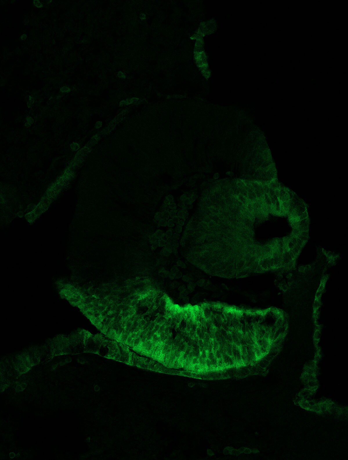
Immunohistochemistry-Frozen: ALDH1A3 Antibody [NBP1-91657]
Immunohistochemistry-Frozen: ALDH1A3 Antibody [NBP1-91657] - Aldh1a3 expression on E11.5 mouse embryo eye. Heat induced antigen retrieval at pH 6.0 was done for 20 minutes with mouse E11.5 coronal cryosection. Image submitted by a verified customer review.![Immunohistochemistry-Paraffin: ALDH1A3 Antibody [NBP1-91657]](https://resources.rndsystems.com/images/products/ALDH1A3-Antibody-Immunohistochemistry-Paraffin-NBP1-91657-img0019.jpg "Immunohistochemistry-Paraffin: ALDH1A3 Antibody [NBP1-91657]")
Immunohistochemistry-Paraffin: ALDH1A3 Antibody [NBP1-91657]
Immunohistochemistry-Paraffin: ALDH1A3 Antibody [NBP1-91657] - Staining of human colon shows moderate cytoplasmic positivity in glandular cells.![Immunohistochemistry-Paraffin: ALDH1A3 Antibody [NBP1-91657]](https://resources.rndsystems.com/images/products/ALDH1A3-Antibody-Immunohistochemistry-Paraffin-NBP1-91657-img0020.jpg "Immunohistochemistry-Paraffin: ALDH1A3 Antibody [NBP1-91657]")
Immunohistochemistry-Paraffin: ALDH1A3 Antibody [NBP1-91657]
Immunohistochemistry-Paraffin: ALDH1A3 Antibody [NBP1-91657] - Staining of human testis shows moderate cytoplasmic positivity in cells in seminiferous ducts.
Immunocytochemistry/ Immunofluorescence: ALDH1A3 Antibody [NBP1-91657] -
Immunocytochemistry/ Immunofluorescence: ALDH1A3 Antibody [NBP1-91657] - Cell proliferation during the oestrus cycle.(a) Protocol: adult (≥10-week-old) C57Bl6/J mice were administered CldU during pro-oestrus/early oestrus (blue arrows) & then IdU at metoestrus (red arrows) to detect sequential proliferation within the mammary gland. Immunofluorescent staining of adult mammary glands stained with CldU & IdU (b), & with ER, Aldh1a3 or K5 (c). Arrows indicate CldU+IdU+ cells, which demonstrate sequential cell division among mammary epithelial cells during the oestrus cycle. Representative image from four independent samples. Scale bars, 20 μm. (d) Representative flow cytometry dot plot showing the incorporation of EdU by NCL cells following injection of either oestrogen & progesterone or oil. A total of six independent adult (≥10-week-old) mice were analysed (four injected with oestrogen & progesterone, two injected with oil only). (e) NCL cells from adult (≥10-week-old) mice that are either in G0/G1 versus S/G2/M stages of the cell cycle were analysed by quantitative RT–PCR for ER alpha mRNA expression relative to Gapdh mRNA. Expression is relative to that of NCL G0/G1 cells. Mean±s.e.m of five independent mice. Image collected & cropped by CiteAb from the following publication (https://pubmed.ncbi.nlm.nih.gov/26511661), licensed under a CC-BY license. Not internally tested by Novus Biologicals.![ALDH1A3 Antibody - BSA Free Immunohistochemistry: ALDH1A3 Antibody - BSA Free [NBP1-91657]](https://resources.rndsystems.com/images/products/nbp1-91657_rabbit-polyclonal-aldh1a3-antibody-295202516464811.jpg "Immunohistochemistry: ALDH1A3 Antibody - BSA Free [NBP1-91657]")
Immunohistochemistry: ALDH1A3 Antibody - BSA Free [NBP1-91657]
Staining of human colon shows moderate cytoplasmic positivity in glandular cells.![ALDH1A3 Antibody - BSA Free Immunohistochemistry: ALDH1A3 Antibody - BSA Free [NBP1-91657]](https://resources.rndsystems.com/images/products/nbp1-91657_rabbit-polyclonal-aldh1a3-antibody-295202516495914.jpg "Immunohistochemistry: ALDH1A3 Antibody - BSA Free [NBP1-91657]")
Immunohistochemistry: ALDH1A3 Antibody - BSA Free [NBP1-91657]
Staining of human testis shows moderate cytoplasmic positivity in cells in seminiferous ducts.![ALDH1A3 Antibody - BSA Free Immunohistochemistry: ALDH1A3 Antibody - BSA Free [NBP1-91657]](https://resources.rndsystems.com/images/products/nbp1-91657_rabbit-polyclonal-aldh1a3-antibody-30520257552918.jpg "Immunohistochemistry: ALDH1A3 Antibody - BSA Free [NBP1-91657]")
![ALDH1A3 Antibody - BSA Free Immunocytochemistry/ Immunofluorescence: ALDH1A3 Antibody - BSA Free [NBP1-91657]](https://resources.rndsystems.com/images/products/nbp1-91657_rabbit-polyclonal-aldh1a3-antibody-29520251647363.jpg "Immunocytochemistry/ Immunofluorescence: ALDH1A3 Antibody - BSA Free [NBP1-91657]")
Immunocytochemistry/ Immunofluorescence: ALDH1A3 Antibody - BSA Free [NBP1-91657]
Staining of human cell line A-431 shows positivity in plasma membrane & cytoplasm.![ALDH1A3 Antibody - BSA Free Immunohistochemistry: ALDH1A3 Antibody - BSA Free [NBP1-91657]](https://resources.rndsystems.com/images/products/nbp1-91657_rabbit-polyclonal-aldh1a3-antibody-29520251613467.jpg "Immunohistochemistry: ALDH1A3 Antibody - BSA Free [NBP1-91657]")
Immunohistochemistry: ALDH1A3 Antibody - BSA Free [NBP1-91657]
Staining of human prostate shows strong cytoplasmic positivity in glandular cells.Applications for ALDH1A3 Antibody - BSA Free
Application
Recommended Usage
Immunocytochemistry/ Immunofluorescence
0.25-2 ug/ml
Immunohistochemistry
1:20 - 1:50
Immunohistochemistry-Frozen
Validated from a verified customer review
Immunohistochemistry-Paraffin
1:20-1:50
Western Blot
0.04-0.4 ug/ml
Application Notes
For IHC-Paraffin, HIER pH 6 retrieval method is recommended. ICC/IF Fixation Permeabilization, Use PFA/Triton X-100.
Reviewed Applications
Read 1 review rated 5 using NBP1-91657 in the following applications:
Formulation, Preparation, and Storage
Purification
Affinity purified
Formulation
PBS (pH 7.2) and 40% Glycerol
Format
BSA Free
Preservative
0.02% Sodium Azide
Concentration
Concentrations vary lot to lot. See vial label for concentration. If unlisted please contact technical services.
Shipping
The product is shipped with polar packs. Upon receipt, store it immediately at the temperature recommended below.
Stability & Storage
Store at 4C short term. Aliquot and store at -20C long term. Avoid freeze-thaw cycles.
Background: Aldehyde Dehydrogenase 1-A3/ALDH1A3
Long Name
Aldehyde Dehydrogenase 1 Family Member A3
Alternate Names
ALDH1A3, ALDH6, MCOP8, RALDH3
Entrez Gene IDs
220 (Human)
Gene Symbol
ALDH1A3
UniProt
Additional Aldehyde Dehydrogenase 1-A3/ALDH1A3 Products
- All Products for Aldehyde Dehydrogenase 1-A3/ALDH1A3
- Aldehyde Dehydrogenase 1-A3/ALDH1A3 cDNA Clones
- Aldehyde Dehydrogenase 1-A3/ALDH1A3 ELISA Kits
- Aldehyde Dehydrogenase 1-A3/ALDH1A3 Lysates
- Aldehyde Dehydrogenase 1-A3/ALDH1A3 Primary Antibodies
- Aldehyde Dehydrogenase 1-A3/ALDH1A3 Proteins and Enzymes
Product Documents for ALDH1A3 Antibody - BSA Free
Certificate of Analysis
To download a Certificate of Analysis, please enter a lot or batch number in the search box below.
Product Specific Notices for ALDH1A3 Antibody - BSA Free
This product is for research use only and is not approved for use in humans or in clinical diagnosis. Primary Antibodies are guaranteed for 1 year from date of receipt.
Related Research Areas
Citations for ALDH1A3 Antibody - BSA Free
 Powered by Bioz
Customer Reviews for ALDH1A3 Antibody - BSA Free (1)
5 out of 5
1 Customer Rating
Have you used ALDH1A3 Antibody - BSA Free?
Submit a review and receive an Amazon gift card!
$25/€18/£15/$25CAN/¥2500 Yen for a review with an image
$10/€7/£6/$10CAN/¥1110 Yen for a review without an image
Submit a review
Customer Images
Showing
1
-
1 of
1 review
Showing All
Filter By:
-
Application: Immunohistochemistry-FrozenSample Tested: E11.5 mouse embryoSpecies: MouseVerified Customer | Posted 08/07/2018Aldh1a3 expression on E11.5 mouse embryo eye.Heat induced Antigen Retrieval at pH 6.0 was done for 20 minutes with mouse E11.5 coronal cryosection.

There are no reviews that match your criteria.
Protocols
Find general support by application which include: protocols, troubleshooting, illustrated assays, videos and webinars.
- Antigen Retrieval Protocol (PIER)
- Antigen Retrieval for Frozen Sections Protocol
- Appropriate Fixation of IHC/ICC Samples
- Cellular Response to Hypoxia Protocols
- Chromogenic IHC Staining of Formalin-Fixed Paraffin-Embedded (FFPE) Tissue Protocol
- Chromogenic Immunohistochemistry Staining of Frozen Tissue
- ClariTSA™ Fluorophore Kits
- Detection & Visualization of Antibody Binding
- Fluorescent IHC Staining of Frozen Tissue Protocol
- Graphic Protocol for Heat-induced Epitope Retrieval
- Graphic Protocol for the Preparation and Fluorescent IHC Staining of Frozen Tissue Sections
- Graphic Protocol for the Preparation and Fluorescent IHC Staining of Paraffin-embedded Tissue Sections
- Graphic Protocol for the Preparation of Gelatin-coated Slides for Histological Tissue Sections
- ICC Cell Smear Protocol for Suspension Cells
- ICC Immunocytochemistry Protocol Videos
- ICC for Adherent Cells
- IHC Sample Preparation (Frozen sections vs Paraffin)
- Immunocytochemistry (ICC) Protocol
- Immunocytochemistry Troubleshooting
- Immunofluorescence of Organoids Embedded in Cultrex Basement Membrane Extract
- Immunofluorescent IHC Staining of Formalin-Fixed Paraffin-Embedded (FFPE) Tissue Protocol
- Immunohistochemistry (IHC) and Immunocytochemistry (ICC) Protocols
- Immunohistochemistry Frozen Troubleshooting
- Immunohistochemistry Paraffin Troubleshooting
- Preparing Samples for IHC/ICC Experiments
- Preventing Non-Specific Staining (Non-Specific Binding)
- Primary Antibody Selection & Optimization
- Protocol for Heat-Induced Epitope Retrieval (HIER)
- Protocol for Making a 4% Formaldehyde Solution in PBS
- Protocol for VisUCyte™ HRP Polymer Detection Reagent
- Protocol for the Fluorescent ICC Staining of Cell Smears - Graphic
- Protocol for the Fluorescent ICC Staining of Cultured Cells on Coverslips - Graphic
- Protocol for the Preparation & Fixation of Cells on Coverslips
- Protocol for the Preparation and Chromogenic IHC Staining of Frozen Tissue Sections
- Protocol for the Preparation and Chromogenic IHC Staining of Frozen Tissue Sections - Graphic
- Protocol for the Preparation and Chromogenic IHC Staining of Paraffin-embedded Tissue Sections
- Protocol for the Preparation and Chromogenic IHC Staining of Paraffin-embedded Tissue Sections - Graphic
- Protocol for the Preparation and Fluorescent ICC Staining of Cells on Coverslips
- Protocol for the Preparation and Fluorescent ICC Staining of Non-adherent Cells
- Protocol for the Preparation and Fluorescent ICC Staining of Stem Cells on Coverslips
- Protocol for the Preparation and Fluorescent IHC Staining of Frozen Tissue Sections
- Protocol for the Preparation and Fluorescent IHC Staining of Paraffin-embedded Tissue Sections
- Protocol for the Preparation of Gelatin-coated Slides for Histological Tissue Sections
- Protocol for the Preparation of a Cell Smear for Non-adherent Cell ICC - Graphic
- R&D Systems Quality Control Western Blot Protocol
- TUNEL and Active Caspase-3 Detection by IHC/ICC Protocol
- The Importance of IHC/ICC Controls
- Troubleshooting Guide: Immunohistochemistry
- Troubleshooting Guide: Western Blot Figures
- Western Blot Conditions
- Western Blot Protocol
- Western Blot Protocol for Cell Lysates
- Western Blot Troubleshooting
- Western Blot Troubleshooting Guide
- View all Protocols, Troubleshooting, Illustrated assays and Webinars
Loading...