ALDH1A3 Antibody - BSA Free
Novus Biologicals | Catalog # NBP2-15339
![Western Blot: ALDH1A3 AntibodyBSA Free [NBP2-15339]](https://resources.rndsystems.com/images/products/ALDH1A3-Antibody---BSA-Free-Western-Blot-NBP2-15339-img0021.jpg "Western Blot: ALDH1A3 AntibodyBSA Free [NBP2-15339]")
Key Product Details
Validated by
Knockout/Knockdown
Species Reactivity
Validated:
Human, Mouse, Rat
Cited:
Human, Mouse, Primate - Macaca mulatta (Rhesus Macaque)
Predicted:
Rhesus Macaque (98%). Backed by our 100% Guarantee.
Applications
Validated:
Immunohistochemistry, Immunohistochemistry-Paraffin, Immunohistochemistry-Frozen, Western Blot, Immunoblotting, Immunocytochemistry/ Immunofluorescence, Simple Western
Cited:
Immunohistochemistry, Immunohistochemistry-Paraffin, Immunohistochemistry-Frozen, Western Blot, Immunoblotting, Immunocytochemistry/ Immunofluorescence, Simple Western, IF/IHC
Label
Unconjugated
Antibody Source
Polyclonal Rabbit IgG
Format
BSA Free
Loading...
Product Specifications
Immunogen
Recombinant protein encompassing a sequence within the center region of human ALDH1A3. The exact sequence is proprietary.
Reactivity Notes
Use in Mouse reported in scientific literature (PMID:34572374). Mouse reactivity reported in scientific literature (PMID: 28249000). Chicken (88%)., Xenopus laevis (83%)..
Localization
Cytoplasm
Clonality
Polyclonal
Host
Rabbit
Isotype
IgG
Theoretical MW
56 kDa.
Disclaimer note: The observed molecular weight of the protein may vary from the listed predicted molecular weight due to post translational modifications, post translation cleavages, relative charges, and other experimental factors.
Disclaimer note: The observed molecular weight of the protein may vary from the listed predicted molecular weight due to post translational modifications, post translation cleavages, relative charges, and other experimental factors.
Scientific Data Images for ALDH1A3 Antibody - BSA Free
Western Blot: ALDH1A3 AntibodyBSA Free [NBP2-15339]
Western Blot: ALDH1A3 Antibody - BSA Free [NBP2-15339] - Total protein from human Lymph node, mouse Stomach and A431 cells was separated on a 7.5% gel by SDS-PAGE, transferred to PVDF membrane and blocked in 5% non-fat milk in TBST. The membrane was probed with 2.0 ug/ml anti-ALDH1A3 (NBP2-15339) in blocking buffer and detected with an anti-rabbit HRP secondary antibody using chemiluminescence.![Immunocytochemistry/ Immunofluorescence: ALDH1A3 Antibody - BSA Free [NBP2-15339]](https://resources.rndsystems.com/images/products/ALDH1A3-Antibody---BSA-Free-Immunocytochemistry-Immunofluorescence-NBP2-15339-img0022.jpg "Immunocytochemistry/ Immunofluorescence: ALDH1A3 Antibody - BSA Free [NBP2-15339]")
Immunocytochemistry/ Immunofluorescence: ALDH1A3 Antibody - BSA Free [NBP2-15339]
Immunocytochemistry/Immunofluorescence: ALDH1A3 Antibody - BSA Free [NBP2-15339] - A431 cells were fixed for 10 minutes using 4% PFA and then permeabilized for 5 minutes using 1X PBS + 0.05% Triton-X100. The cells were incubated with anti-ALDH1A3 at 2 ug/ml overnight at 4C and detected with an anti-rabbit Dylight 488 (Green) at a 1:500 dilution. Alpha tubulin (DM1A) NB100-690 was used as a co-stain at a 1:1000 dilution and detected with an anti-mouse Dylight 550 (Red) at a 1:500 dilution. Nuclei were counterstained with DAPI (Blue). Cells were imaged using a 40X objective.![Immunohistochemistry: ALDH1A3 Antibody - BSA Free [NBP2-15339]](https://resources.rndsystems.com/images/products/ALDH1A3-Antibody---BSA-Free-Immunohistochemistry-NBP2-15339-img0020.jpg "Immunohistochemistry: ALDH1A3 Antibody - BSA Free [NBP2-15339]")
Immunohistochemistry: ALDH1A3 Antibody - BSA Free [NBP2-15339]
ALDH1A3-Antibody---BSA-Free-Immunohistochemistry-NBP2-15339-img0020.jpg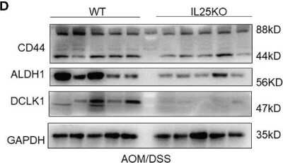
ALDH1A3 Antibody - BSA Free-Western Blot-NBP2-15339-img0023.jpg
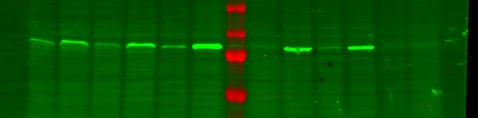
![Western Blot: ALDH1A3 AntibodyBSA Free [NBP2-15339]](https://resources.rndsystems.com/images/products/ALDH1A3-Antibody---BSA-Free-Western-Blot-NBP2-15339-img0010.jpg "Western Blot: ALDH1A3 AntibodyBSA Free [NBP2-15339]")
Western Blot: ALDH1A3 AntibodyBSA Free [NBP2-15339]
Western Blot: ALDH1A3 Antibody - BSA Free [NBP2-15339] - Whole cell extract (30 ug) was separated by 10% SDS-PAGE, and the membrane was blotted with ALDH1A3 antibody [N2C2], diluted at 1:1000. The HRP-conjugated anti-rabbit IgG antibody (NBP2-19301) was used to detect the primary antibody.![Western Blot: ALDH1A3 AntibodyBSA Free [NBP2-15339]](https://resources.rndsystems.com/images/products/ALDH1A3-Antibody---BSA-Free-Western-Blot-NBP2-15339-img0011.jpg "Western Blot: ALDH1A3 AntibodyBSA Free [NBP2-15339]")
Western Blot: ALDH1A3 AntibodyBSA Free [NBP2-15339]
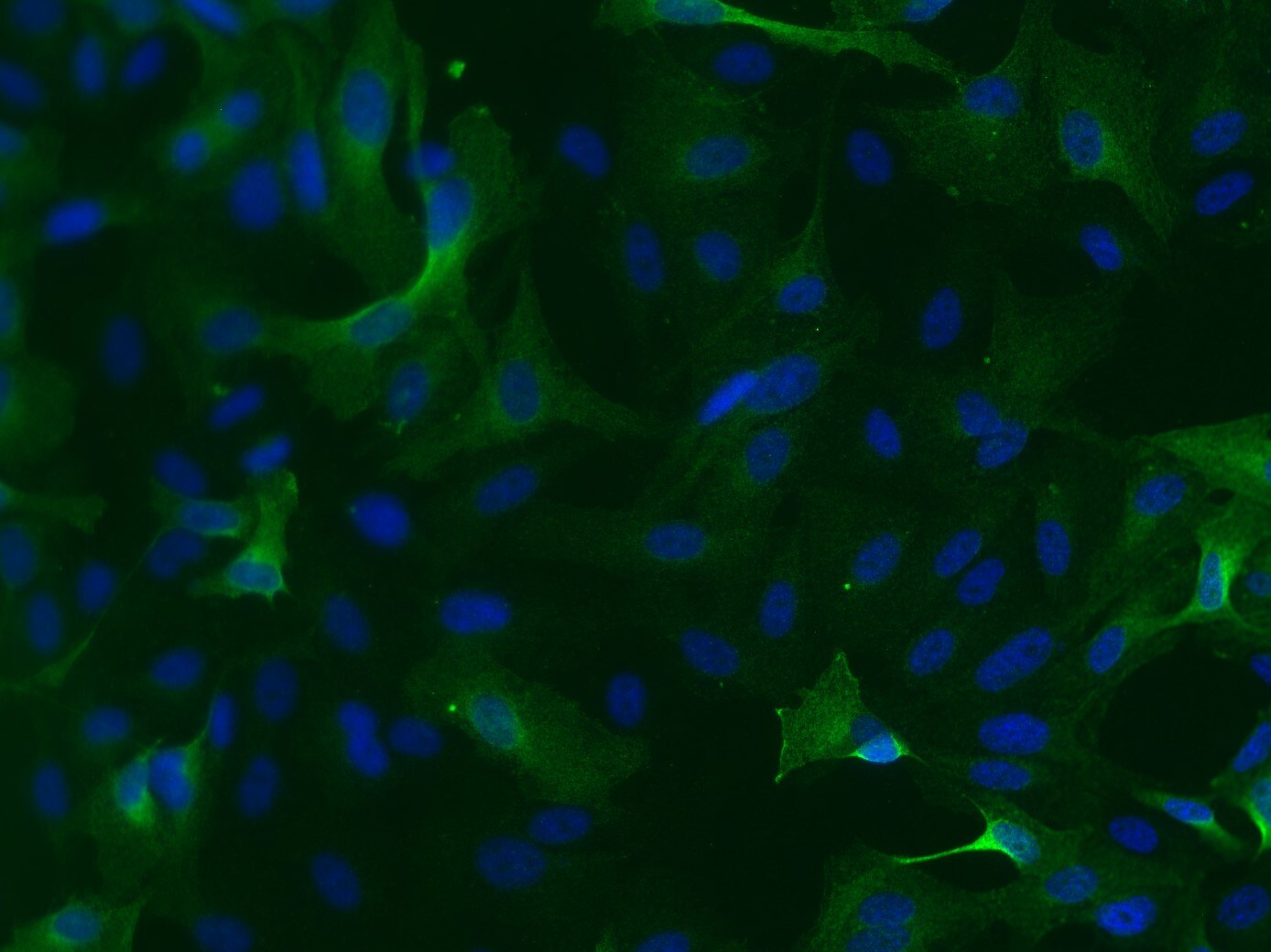
Western Blot: ALDH1A3 Antibody - BSA Free [NBP2-15339] - Whole cell extract (30 ug) was separated by 7.5% SDS-PAGE, and the membrane was blotted with ALDH1A3 antibody [N2C2], diluted at 1:2000.![Immunocytochemistry/ Immunofluorescence: ALDH1A3 Antibody - BSA Free [NBP2-15339]](https://resources.rndsystems.com/images/products/ALDH1A3-Antibody---BSA-Free-Immunocytochemistry-Immunofluorescence-NBP2-15339-img0014.jpg "Immunocytochemistry/ Immunofluorescence: ALDH1A3 Antibody - BSA Free [NBP2-15339]")
Immunocytochemistry/ Immunofluorescence: ALDH1A3 Antibody - BSA Free [NBP2-15339]
Immunocytochemistry/Immunofluorescence: ALDH1A3 Antibody - BSA Free [NBP2-15339] - A431 cells were fixed in 4% paraformaldehyde at RT for 15 min. Green: ALDH1A3 stained by ALDH1A3 antibody [N2C2], diluted at 1:500. Blue: Hoechst 33342 staining.![Immunocytochemistry/ Immunofluorescence: ALDH1A3 Antibody - BSA Free [NBP2-15339]](https://resources.rndsystems.com/images/products/ALDH1A3-Antibody---BSA-Free-Immunocytochemistry-Immunofluorescence-NBP2-15339-img0019.jpg "Immunocytochemistry/ Immunofluorescence: ALDH1A3 Antibody - BSA Free [NBP2-15339]")
Immunocytochemistry/ Immunofluorescence: ALDH1A3 Antibody - BSA Free [NBP2-15339]
Immunocytochemistry/Immunofluorescence: ALDH1A3 Antibody - BSA Free [NBP2-15339] - Human breast cancer cells with Aldh1a3 overexpression were stained at 1:500 in 5% goat serum in PBS-T. Human breast cancer cells were transduced with human Aldh1a3 and probed with antibody at 1:500 overnight at 4C followed by counterstaining with secondary and DAPI. Strong signal observed compared to knockout controls. ICC/IF image submitted by a verified customer review.![Immunohistochemistry-Frozen: ALDH1A3 Antibody - BSA Free [NBP2-15339]](https://resources.rndsystems.com/images/products/ALDH1A3-Antibody---BSA-Free-Immunohistochemistry-Frozen-NBP2-15339-img0007.jpg "Immunohistochemistry-Frozen: ALDH1A3 Antibody - BSA Free [NBP2-15339]")
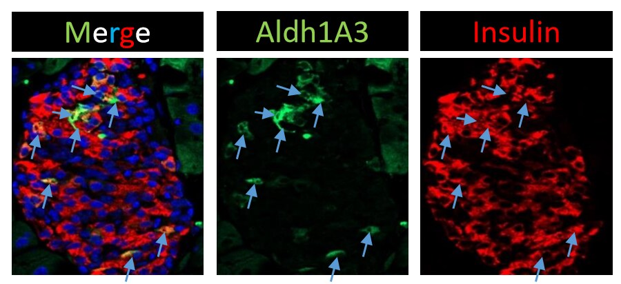
Immunohistochemistry-Frozen: ALDH1A3 Antibody - BSA Free [NBP2-15339]
Immunohistochemistry-Frozen: ALDH1A3 Antibody - BSA Free [NBP2-15339] - 8-week old mouse pancreas cryosections with beta cell dedifferentiation properties were stained with ALDH1A3 (1:500 dilution) and Insulin (1:1000 dilution) antibodies. Blue arrows indicate insulin+Aldh1A3+ cells. IHC-Fr image submitted by a verified customer review.![Immunohistochemistry-Paraffin: ALDH1A3 Antibody - BSA Free [NBP2-15339]](https://resources.rndsystems.com/images/products/ALDH1A3-Antibody---BSA-Free-Immunohistochemistry-Paraffin-NBP2-15339-img0015.jpg "Immunohistochemistry-Paraffin: ALDH1A3 Antibody - BSA Free [NBP2-15339]")
Immunohistochemistry-Paraffin: ALDH1A3 Antibody - BSA Free [NBP2-15339]
Immunohistochemistry-Paraffin: ALDH1A3 Antibody - BSA Free [NBP2-15339] - Mouse prostate. ALDH1A3 stained by ALDH1A3 antibody [N2C2], diluted at 1:500. Antigen Retrieval: Citrate buffer, pH 6.0, 15 min.![Immunohistochemistry-Paraffin: ALDH1A3 Antibody - BSA Free [NBP2-15339]](https://resources.rndsystems.com/images/products/ALDH1A3-Antibody---BSA-Free-Immunohistochemistry-Paraffin-NBP2-15339-img0016.jpg "Immunohistochemistry-Paraffin: ALDH1A3 Antibody - BSA Free [NBP2-15339]")
Immunohistochemistry-Paraffin: ALDH1A3 Antibody - BSA Free [NBP2-15339]
Immunohistochemistry-Paraffin: ALDH1A3 Antibody - BSA Free [NBP2-15339] - Rat prostate. ALDH1A3 stained by ALDH1A3 antibody [N2C2], diluted at 1:500. Antigen Retrieval: Citrate buffer, pH 6.0, 15 min.
Western Blot: ALDH1A3 Antibody - BSA Free [NBP2-15339] -
Western Blot: ALDH1A3 Antibody - BSA Free [NBP2-15339] - Palbociclib induces a potential transition of PN GSCs to MES GSCs(A) qPCR analysis of markers of PN & MES subtypes in G44 & G559 treated with the indicated doses of palbociclib for 5 days. n = 3. Differences were analyzed between the control & the palbociclib-treated groups. NS: not statistically significant; #p < 0.05; *p < 0.01; **p < 0.001. (B) qPCR & immunoblot analysis of ALDH1A3 in three independent PN GSC lines that were treated with 10 nM of palbociclib for 5 days. n = 3. Differences were analyzed between the control & the palbociclib-treated groups. NS: not statistically significant; #p < 0.05; *p < 0.01; **p < 0.001. (C) Cell proliferation analysis of three independent PN GSC lines that were treated with palbociclib (10 nM), DEAB (25 μM), or both for 5 days. The cells were cultured on a laminin (10 μg/ml in poly ornithine)-coated 96-well plate. Cell number was determined by CyQUANT Direct Cell Proliferation assay. n = 3. *p < 0.05, comparison between palbociclib or DEAB treatment with the vehicle control; #p < 0.05, comparison between combination group with palbociclib treatment. G44, CDI = 0.79; G559, CDI = 0.565; G816, CDI = 0.677. Shown are representative data of three independent experiments with similar results. Image collected & cropped by CiteAb from the following publication (https://www.oncotarget.com/lookup/doi/10.18632/oncotarget.19429), licensed under a CC-BY license. Not internally tested by Novus Biologicals.
Immunocytochemistry/ Immunofluorescence: ALDH1A3 Antibody - BSA Free [NBP2-15339] -
Immunocytochemistry/ Immunofluorescence: ALDH1A3 Antibody - BSA Free [NBP2-15339] - Markers of dedifferentiation are present in islets of db/db, but not weight-matched WD-fed mice. (a) Immunofluorescent analysis of islets from weight-matched WD-fed mice (top row) & db/db mice (bottom row) showing Nkx6.1 (red), insulin (green), & DAPI (blue). In the merged image, note the loss of double-positive nuclei (DAPI plus Nkx6.1; purple color) in the db/db mice but not in the WD-fed mice. (b) Expression of the Aldh1a3 gene in islets isolated from mice fed a WD for 4 or 12 weeks normalized to mice fed a control diet compared with db/db mice at 8 weeks of age (normalized to lean db/+ controls). ∗∗∗p < 0.001 versus both WD groups by one-way ANOVA. (c) Staining for Aldh1a3 protein (red) & insulin (green) in islets from weight-matched WD-fed mice (20 weeks on diet) versus db/db mice (14 weeks of age). Image collected & cropped by CiteAb from the following publication (https://pubmed.ncbi.nlm.nih.gov/29038790), licensed under a CC-BY license. Not internally tested by Novus Biologicals.Applications for ALDH1A3 Antibody - BSA Free
Application
Recommended Usage
Immunoblotting
reported in scientific literature (PMID 27572106)
Immunocytochemistry/ Immunofluorescence
1:100 - 1:1000. Use reported by customer review
Immunohistochemistry
1:100 - 1:1000
Immunohistochemistry-Frozen
reported by customer review and in scientific literature (PMID 26713822)
Immunohistochemistry-Paraffin
1:100 - 1:1000
Western Blot
1:500 - 1:3000
Reviewed Applications
Read 3 reviews rated 4.7 using NBP2-15339 in the following applications:
Formulation, Preparation, and Storage
Purification
Immunogen affinity purified
Formulation
PBS
Format
BSA Free
Preservative
0.02% Sodium Azide
Concentration
1.0 mg/ml
Shipping
The product is shipped with polar packs. Upon receipt, store it immediately at the temperature recommended below.
Stability & Storage
Aliquot and store at -20C or -80C. Avoid freeze-thaw cycles.
Background: Aldehyde Dehydrogenase 1-A3/ALDH1A3
Long Name
Aldehyde Dehydrogenase 1 Family Member A3
Alternate Names
ALDH1A3, ALDH6, MCOP8, RALDH3
Gene Symbol
ALDH1A3
UniProt
Additional Aldehyde Dehydrogenase 1-A3/ALDH1A3 Products
- All Products for Aldehyde Dehydrogenase 1-A3/ALDH1A3
- Aldehyde Dehydrogenase 1-A3/ALDH1A3 cDNA Clones
- Aldehyde Dehydrogenase 1-A3/ALDH1A3 ELISA Kits
- Aldehyde Dehydrogenase 1-A3/ALDH1A3 Lysates
- Aldehyde Dehydrogenase 1-A3/ALDH1A3 Primary Antibodies
- Aldehyde Dehydrogenase 1-A3/ALDH1A3 Proteins and Enzymes
Product Documents for ALDH1A3 Antibody - BSA Free
Certificate of Analysis
To download a Certificate of Analysis, please enter a lot or batch number in the search box below.
Product Specific Notices for ALDH1A3 Antibody - BSA Free
This product is for research use only and is not approved for use in humans or in clinical diagnosis. Primary Antibodies are guaranteed for 1 year from date of receipt.
Related Research Areas
Citations for ALDH1A3 Antibody - BSA Free
 Powered by Bioz
Customer Reviews for ALDH1A3 Antibody - BSA Free (3)
4.7 out of 5
3 Customer Ratings
Have you used ALDH1A3 Antibody - BSA Free?
Submit a review and receive an Amazon gift card!
$25/€18/£15/$25CAN/¥2500 Yen for a review with an image
$10/€7/£6/$10CAN/¥1110 Yen for a review without an image
Submit a review
Customer Images
Showing
1
-
3 of
3 reviews
Showing All
Filter By:
-
Application: Western BlotSample Tested: T47D human breast cancer cell lineSpecies: HumanVerified Customer | Posted 03/08/2020Left 6 lanes, empty vector and overexpression vectors of ALDH1A3 in T47D cell lines Right 6 lanes, empty vector and overexpression vectors of ALDH1A3 in MDA-MB-468 cell lines

-
Application: ImmunocytochemistrySample Tested: Human breast cancer cellsSpecies: HumanVerified Customer | Posted 01/14/2020Human breast cancer cells were transduced with human Aldh1a3 and then were probed with antibody at 1:500 overnight at 4C followed by counterstaining with secondary and DAPI. Strong signal observed compared to knockout controlsHuman breast cancer cells with Aldh1a3 overexpression were stained at 1:500 in 5% goat serum in PBS-T.
-
Application: Immunohistochemistry-FrozenSample Tested: Mouse PancreasSpecies: MouseVerified Customer | Posted 11/09/20168 Week old mouse pancreas cryosections with beta cell dedifferentiation properties were stained with Aldh1A3 (1:500), Insulin (1:1000). Blue arrows indicate insulin+Aldh1A3+ cells.
There are no reviews that match your criteria.
Protocols
Find general support by application which include: protocols, troubleshooting, illustrated assays, videos and webinars.
- Antigen Retrieval Protocol (PIER)
- Antigen Retrieval for Frozen Sections Protocol
- Appropriate Fixation of IHC/ICC Samples
- Cellular Response to Hypoxia Protocols
- Chromogenic IHC Staining of Formalin-Fixed Paraffin-Embedded (FFPE) Tissue Protocol
- Chromogenic Immunohistochemistry Staining of Frozen Tissue
- ClariTSA™ Fluorophore Kits
- Detection & Visualization of Antibody Binding
- Fluorescent IHC Staining of Frozen Tissue Protocol
- Graphic Protocol for Heat-induced Epitope Retrieval
- Graphic Protocol for the Preparation and Fluorescent IHC Staining of Frozen Tissue Sections
- Graphic Protocol for the Preparation and Fluorescent IHC Staining of Paraffin-embedded Tissue Sections
- Graphic Protocol for the Preparation of Gelatin-coated Slides for Histological Tissue Sections
- ICC Cell Smear Protocol for Suspension Cells
- ICC Immunocytochemistry Protocol Videos
- ICC for Adherent Cells
- IHC Sample Preparation (Frozen sections vs Paraffin)
- Immunocytochemistry (ICC) Protocol
- Immunocytochemistry Troubleshooting
- Immunofluorescence of Organoids Embedded in Cultrex Basement Membrane Extract
- Immunofluorescent IHC Staining of Formalin-Fixed Paraffin-Embedded (FFPE) Tissue Protocol
- Immunohistochemistry (IHC) and Immunocytochemistry (ICC) Protocols
- Immunohistochemistry Frozen Troubleshooting
- Immunohistochemistry Paraffin Troubleshooting
- Preparing Samples for IHC/ICC Experiments
- Preventing Non-Specific Staining (Non-Specific Binding)
- Primary Antibody Selection & Optimization
- Protocol for Heat-Induced Epitope Retrieval (HIER)
- Protocol for Making a 4% Formaldehyde Solution in PBS
- Protocol for VisUCyte™ HRP Polymer Detection Reagent
- Protocol for the Fluorescent ICC Staining of Cell Smears - Graphic
- Protocol for the Fluorescent ICC Staining of Cultured Cells on Coverslips - Graphic
- Protocol for the Preparation & Fixation of Cells on Coverslips
- Protocol for the Preparation and Chromogenic IHC Staining of Frozen Tissue Sections
- Protocol for the Preparation and Chromogenic IHC Staining of Frozen Tissue Sections - Graphic
- Protocol for the Preparation and Chromogenic IHC Staining of Paraffin-embedded Tissue Sections
- Protocol for the Preparation and Chromogenic IHC Staining of Paraffin-embedded Tissue Sections - Graphic
- Protocol for the Preparation and Fluorescent ICC Staining of Cells on Coverslips
- Protocol for the Preparation and Fluorescent ICC Staining of Non-adherent Cells
- Protocol for the Preparation and Fluorescent ICC Staining of Stem Cells on Coverslips
- Protocol for the Preparation and Fluorescent IHC Staining of Frozen Tissue Sections
- Protocol for the Preparation and Fluorescent IHC Staining of Paraffin-embedded Tissue Sections
- Protocol for the Preparation of Gelatin-coated Slides for Histological Tissue Sections
- Protocol for the Preparation of a Cell Smear for Non-adherent Cell ICC - Graphic
- R&D Systems Quality Control Western Blot Protocol
- TUNEL and Active Caspase-3 Detection by IHC/ICC Protocol
- The Importance of IHC/ICC Controls
- Troubleshooting Guide: Immunohistochemistry
- Troubleshooting Guide: Western Blot Figures
- Western Blot Conditions
- Western Blot Protocol
- Western Blot Protocol for Cell Lysates
- Western Blot Troubleshooting
- Western Blot Troubleshooting Guide
- View all Protocols, Troubleshooting, Illustrated assays and Webinars
Loading...