Alix Antibody - BSA Free
Novus Biologicals | Catalog # NBP1-90201
![Immunohistochemistry-Paraffin: Alix Antibody [NBP1-90201]](https://resources.rndsystems.com/images/products/Alix-Antibody-Immunohistochemistry-Paraffin-NBP1-90201-img0014.jpg "Immunohistochemistry-Paraffin: Alix Antibody [NBP1-90201]")
Loading...
Key Product Details
Validated by
Knockout/Knockdown
Species Reactivity
Validated:
Human
Cited:
Human
Predicted:
Mouse (94%), Rat (94%). Backed by our 100% Guarantee.
Applications
Validated:
Immunohistochemistry, Immunohistochemistry-Paraffin, Western Blot, Immunocytochemistry/ Immunofluorescence, Simple Western, Knockdown Validated
Cited:
Western Blot
Label
Unconjugated
Antibody Source
Polyclonal Rabbit IgG
Format
BSA Free
Loading...
Product Specifications
Immunogen
This antibody was developed against Recombinant Protein corresponding to amino acids: VPVSVQQSLAAYNQRKADLVNRSIAQMREATTLANGVLASLNLPAAIEDVSGDTVPQSILTKSRSVIEQGGIQTVDQLIKELPELLQRNREILDESLRLLDEEEATDND
Marker
Exosome Marker
Clonality
Polyclonal
Host
Rabbit
Isotype
IgG
Scientific Data Images for Alix Antibody - BSA Free
Immunohistochemistry-Paraffin: Alix Antibody [NBP1-90201]
Immunohistochemistry-Paraffin: Alix Antibody [NBP1-90201] - Staining of human fallopian tube shows strong cytoplasmic positivity in glandular cells.![Alix Antibody - BSA Free Immunohistochemistry: Alix Antibody - BSA Free [NBP1-90201]](https://resources.rndsystems.com/images/products/nbp1-90201_rabbit-polyclonal-alix-antibody-3052025101916.jpg "Immunohistochemistry: Alix Antibody - BSA Free [NBP1-90201]")
Immunohistochemistry: Alix Antibody - BSA Free [NBP1-90201]
Staining of human kidney shows moderate cytoplasmic positivity in cells in tubules.![Alix Antibody - BSA Free Immunohistochemistry: Alix Antibody - BSA Free [NBP1-90201]](https://resources.rndsystems.com/images/products/nbp1-90201_rabbit-polyclonal-alix-antibody-29520251614716.jpg "Immunohistochemistry: Alix Antibody - BSA Free [NBP1-90201]")
Immunohistochemistry: Alix Antibody - BSA Free [NBP1-90201]
Staining of human testis shows moderate cytoplasmic positivity in cells in seminiferous ducts.![Alix Antibody - BSA Free Immunohistochemistry: Alix Antibody - BSA Free [NBP1-90201]](https://resources.rndsystems.com/images/products/nbp1-90201_rabbit-polyclonal-alix-antibody-3052025723218.jpg "Immunohistochemistry: Alix Antibody - BSA Free [NBP1-90201]")
Immunohistochemistry: Alix Antibody - BSA Free [NBP1-90201]
Staining of human duodenum shows moderate cytoplasmic positivity in glandular cells.![Alix Antibody - BSA Free Western Blot: Alix Antibody - BSA Free [NBP1-90201]](https://resources.rndsystems.com/images/products/nbp1-90201_rabbit-polyclonal-alix-antibody-3052025748326.jpg "Western Blot: Alix Antibody - BSA Free [NBP1-90201]")

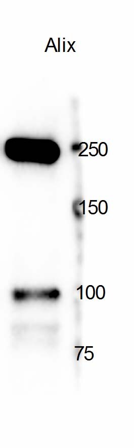
Western Blot: Alix Antibody - BSA Free [NBP1-90201] -
(A–D) Representative NTA of EV and CM samples derived from ASCs and DFs. Each graph shows the size distribution of 3 consecutive 1 min runs for each sample. (E–F) Size distribution and vesicular yield deriving from 6 NTA measurements/group shown as mean +/- SD. (G) Western Blot of CM and EV samples from ASCs and DFs, showing the expression of the vesicular markers Alix, FLOT-1, TSG101 and CD9. In each lane, 10μg of CM or EV deriving from 1.5 × 10⁶ cells were loaded. Image collected and cropped by CiteAb from the following open publication (https://pubmed.ncbi.nlm.nih.gov/33928071), licensed under a CC-BY license. Not internally tested by Novus Biologicals.![Alix Antibody - BSA Free Immunocytochemistry/ Immunofluorescence: Alix Antibody [NBP1-90201]](https://resources.rndsystems.com/images/products/nbp1-90201_-immunocytochemistry-immunofluorescence-639174076813832429.jpg "Immunocytochemistry/ Immunofluorescence: Alix Antibody [NBP1-90201]")
Immunocytochemistry/ Immunofluorescence: Alix Antibody [NBP1-90201]
Staining of human cell line U-2 OS shows localization to cytosol.Applications for Alix Antibody - BSA Free
Application
Recommended Usage
Immunocytochemistry/ Immunofluorescence
0.25-2 ug/ml
Immunohistochemistry
1:500 - 1:1000
Immunohistochemistry-Paraffin
1:500 - 1:1000
Simple Western
1:50
Western Blot
0.04-0.4 ug/ml
Application Notes
IHC-Paraffin, HIER pH 6 retrieval is recommended. ICC/IF, Fixation Permeabilization: Use PFA/Triton X-100. See Simple Western Antibody Database for Simple Western validation: Tested in Typical EV marker, antibody dilution of 1:50
Reviewed Applications
Read 1 review rated 5 using NBP1-90201 in the following applications:
Formulation, Preparation, and Storage
Purification
Affinity purified
Formulation
PBS (pH 7.2) and 40% Glycerol
Format
BSA Free
Preservative
0.02% Sodium Azide
Concentration
Concentrations vary lot to lot. See vial label for concentration. If unlisted please contact technical services.
Shipping
The product is shipped with polar packs. Upon receipt, store it immediately at the temperature recommended below.
Stability & Storage
Store at 4C short term. Aliquot and store at -20C long term. Avoid freeze-thaw cycles.
Background: Alix
Alternate Names
AIP1DRIP4, ALG-2 interacting protein 1, ALG-2 interacting protein X, ALG-2-interacting protein 1, alinx, ALIX, apoptosis-linked gene 2-interacting protein X, dopamine receptor interacting protein 4, HP95, KIAA1375, MGC17003, PDCD6-interacting protein, programmed cell death 6 interacting protein, programmed cell death 6-interacting protein
Gene Symbol
PDCD6IP
Additional Alix Products
Product Documents for Alix Antibody - BSA Free
Certificate of Analysis
To download a Certificate of Analysis, please enter a lot or batch number in the search box below.
Product Specific Notices for Alix Antibody - BSA Free
This product is for research use only and is not approved for use in humans or in clinical diagnosis. Primary Antibodies are guaranteed for 1 year from date of receipt.
Citations for Alix Antibody - BSA Free
 Powered by Bioz
Customer Reviews for Alix Antibody - BSA Free (1)
5 out of 5
1 Customer Rating
Have you used Alix Antibody - BSA Free?
Submit a review and receive an Amazon gift card!
$25/€18/£15/$25CAN/¥2500 Yen for a review with an image
$10/€7/£6/$10CAN/¥1110 Yen for a review without an image
Submit a review
Customer Images
Showing
1
-
1 of
1 review
Showing All
Filter By:
-
Application: Western BlotSample Tested: Rat astrocytesSpecies: RatVerified Customer | Posted 01/17/2018

There are no reviews that match your criteria.
Protocols
Find general support by application which include: protocols, troubleshooting, illustrated assays, videos and webinars.
- Antigen Retrieval Protocol (PIER)
- Antigen Retrieval for Frozen Sections Protocol
- Appropriate Fixation of IHC/ICC Samples
- Cellular Response to Hypoxia Protocols
- Chromogenic IHC Staining of Formalin-Fixed Paraffin-Embedded (FFPE) Tissue Protocol
- Chromogenic Immunohistochemistry Staining of Frozen Tissue
- ClariTSA™ Fluorophore Kits
- Detection & Visualization of Antibody Binding
- Fluorescent IHC Staining of Frozen Tissue Protocol
- Graphic Protocol for Heat-induced Epitope Retrieval
- Graphic Protocol for the Preparation and Fluorescent IHC Staining of Frozen Tissue Sections
- Graphic Protocol for the Preparation and Fluorescent IHC Staining of Paraffin-embedded Tissue Sections
- Graphic Protocol for the Preparation of Gelatin-coated Slides for Histological Tissue Sections
- ICC Cell Smear Protocol for Suspension Cells
- ICC Immunocytochemistry Protocol Videos
- ICC for Adherent Cells
- IHC Sample Preparation (Frozen sections vs Paraffin)
- Immunocytochemistry (ICC) Protocol
- Immunocytochemistry Troubleshooting
- Immunofluorescence of Organoids Embedded in Cultrex Basement Membrane Extract
- Immunofluorescent IHC Staining of Formalin-Fixed Paraffin-Embedded (FFPE) Tissue Protocol
- Immunohistochemistry (IHC) and Immunocytochemistry (ICC) Protocols
- Immunohistochemistry Frozen Troubleshooting
- Immunohistochemistry Paraffin Troubleshooting
- Preparing Samples for IHC/ICC Experiments
- Preventing Non-Specific Staining (Non-Specific Binding)
- Primary Antibody Selection & Optimization
- Protocol for Heat-Induced Epitope Retrieval (HIER)
- Protocol for Making a 4% Formaldehyde Solution in PBS
- Protocol for VisUCyte™ HRP Polymer Detection Reagent
- Protocol for the Fluorescent ICC Staining of Cell Smears - Graphic
- Protocol for the Fluorescent ICC Staining of Cultured Cells on Coverslips - Graphic
- Protocol for the Preparation & Fixation of Cells on Coverslips
- Protocol for the Preparation and Chromogenic IHC Staining of Frozen Tissue Sections
- Protocol for the Preparation and Chromogenic IHC Staining of Frozen Tissue Sections - Graphic
- Protocol for the Preparation and Chromogenic IHC Staining of Paraffin-embedded Tissue Sections
- Protocol for the Preparation and Chromogenic IHC Staining of Paraffin-embedded Tissue Sections - Graphic
- Protocol for the Preparation and Fluorescent ICC Staining of Cells on Coverslips
- Protocol for the Preparation and Fluorescent ICC Staining of Non-adherent Cells
- Protocol for the Preparation and Fluorescent ICC Staining of Stem Cells on Coverslips
- Protocol for the Preparation and Fluorescent IHC Staining of Frozen Tissue Sections
- Protocol for the Preparation and Fluorescent IHC Staining of Paraffin-embedded Tissue Sections
- Protocol for the Preparation of Gelatin-coated Slides for Histological Tissue Sections
- Protocol for the Preparation of a Cell Smear for Non-adherent Cell ICC - Graphic
- R&D Systems Quality Control Western Blot Protocol
- TUNEL and Active Caspase-3 Detection by IHC/ICC Protocol
- The Importance of IHC/ICC Controls
- Troubleshooting Guide: Immunohistochemistry
- Troubleshooting Guide: Western Blot Figures
- Western Blot Conditions
- Western Blot Protocol
- Western Blot Protocol for Cell Lysates
- Western Blot Troubleshooting
- Western Blot Troubleshooting Guide
- View all Protocols, Troubleshooting, Illustrated assays and Webinars
Loading...