alpha-Smooth Muscle Actin Antibody (SPM332) [Allophycocyanin]
Novus Biologicals | Catalog # NBP2-34760APC
Key Product Details
Species Reactivity
Validated:
Human, Mouse, Rat, Porcine, Baboon, Bovine, Canine, Chicken, Feline, Goat, Guinea Pig, Monkey, Rabbit, Sheep
Cited:
Human
Applications
Validated:
Immunohistochemistry, Immunohistochemistry-Paraffin, Western Blot, Flow Cytometry, Flow (Intracellular), Immunocytochemistry/ Immunofluorescence, CyTOF-ready
Cited:
Flow Cytometry
Label
Allophycocyanin (Excitation = 620-650 nm, Emission = 660-670 nm)
Antibody Source
Monoclonal Mouse IgG2a Kappa Clone # SPM332
Loading...
Product Specifications
Immunogen
N-Terminal decapeptide of alpha smooth muscle isoform of actin and conjugated to KLH. (Uniprot: P62736)
Localization
Cytoplasmic
Marker
Leiomyosarcoma Marker
Specificity
Actin is a major component of the cytoskeleton and is present in most cell types. This monoclonal antibody is highly specific to actin from smooth muscles. Its epitope lies in the first four N-terminal amino acids. This monoclonal antibody does not stain cardiac or skeletal muscle; however, it does stain myofibroblasts and myoepithelial cells. This antibody could be used together with anti-muscle specific actin and myogenin in making a diagnosis of smooth muscle and skeletal muscle tumors. In most cases of rhabdomyosarcoma, this antibody yields negative results whereas anti-muscle specific actin and myogenin are positive. Leiomyosarcomas are positive only with anti-muscle specific actin and anti-smooth muscle actin and are negative with anti-myogenin.
Clonality
Monoclonal
Host
Mouse
Isotype
IgG2a Kappa
Description
This conjugate is made on demand. Actual recovery may vary from the stated volume of this product. The volume will be greater than or equal to the unit size stated on the datasheet.
Scientific Data Images for alpha-Smooth Muscle Actin Antibody (SPM332) [Allophycocyanin]
alpha-Smooth-Muscle-Actin-Antibody-SPM332-Allophycocyanin-Immunohistochemistry-NBP2-34760APC-img0004.jpg
alpha-Smooth-Muscle-Actin-Antibody-SPM332-Allophycocyanin-Immunohistochemistry-NBP2-34760APC-img0002.jpg
alpha-Smooth-Muscle-Actin-Antibody-SPM332-Allophycocyanin-Immunohistochemistry-NBP2-34760APC-img0003.jpg
![alpha-Smooth Muscle Actin Antibody (SPM332) [Allophycocyanin]](https://resources.rndsystems.com/images/products/nbp2-34760apc_mouse-monoclonal-alpha-smooth-muscle-actin-antibody-spm332-allophycocyanin-210202423454881.jpg "Immunocytochemistry/ Immunofluorescence: alpha-Smooth Muscle Actin Antibody (SPM332) [Allophycocyanin] [NBP2-34760APC] -")
Immunocytochemistry/ Immunofluorescence: alpha-Smooth Muscle Actin Antibody (SPM332) [Allophycocyanin] [NBP2-34760APC] -
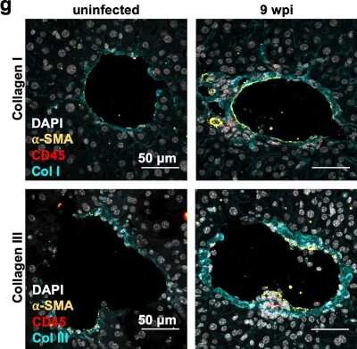
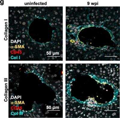
Cells expressing IL-1⍺ & IL-1R are observed in the fibrotic liver environment. (a) Cytokines in tissue lysates from mice at 9 wpi were measured by ELISA in liver. Data are presented as fold change relative to the mean of uninfected levels. N = 11–12 mice per group, pooled from three independent experiments. (b) IL-1 alpha levels in the sera at 9 wpi measured by ELISA. N = 9–14 mice per group, pooled from four independent experiments. (c) Immunofluorescence labeling of nuclei (DAPI white), IL-1⍺ (green), CD45 (red), & collagen1⍺1 (blue) in the liver of UI or 9 wpi WT mice. Number of cells staining positive for CD45 and/or IL-1⍺, average 2–3 fields of view where immune infiltrate was present from 3 mice per condition are quantified on the right. Error bars are standard deviation. (d) Immunofluorescent labeling of nuclei (DAPI white), ⍺-smooth muscle actin (green), IL-1R (red), & collagen1⍺ (blue) in the liver of uninfected or 9 wpi WT. Inset, arrow head represents ⍺-smooth muscle actin/IL-1R co-staining cells (arrow heads). Number of cells staining positive for IL-1R and/or ⍺-SMA, average of 4–8 fields of view from 3 mice are quantified on the right. Error bars are standard deviation. (c,d) represent maximum intensity projections of 9–13 μm thick z-stacks. Scale bar represents 50 μm. Error bars are standard error of the mean except where noted otherwise. *P < 0.05; **P < 0.01; ***P < 0.001 by unpaired Student’s t test. Image collected & cropped by CiteAb from the following publication (https://pubmed.ncbi.nlm.nih.gov/32973293), licensed under a CC-BY license. Not internally tested by Novus Biologicals.![alpha-Smooth Muscle Actin Antibody (SPM332) [Allophycocyanin]](https://resources.rndsystems.com/images/products/nbp2-34760apc_mouse-monoclonal-alpha-smooth-muscle-actin-antibody-spm332-allophycocyanin-310202416114746.jpg "Immunocytochemistry/ Immunofluorescence: alpha-Smooth Muscle Actin Antibody (SPM332) [Allophycocyanin] [NBP2-34760APC] -")
Immunocytochemistry/ Immunofluorescence: alpha-Smooth Muscle Actin Antibody (SPM332) [Allophycocyanin] [NBP2-34760APC] -
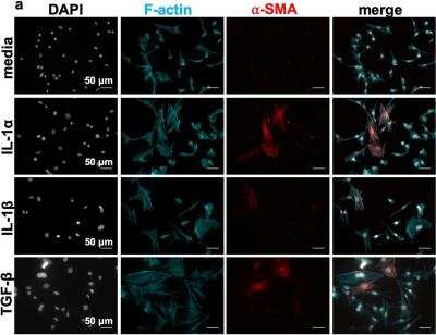
Immunocytochemistry/ Immunofluorescence: alpha-Smooth Muscle Actin Antibody (SPM332) [Allophycocyanin] [NBP2-34760APC] - IL-1 induces contractility & alpha smooth muscle actin expression in murine embryonic fibroblasts & primary hepatic stellate cells. (a–c) MEFs were incubated with media, 10 ng/mL IL-1 alpha, 150 pg/mL IL-1 beta or 10 ng/mL TGF beta -1 for 48 h. After fixation, MEFs were stained for F-actin, & alpha-smooth muscle actin ( alpha -SMA). Cell spreading was quantified in (b), & levels of alpha -SMA expression were quantified in (c) as the mean relative to untreated for each biological replicate (left panels) or the single cell data pooled from three biological replicate experiments (right panels). For each experiment, 50–200 cells/group were analyzed. (d–f) Primary hepatic stellate cells (HSCs) were isolated from uninfected mouse livers, & FACS-sorted based on endogenous retinoid fluorescence (d). (e,f) HSCs were seeded onto 4 kPa hydrogels coated with 10 μg/mL of fibronectin & cultured with 10 ng/mL IL-1 alpha, 10 ng/mL TGF-beta, or media alone for 48 h & then fixed & stained for F-actin & alpha -SMA & imaged by confocal microscopy. Scale bar represents 50 μm. Total cell area quantified in (e) & levels of alpha -SMA expression were quantified in terms of pixels/cell in (f) as the mean relative to untreated for each biological replicate (left panels) or the single cell data pooled from four biological replicate experiments (right panels). Error bars are standard error of the mean. For the left panels, data were compared by one-way ANOVA with Bonferroni’s multiple comparisons test. Image collected & cropped by CiteAb from the following publication (https://pubmed.ncbi.nlm.nih.gov/32973293), licensed under a CC-BY license. Not internally tested by Novus Biologicals.Applications for alpha-Smooth Muscle Actin Antibody (SPM332) [Allophycocyanin]
Application
Recommended Usage
CyTOF-ready
Optimal dilutions of this antibody should be experimentally determined.
Flow (Intracellular)
Optimal dilutions of this antibody should be experimentally determined.
Flow Cytometry
Optimal dilutions of this antibody should be experimentally determined.
Immunocytochemistry/ Immunofluorescence
Optimal dilutions of this antibody should be experimentally determined.
Immunohistochemistry
Optimal dilutions of this antibody should be experimentally determined.
Immunohistochemistry-Paraffin
Optimal dilutions of this antibody should be experimentally determined.
Western Blot
Optimal dilutions of this antibody should be experimentally determined.
Application Notes
Use in Immunofluorescence reported by customer review.
Reviewed Applications
Read 1 review rated 5 using NBP2-34760APC in the following applications:
Spectra Viewer
Plan Your Experiments
Use our spectra viewer to interactively plan your experiments, assessing multiplexing options. View the excitation and emission spectra for our fluorescent dye range and other commonly used dyes.
Spectra Viewer
Flow Cytometry Panel Builder
Bio-Techne Knows Flow Cytometry
Save time and reduce costly mistakes by quickly finding compatible reagents using the Panel Builder Tool.
Advanced Features
- Spectra Viewer - Custom analysis of spectra from multiple fluorochromes
- Spillover Popups - Visualize the spectra of individual fluorochromes
- Antigen Density Selector - Match fluorochrome brightness with antigen density

Formulation, Preparation, and Storage
Purification
Protein A or G purified
Formulation
PBS
Preservative
0.05% Sodium Azide
Concentration
Please see the vial label for concentration. If unlisted please contact technical services.
Shipping
The product is shipped with polar packs. Upon receipt, store it immediately at the temperature recommended below.
Stability & Storage
Store at 4C in the dark.
Background: alpha-Smooth Muscle Actin
ACTA2 encodes alpha Smooth Muscle Actin and is also known as alpha-actin, alpha-actin-2, aortic smooth muscle or alpha smooth muscle actin (alpha-SMA, SMactin, alpha-SM-actin, ASMA). Alpha Smooth Muscle Actin is frequently used as a marker of smooth muscle differentiation and has a theoretical molecular weight of 42 kDa. Smooth muscle alpha actin is one of a few genes whose expression is predominantly expressed in vascular smooth muscle cells that is used to study myofibroblasts and fibrosis (1). Excessive accumulation of alpha smooth muscle actin-positive (ACTA2+) activated myofibroblasts is observed in Idiopathic pulmonary fibrosis (2). Expression of smooth muscle alpha actin is regulated by cell proliferation and is altered in pathological conditions including atherosclerosis and is common in metastatic cancers (1,3).
References
1. Chakraborty, R., Saddouk, F. Z., Carrao, A. C., Krause, D. S., Greif, D. M., & Martin, K. A. (2019). Promoters to Study Vascular Smooth Muscle. Arterioscler Thromb Vasc Biol, 39(4), 603-612. doi:10.1161/atvbaha.119.312449
2.El Agha, E., Moiseenko, A., Kheirollahi, V., De Langhe, S., Crnkovic, S., Kwapiszewska, G.,... Bellusci, S. (2017). Two-Way Conversion between Lipogenic and Myogenic Fibroblastic Phenotypes Marks the Progression and Resolution of Lung Fibrosis. Cell Stem Cell, 20(2), 261-273.e263. doi:10.1016/j.stem.2016.10.004
3. Chen, Y. C., Gonzalez, M. E., Burman, B., Zhao, X., Anwar, T., Tran, M.,... Kleer, C. G. (2019). Mesenchymal Stem/Stromal Cell Engulfment Reveals Metastatic Advantage in Breast Cancer. Cell Rep, 27(13), 3916-3926.e3915. doi:10.1016/j.celrep.2019.05.084
Long Name
Actin, Alpha 2, Smooth Muscle, Aorta
Alternate Names
AAT6, ACTA2, Actin alpha 2, ACTSA, ACTVS, alphaSmooth Muscle Actin, MYMY5, SMA
Gene Symbol
ACTA2
Additional alpha-Smooth Muscle Actin Products
Product Documents for alpha-Smooth Muscle Actin Antibody (SPM332) [Allophycocyanin]
Certificate of Analysis
To download a Certificate of Analysis, please enter a lot or batch number in the search box below.
Product Specific Notices for alpha-Smooth Muscle Actin Antibody (SPM332) [Allophycocyanin]
This product is for research use only and is not approved for use in humans or in clinical diagnosis. Primary Antibodies are guaranteed for 1 year from date of receipt.
Related Research Areas
Citations for alpha-Smooth Muscle Actin Antibody (SPM332) [Allophycocyanin]
 Powered by Bioz
Customer Reviews for alpha-Smooth Muscle Actin Antibody (SPM332) [Allophycocyanin] (1)
5 out of 5
1 Customer Rating
Have you used alpha-Smooth Muscle Actin Antibody (SPM332) [Allophycocyanin]?
Submit a review and receive an Amazon gift card!
$25/€18/£15/$25CAN/¥2500 Yen for a review with an image
$10/€7/£6/$10CAN/¥1110 Yen for a review without an image
Submit a review
Customer Images
![alpha-Smooth Muscle Actin Antibody (SPM332) [Allophycocyanin] NBP2-34760APC](https://resources.rndsystems.com/images/reviews/review_nbp2-34760apc_47966_0_0_0_0.jpg)
Showing
1
-
1 of
1 review
Showing All
Filter By:
-
Application: Immunofluorescence-ParaffinSample Tested: Mammary gland tissueSpecies: MouseVerified Customer | Posted 05/16/2019Red: alpha-smooth muscle actin, Blue: DAPI; Green: CD140alpha; mouse mammary gland
![alpha-Smooth Muscle Actin Antibody (SPM332) [Allophycocyanin] NBP2-34760APC](data:image/png;base64,R0lGODlhAQABAAD/ACwAAAAAAQABAAACADs=)
There are no reviews that match your criteria.
Protocols
Find general support by application which include: protocols, troubleshooting, illustrated assays, videos and webinars.
- 7-Amino Actinomycin D (7-AAD) Cell Viability Flow Cytometry Protocol
- Antigen Retrieval Protocol (PIER)
- Antigen Retrieval for Frozen Sections Protocol
- Appropriate Fixation of IHC/ICC Samples
- Cellular Response to Hypoxia Protocols
- Chromogenic IHC Staining of Formalin-Fixed Paraffin-Embedded (FFPE) Tissue Protocol
- Chromogenic Immunohistochemistry Staining of Frozen Tissue
- ClariTSA™ Fluorophore Kits
- Detection & Visualization of Antibody Binding
- Extracellular Membrane Flow Cytometry Protocol
- Flow Cytometry Protocol for Cell Surface Markers
- Flow Cytometry Protocol for Staining Membrane Associated Proteins
- Flow Cytometry Staining Protocols
- Flow Cytometry Troubleshooting Guide
- Fluorescent IHC Staining of Frozen Tissue Protocol
- Graphic Protocol for Heat-induced Epitope Retrieval
- Graphic Protocol for the Preparation and Fluorescent IHC Staining of Frozen Tissue Sections
- Graphic Protocol for the Preparation and Fluorescent IHC Staining of Paraffin-embedded Tissue Sections
- Graphic Protocol for the Preparation of Gelatin-coated Slides for Histological Tissue Sections
- ICC Cell Smear Protocol for Suspension Cells
- ICC Immunocytochemistry Protocol Videos
- ICC for Adherent Cells
- IHC Sample Preparation (Frozen sections vs Paraffin)
- Immunocytochemistry (ICC) Protocol
- Immunocytochemistry Troubleshooting
- Immunofluorescence of Organoids Embedded in Cultrex Basement Membrane Extract
- Immunofluorescent IHC Staining of Formalin-Fixed Paraffin-Embedded (FFPE) Tissue Protocol
- Immunohistochemistry (IHC) and Immunocytochemistry (ICC) Protocols
- Immunohistochemistry Frozen Troubleshooting
- Immunohistochemistry Paraffin Troubleshooting
- Intracellular Flow Cytometry Protocol Using Alcohol (Methanol)
- Intracellular Flow Cytometry Protocol Using Detergents
- Intracellular Nuclear Staining Flow Cytometry Protocol Using Detergents
- Intracellular Staining Flow Cytometry Protocol Using Alcohol Permeabilization
- Intracellular Staining Flow Cytometry Protocol Using Detergents to Permeabilize Cells
- Preparing Samples for IHC/ICC Experiments
- Preventing Non-Specific Staining (Non-Specific Binding)
- Primary Antibody Selection & Optimization
- Propidium Iodide Cell Viability Flow Cytometry Protocol
- Protocol for Heat-Induced Epitope Retrieval (HIER)
- Protocol for Liperfluo
- Protocol for Making a 4% Formaldehyde Solution in PBS
- Protocol for VisUCyte™ HRP Polymer Detection Reagent
- Protocol for the Characterization of Human Th22 Cells
- Protocol for the Characterization of Human Th9 Cells
- Protocol for the Fluorescent ICC Staining of Cell Smears - Graphic
- Protocol for the Fluorescent ICC Staining of Cultured Cells on Coverslips - Graphic
- Protocol for the Preparation & Fixation of Cells on Coverslips
- Protocol for the Preparation and Chromogenic IHC Staining of Frozen Tissue Sections
- Protocol for the Preparation and Chromogenic IHC Staining of Frozen Tissue Sections - Graphic
- Protocol for the Preparation and Chromogenic IHC Staining of Paraffin-embedded Tissue Sections
- Protocol for the Preparation and Chromogenic IHC Staining of Paraffin-embedded Tissue Sections - Graphic
- Protocol for the Preparation and Fluorescent ICC Staining of Cells on Coverslips
- Protocol for the Preparation and Fluorescent ICC Staining of Non-adherent Cells
- Protocol for the Preparation and Fluorescent ICC Staining of Stem Cells on Coverslips
- Protocol for the Preparation and Fluorescent IHC Staining of Frozen Tissue Sections
- Protocol for the Preparation and Fluorescent IHC Staining of Paraffin-embedded Tissue Sections
- Protocol for the Preparation of Gelatin-coated Slides for Histological Tissue Sections
- Protocol for the Preparation of a Cell Smear for Non-adherent Cell ICC - Graphic
- Protocol: Annexin V and PI Staining by Flow Cytometry
- Protocol: Annexin V and PI Staining for Apoptosis by Flow Cytometry
- R&D Systems Quality Control Western Blot Protocol
- TUNEL and Active Caspase-3 Detection by IHC/ICC Protocol
- The Importance of IHC/ICC Controls
- Troubleshooting Guide: Fluorokine Flow Cytometry Kits
- Troubleshooting Guide: Immunohistochemistry
- Troubleshooting Guide: Western Blot Figures
- Western Blot Conditions
- Western Blot Protocol
- Western Blot Protocol for Cell Lysates
- Western Blot Troubleshooting
- Western Blot Troubleshooting Guide
- View all Protocols, Troubleshooting, Illustrated assays and Webinars
FAQs for alpha-Smooth Muscle Actin Antibody (SPM332) [Allophycocyanin]
Showing
1
-
1 of
1 FAQ
Showing All
-
Q: I do not see horse listed as a predicted or confirmed species. Would you be able to confirm if any of your alpha-SMA antibodies will react with equine samples?
A: We have not directly tested equine samples nor do we have cited publications that have tested equine samples. Since alpha-Smooth Muscle Actin is a highly conserved sequence, these antibodies should react with most mammal species.
Loading...
Associated Pathways
