alpha-Synuclein Antibody - Azide Free
Novus Biologicals | Catalog # NBP2-15365
![Western Blot: alpha-Synuclein Antibody [NBP2-15365]](https://resources.rndsystems.com/images/products/alpha-Synuclein-Antibody-Western-Blot-NBP2-15365-img0027.jpg "Western Blot: alpha-Synuclein Antibody [NBP2-15365]")
Loading...
Key Product Details
Validated by
Independent Antibodies
Species Reactivity
Validated:
Human, Mouse, Rat
Cited:
Human, Mouse
Predicted:
Bovine (94%), Chimpanzee (100%), Porcine (97%), Sheep (95%). Backed by our 100% Guarantee.
Applications
Validated:
Immunohistochemistry, Immunohistochemistry-Paraffin, Immunohistochemistry-Frozen, Western Blot, Immunocytochemistry/ Immunofluorescence
Cited:
Western Blot, ELISA, Immunocytochemistry/ Immunofluorescence, IF/IHC
Label
Unconjugated
Antibody Source
Polyclonal Rabbit IgG
Format
Azide Free
Loading...
Product Specifications
Immunogen
Full length human alpha-Synuclein Recombinant protein.
Reactivity Notes
Chicken (88%), Xenopus laevis (85%).
Localization
Cytoplasm; Membrane; Nucleus
Clonality
Polyclonal
Host
Rabbit
Isotype
IgG
Theoretical MW
14 kDa.
Disclaimer note: The observed molecular weight of the protein may vary from the listed predicted molecular weight due to post translational modifications, post translation cleavages, relative charges, and other experimental factors.
Disclaimer note: The observed molecular weight of the protein may vary from the listed predicted molecular weight due to post translational modifications, post translation cleavages, relative charges, and other experimental factors.
Scientific Data Images for alpha-Synuclein Antibody - Azide Free
Western Blot: alpha-Synuclein Antibody [NBP2-15365]
Western Blot: alpha-Synuclein Antibody [NBP2-15365] - Various tissue extracts (50 ug) were separated by 15% SDS-PAGE, and the membranes were blotted with alpha Synuclein antibody diluted at 1:2000 and competitor's antibody diluted at 1:2000. The HRP-conjugated anti-rabbit IgG antibody was used to detect the primary antibody.![Immunohistochemistry-Paraffin: alpha-Synuclein Antibody [NBP2-15365]](https://resources.rndsystems.com/images/products/Synuclein-alpha-Antibody-Immunohistochemistry-Paraffin-NBP2-15365-img0009.jpg "Immunohistochemistry-Paraffin: alpha-Synuclein Antibody [NBP2-15365]")
Immunohistochemistry-Paraffin: alpha-Synuclein Antibody [NBP2-15365]
Immunohistochemistry-Paraffin: alpha-Synuclein Antibody [NBP2-15365] - Rat brain. alpha Synuclein antibody diluted at 1:500. Antigen Retrieval: Citrate buffer, pH 6.0, 15 min.![Immunohistochemistry-Paraffin: alpha-Synuclein Antibody [NBP2-15365]](https://resources.rndsystems.com/images/products/alpha-Synuclein-Antibody-Immunohistochemistry-Paraffin-NBP2-15365-img0033.jpg "Immunohistochemistry-Paraffin: alpha-Synuclein Antibody [NBP2-15365]")
Immunohistochemistry-Paraffin: alpha-Synuclein Antibody [NBP2-15365]
Immunohistochemistry-Paraffin: alpha-Synuclein Antibody [NBP2-15365] - Mouse hippocampus. alpha Synuclein stained by alpha Synuclein antibody diluted at 1:500. Antigen Retrieval: Citrate buffer, pH 6.0, 15 min.![Immunohistochemistry-Paraffin: alpha-Synuclein Antibody [NBP2-15365]](https://resources.rndsystems.com/images/products/Synuclein-alpha-Antibody-Immunohistochemistry-Paraffin-NBP2-15365-img0006.jpg "Immunohistochemistry-Paraffin: alpha-Synuclein Antibody [NBP2-15365]")
Immunohistochemistry-Paraffin: alpha-Synuclein Antibody [NBP2-15365]
Immunohistochemistry-Paraffin: alpha-Synuclein Antibody [NBP2-15365] - Human colon carcinoma, using alpha Synuclein antibody at 1:250 dilution. Antigen Retrieval: Trilogy™ (EDTA based, pH 8.0) buffer, 15min.
Western Blot: alpha-Synuclein Antibody [NBP2-15365] -
Western Blot: alpha-Synuclein Antibody [NBP2-15365] - Various tissue extracts (50 ug) were separated by 15% SDS-PAGE, and the membrane was blotted with alpha-Synuclein antibody (NBP2-15365) diluted at 1:1000. The HRP-conjugated anti-rabbit IgG antibody was used to detect the primary antibody.
Immunohistochemistry-Paraffin: alpha-Synuclein Antibody [NBP2-15365] -
Immunohistochemistry-Paraffin: alpha-Synuclein Antibody [NBP2-15365] - alpha-Synuclein antibody detects alpha-Synuclein protein by immunohistochemical analysis.Sample: Paraffin-embedded mouse tissues.
alpha-Synuclein stained by alpha-Synuclein antibody (NBP2-15365) diluted at 1:500.
Antigen Retrieval: Citrate buffer, pH 6.0, 15 min

Immunohistochemistry-Paraffin: alpha-Synuclein Antibody [NBP2-15365] -
Immunohistochemistry-Paraffin: alpha-Synuclein Antibody [NBP2-15365] - alpha-Synuclein antibody detects alpha-Synuclein protein by immunohistochemical analysis.Sample: Paraffin-embedded rat tissues.
alpha-Synuclein stained by alpha-Synuclein antibody (NBP2-15365) diluted at 1:500.
Antigen Retrieval: Citrate buffer, pH 6.0, 15 min

Immunohistochemistry-Frozen: alpha-Synuclein Antibody [NBP2-15365] -
alpha Synuclein antibody detects alpha Synuclein protein expression by immunohistochemical analysis.Sample: Frozen-sectioned adult mouse hippocampus.
Green: alpha Synuclein protein stained by alpha Synuclein antibody (NBP2-15365) diluted at 1:250.
Red: NeuN, stained by NeuN antibody [2Q158] diluted at 1:500.

Immunocytochemistry/ Immunofluorescence: alpha-Synuclein Antibody [NBP2-15365] -
alpha Synuclein antibody detects alpha Synuclein protein by immunofluorescent analysis.Sample: DIV14 rat E18 primary cortical neurons were fixed in 4% paraformaldehyde at RT for 15 min.
Green: alpha Synuclein protein stained by alpha Synuclein antibody (NBP2-15365) diluted at 1:500.
Red: beta Tubulin 3/ Tuj1, stained by beta Tubulin 3/ Tuj1 antibody [GT1338] diluted at 1:500.
Blue: Fluoroshield with DAPI.

Immunohistochemistry-Frozen: alpha-Synuclein Antibody [NBP2-15365] -
alpha Synuclein antibody detects alpha Synuclein protein expression by immunohistochemical analysis.Sample: Frozen sectioned E13.5 Rat brain.
Green: alpha Synuclein protein stained by alpha Synuclein antibody (NBP2-15365) diluted at 1:250.
Blue: Fluoroshield with DAPI.

Western Blot: alpha-Synuclein Antibody [NBP2-15365] -
Various whole cell extracts (30 ug) were separated by 15% SDS-PAGE, and the membrane was blotted with alpha Synuclein antibody (NBP2-15365) diluted at 1:1000. The HRP-conjugated anti-rabbit IgG antibody was used to detect the primary antibody. Corresponding RNA expression data for the same cell lines are based on Human Protein Atlas program.
Immunohistochemistry-Frozen: alpha-Synuclein Antibody [NBP2-15365] -
alpha Synuclein antibody detects alpha Synuclein protein expression by immunohistochemical analysis.Sample: Frozen-sectioned adult mouse cerebellum.
Green: alpha Synuclein protein stained by alpha Synuclein antibody (NBP2-15365) diluted at 1:250.
Red: beta Tubulin 3/ TUJ1, stained by beta Tubulin 3/ TUJ1 antibody [GT11710] diluted at 1:500.
Blue: Fluoroshield with DAPI.

Western Blot: alpha-Synuclein Antibody [NBP2-15365] -
Various whole cell extracts (30 ug) were separated by 15% SDS-PAGE, and the membrane was blotted with alpha Synuclein antibody (NBP2-15365) diluted at 1:1000. The HRP-conjugated anti-rabbit IgG antibody was used to detect the primary antibody.
Western Blot: alpha-Synuclein Antibody [NBP2-15365] -
Various tissue extracts (50 ug) were separated by 15% SDS-PAGE, and the membrane was blotted with alpha Synuclein antibody (NBP2-15365) diluted at 1:1000. The HRP-conjugated anti-rabbit IgG antibody was used to detect the primary antibody.
Immunohistochemistry-Paraffin: alpha-Synuclein Antibody [NBP2-15365] -
alpha Synuclein antibody detects alpha Synuclein protein at cytoplasm and nucleus in rat brain by immunohistochemical analysis.Sample: Paraffin-embedded rat brain.
Green: alpha Synuclein antibody (NBP2-15365) diluted at 1:200. The signal was developed using goat anti-rabbit IgG antibody (Dylight488).
Blue: Nuclear staining with Hoechst 33342.
br>Antigen Retrieval: Citrate buffer, pH 6.0, 15 min
Applications for alpha-Synuclein Antibody - Azide Free
Application
Recommended Usage
Immunocytochemistry/ Immunofluorescence
1:100-1:1000
Immunohistochemistry
1:100-1:1000
Immunohistochemistry-Frozen
1:100-1:1000
Immunohistochemistry-Paraffin
1:100-1:1000
Western Blot
1:500-1:3000
Reviewed Applications
Read 1 review rated 5 using NBP2-15365 in the following applications:
Formulation, Preparation, and Storage
Purification
Antigen Affinity-purified
Formulation
PBS, 1% BSA, 20% Glycerol
Format
Azide Free
Preservative
0.025% Proclin 300
Concentration
Concentrations vary lot to lot. See vial label for concentration. If unlisted please contact technical services.
Shipping
The product is shipped with polar packs. Upon receipt, store it immediately at the temperature recommended below.
Stability & Storage
Aliquot and store at -20C or -80C. Avoid freeze-thaw cycles.
Background: alpha-Synuclein
A number of studies have revealed that alpha-synuclein aggregation is a hallmark feature in a number of neurodegenerative diseases, referred to as synucleinopathies (2-4). Alpha-synuclein protein aggregates are a large component of Lewy bodies that are present in Parkinson's disease (PD), Lewy body dementia (LBD), and multiple system atrophy (1-6). Research has shown phosphorylation of alpha-synuclein at Ser129 moves the protein from the nucleus to the cytoplasm and promotes fibril formation associated with synucleinopathies (1,2,5). Recent studies also suggest that alpha-synuclein accumulation can prevent mitochondrial import machinery causing mitochondrial dysfunction that is often observed in neurodegeneration (5). It is thought that preventing alpha-synuclein aggregation may prevent PD, thus alpha-synuclein is a target for many potential therapeutic interventions aimed at decreasing aggregate formation or increasing clearance (1,2,4-6).
References
1. Villar-Pique, A., Lopes da Fonseca, T., & Outeiro, T. F. (2016). Structure, function and toxicity of alpha-synuclein: the Bermuda triangle in synucleinopathies. Journal of neurochemistry. https://doi.org/10.1111/jnc.13249
2. Emamzadeh F. N. (2016). Alpha-synuclein structure, functions, and interactions. Journal of research in medical sciences : the official journal of Isfahan University of Medical Sciences. https://doi.org/10.4103/1735-1995.181989
3. Burre J. (2015). The Synaptic Function of alpha-Synuclein. Journal of Parkinson's disease. https://doi.org/10.3233/JPD-150642
4. Lashuel, H. A., Overk, C. R., Oueslati, A., & Masliah, E. (2013). The many faces of alpha-synuclein: from structure and toxicity to therapeutic target. Nature reviews. Neuroscience. https://doi.org/10.1038/nrn3406
5. Rocha, E. M., De Miranda, B., & Sanders, L. H. (2018). Alpha-synuclein: Pathology, mitochondrial dysfunction and neuroinflammation in Parkinson's disease. Neurobiology of disease. https://doi.org/10.1016/j.nbd.2017.04.004
6. O'Leary, E. I., & Lee, J. C. (2019). Interplay between alpha-synuclein amyloid formation and membrane structure. Biochimica et biophysica acta. Proteins and proteomics. https://doi.org/10.1016/j.bbapap.2018.09.012
Alternate Names
NACP, PARK1, PARK4, SNCA, Synuclein-alpha
Gene Symbol
SNCA
Additional alpha-Synuclein Products
Product Documents for alpha-Synuclein Antibody - Azide Free
Certificate of Analysis
To download a Certificate of Analysis, please enter a lot or batch number in the search box below.
Product Specific Notices for alpha-Synuclein Antibody - Azide Free
This product is for research use only and is not approved for use in humans or in clinical diagnosis. Primary Antibodies are guaranteed for 1 year from date of receipt.
Related Research Areas
Citations for alpha-Synuclein Antibody - Azide Free
 Powered by Bioz
Customer Reviews for alpha-Synuclein Antibody - Azide Free (1)
5 out of 5
1 Customer Rating
Have you used alpha-Synuclein Antibody - Azide Free?
Submit a review and receive an Amazon gift card!
$25/€18/£15/$25CAN/¥2500 Yen for a review with an image
$10/€7/£6/$10CAN/¥1110 Yen for a review without an image
Submit a review
Customer Images
Showing
1
-
1 of
1 review
Showing All
Filter By:
-
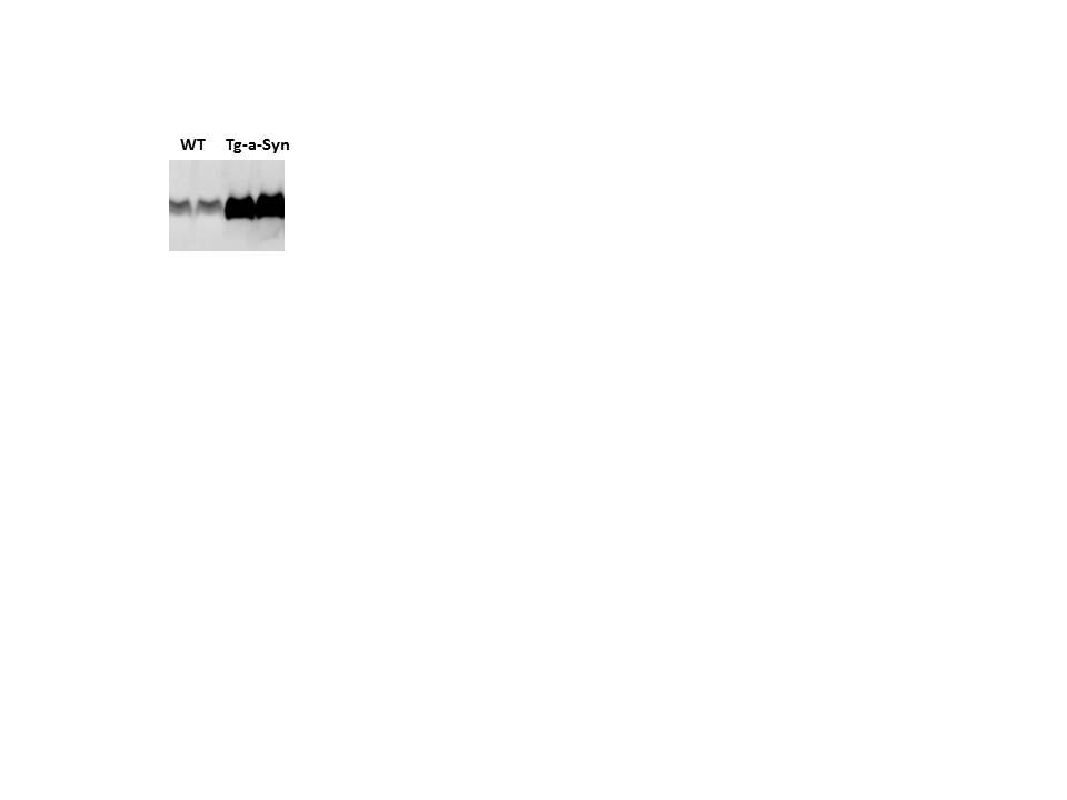
Application: Western BlotSample Tested: Mouse forebrain lysateSpecies: Mouse and HumanVerified Customer | Posted 12/18/2017Alpha-synuclein in forebrain of wildtype (WT) and transgenic (Tg) mice expressing human a-syncuclein

There are no reviews that match your criteria.
Protocols
Find general support by application which include: protocols, troubleshooting, illustrated assays, videos and webinars.
- Antigen Retrieval Protocol (PIER)
- Antigen Retrieval for Frozen Sections Protocol
- Appropriate Fixation of IHC/ICC Samples
- Cellular Response to Hypoxia Protocols
- Chromogenic IHC Staining of Formalin-Fixed Paraffin-Embedded (FFPE) Tissue Protocol
- Chromogenic Immunohistochemistry Staining of Frozen Tissue
- ClariTSA™ Fluorophore Kits
- Detection & Visualization of Antibody Binding
- Fluorescent IHC Staining of Frozen Tissue Protocol
- Graphic Protocol for Heat-induced Epitope Retrieval
- Graphic Protocol for the Preparation and Fluorescent IHC Staining of Frozen Tissue Sections
- Graphic Protocol for the Preparation and Fluorescent IHC Staining of Paraffin-embedded Tissue Sections
- Graphic Protocol for the Preparation of Gelatin-coated Slides for Histological Tissue Sections
- ICC Cell Smear Protocol for Suspension Cells
- ICC Immunocytochemistry Protocol Videos
- ICC for Adherent Cells
- IHC Sample Preparation (Frozen sections vs Paraffin)
- Immunocytochemistry (ICC) Protocol
- Immunocytochemistry Troubleshooting
- Immunofluorescence of Organoids Embedded in Cultrex Basement Membrane Extract
- Immunofluorescent IHC Staining of Formalin-Fixed Paraffin-Embedded (FFPE) Tissue Protocol
- Immunohistochemistry (IHC) and Immunocytochemistry (ICC) Protocols
- Immunohistochemistry Frozen Troubleshooting
- Immunohistochemistry Paraffin Troubleshooting
- Preparing Samples for IHC/ICC Experiments
- Preventing Non-Specific Staining (Non-Specific Binding)
- Primary Antibody Selection & Optimization
- Protocol for Heat-Induced Epitope Retrieval (HIER)
- Protocol for Making a 4% Formaldehyde Solution in PBS
- Protocol for VisUCyte™ HRP Polymer Detection Reagent
- Protocol for the Fluorescent ICC Staining of Cell Smears - Graphic
- Protocol for the Fluorescent ICC Staining of Cultured Cells on Coverslips - Graphic
- Protocol for the Preparation & Fixation of Cells on Coverslips
- Protocol for the Preparation and Chromogenic IHC Staining of Frozen Tissue Sections
- Protocol for the Preparation and Chromogenic IHC Staining of Frozen Tissue Sections - Graphic
- Protocol for the Preparation and Chromogenic IHC Staining of Paraffin-embedded Tissue Sections
- Protocol for the Preparation and Chromogenic IHC Staining of Paraffin-embedded Tissue Sections - Graphic
- Protocol for the Preparation and Fluorescent ICC Staining of Cells on Coverslips
- Protocol for the Preparation and Fluorescent ICC Staining of Non-adherent Cells
- Protocol for the Preparation and Fluorescent ICC Staining of Stem Cells on Coverslips
- Protocol for the Preparation and Fluorescent IHC Staining of Frozen Tissue Sections
- Protocol for the Preparation and Fluorescent IHC Staining of Paraffin-embedded Tissue Sections
- Protocol for the Preparation of Gelatin-coated Slides for Histological Tissue Sections
- Protocol for the Preparation of a Cell Smear for Non-adherent Cell ICC - Graphic
- R&D Systems Quality Control Western Blot Protocol
- TUNEL and Active Caspase-3 Detection by IHC/ICC Protocol
- The Importance of IHC/ICC Controls
- Troubleshooting Guide: Immunohistochemistry
- Troubleshooting Guide: Western Blot Figures
- Western Blot Conditions
- Western Blot Protocol
- Western Blot Protocol for Cell Lysates
- Western Blot Troubleshooting
- Western Blot Troubleshooting Guide
- View all Protocols, Troubleshooting, Illustrated assays and Webinars
FAQs for alpha-Synuclein Antibody - Azide Free
Showing
1
-
1 of
1 FAQ
Showing All
-
Q: I'm looking for an alpha-Synuclein antibody with an epitope located in the first half (N-terminus) of the protein - preferably a monoclonal antibody. Can you help me with that?
A:
Please take a look at NB110-57475. It has been validated for human, rat and mouse and the applications ICC and WB and the epitope it detects is in the N terminal.
Loading...