Anillin Antibody (CL0303) - BSA Free
Novus Biologicals | Catalog # NBP2-52908
Key Product Details
Validated by
Knockout/Knockdown, Orthogonal Validation
Species Reactivity
Human
Applications
Immunohistochemistry, Immunohistochemistry-Paraffin, Western Blot, Immunocytochemistry/ Immunofluorescence, Knockdown Validated
Label
Unconjugated
Antibody Source
Monoclonal Mouse IgG1 Clone # CL0303
Format
BSA Free
Loading...
Product Specifications
Immunogen
This antibody was developed using a recombinant protein derived from Q9NQW6, with the exact immunogen sequence remaining proprietary.
Clonality
Monoclonal
Host
Mouse
Isotype
IgG1
Scientific Data Images for Anillin Antibody (CL0303) - BSA Free
![Western Blot: Anillin Antibody (CL0303) [NBP2-52908]](https://resources.rndsystems.com/images/products/Anillin-Antibody-CL0303-Western-Blot-NBP2-52908-img0016.jpg "Western Blot: Anillin Antibody (CL0303) [NBP2-52908]")
Western Blot: Anillin Antibody (CL0303) [NBP2-52908]
Western Blot: Anillin Antibody (CL0303) [NBP2-52908] - Lane 1: Marker [kDa] Lane 2:Human cell line U-251 MG![Immunocytochemistry/ Immunofluorescence: Anillin Antibody (CL0303) [NBP2-52908]](https://resources.rndsystems.com/images/products/Anillin-Antibody-CL0303-Immunocytochemistry-Immunofluorescence-NBP2-52908-img0017.jpg "Immunocytochemistry/ Immunofluorescence: Anillin Antibody (CL0303) [NBP2-52908]")
Immunocytochemistry/ Immunofluorescence: Anillin Antibody (CL0303) [NBP2-52908]
Immunocytochemistry/Immunofluorescence: Anillin Antibody (CL0303) [NBP2-52908] - Staining in U251 cell line with Anti-ANLN monoclonal antibody, showing cell cycle dependent nuclear (without nucleoli) staining in green. Microtubule- and nuclear probes are visualized in red and blue respectively (where available). Antibody staining is shown in green.![Immunohistochemistry-Paraffin: Anillin Antibody (CL0303) [NBP2-52908]](https://resources.rndsystems.com/images/products/Anillin-Antibody-CL0303-Immunohistochemistry-Paraffin-NBP2-52908-img0025.jpg "Immunohistochemistry-Paraffin: Anillin Antibody (CL0303) [NBP2-52908]")
![Western Blot: Anillin Antibody (CL0303) [NBP2-52908]](https://resources.rndsystems.com/images/products/Anillin-Antibody-CL0303-Western-Blot-NBP2-52908-img0015.jpg "Western Blot: Anillin Antibody (CL0303) [NBP2-52908]")
Western Blot: Anillin Antibody (CL0303) [NBP2-52908]
Western Blot: Anillin Antibody (CL0303) [NBP2-52908] - Analysis in U-251MG cells transfected with control siRNA, target specific siRNA probe #1 and #2, using Anti-ANLN antibody. Remaining relative intensity is presented. Loading control: Anti-GAPDH.![Western Blot: Anillin Antibody (CL0303) [NBP2-52908]](https://resources.rndsystems.com/images/products/Anillin-Antibody-CL0303-Western-Blot-NBP2-52908-img0018.jpg "Western Blot: Anillin Antibody (CL0303) [NBP2-52908]")
Western Blot: Anillin Antibody (CL0303) [NBP2-52908]
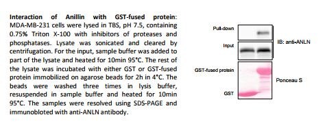
Western Blot: Anillin Antibody (CL0303) [NBP2-52908] - Interaction of Anillin with GST-fused protein: MDA-MB-231 cells were lysed in TBS, pH 7.5, containing 0.75% Triton X-100 with inhibitors of proteases and phosphatases. Lysate was sonicated and cleared by centrifugation. for the input, sample buffer was added to part of the lysate and heated fro 10min 95 C. The rest of the lysate was incubated with either GST or GST-fused protein immobilized on agarose beads for 2h in 4 C. the beads were washed three times in lysis buffer, resuspended in sample buffer and heated for 10min 95 C. The samples were resolved using SDS-PAGE and immunoblotted with anti-ANLN antibody. This image was submitted by customer review.![Immunocytochemistry/ Immunofluorescence: Anillin Antibody (CL0303) [NBP2-52908]](https://resources.rndsystems.com/images/products/Anillin-Antibody-CL0303-Immunocytochemistry-Immunofluorescence-NBP2-52908-img0001.jpg "Immunocytochemistry/ Immunofluorescence: Anillin Antibody (CL0303) [NBP2-52908]")
Immunocytochemistry/ Immunofluorescence: Anillin Antibody (CL0303) [NBP2-52908]
Immunocytochemistry/Immunofluorescence: Anillin Antibody (CL0303) [NBP2-52908] - Staining in A431 cell line with Anti-ANLN monoclonal antibody, showing cell cycle dependent nuclear (without nucleoli) staining in green. Microtubule- and nuclear probes are visualized in red and blue respectively (where available).![Immunocytochemistry/ Immunofluorescence: Anillin Antibody (CL0303) [NBP2-52908]](https://resources.rndsystems.com/images/products/Anillin-Antibody-CL0303-Immunocytochemistry-Immunofluorescence-NBP2-52908-img0002.jpg "Immunocytochemistry/ Immunofluorescence: Anillin Antibody (CL0303) [NBP2-52908]")
Immunocytochemistry/ Immunofluorescence: Anillin Antibody (CL0303) [NBP2-52908]
Immunocytochemistry/Immunofluorescence: Anillin Antibody (CL0303) [NBP2-52908] - Staining in HeLa cell line with Anti-ANLN monoclonal antibody, showing cell cycle dependent nuclear (without nucleoli) staining in green. Microtubule- and nuclear probes are visualized in red and blue respectively (where available).![Immunocytochemistry/ Immunofluorescence: Anillin Antibody (CL0303) [NBP2-52908]](https://resources.rndsystems.com/images/products/Anillin-Antibody-CL0303-Immunocytochemistry-Immunofluorescence-NBP2-52908-img0003.jpg "Immunocytochemistry/ Immunofluorescence: Anillin Antibody (CL0303) [NBP2-52908]")
Immunocytochemistry/ Immunofluorescence: Anillin Antibody (CL0303) [NBP2-52908]
Immunocytochemistry/Immunofluorescence: Anillin Antibody (CL0303) [NBP2-52908] - Staining in MCF7 cell line with Anti-ANLN monoclonal antibody, showing cell cycle dependent nuclear (without nucleoli) staining in green. Microtubule- and nuclear probes are visualized in red and blue respectively (where available).![Immunocytochemistry/ Immunofluorescence: Anillin Antibody (CL0303) [NBP2-52908]](https://resources.rndsystems.com/images/products/Anillin-Antibody-CL0303-Immunocytochemistry-Immunofluorescence-NBP2-52908-img0005.jpg "Immunocytochemistry/ Immunofluorescence: Anillin Antibody (CL0303) [NBP2-52908]")
Immunocytochemistry/ Immunofluorescence: Anillin Antibody (CL0303) [NBP2-52908]
Immunocytochemistry/Immunofluorescence: Anillin Antibody (CL0303) [NBP2-52908] - Staining in U2OS cell line with Anti-ANLN monoclonal antibody, showing cell cycle dependent nuclear (without nucleoli) staining in green. Microtubule- and nuclear probes are visualized in red and blue respectively (where available).![Immunohistochemistry-Paraffin: Anillin Antibody (CL0303) [NBP2-52908]](https://resources.rndsystems.com/images/products/Anillin-Antibody-CL0303-Immunohistochemistry-Paraffin-NBP2-52908-img0020.jpg "Immunohistochemistry-Paraffin: Anillin Antibody (CL0303) [NBP2-52908]")
Immunohistochemistry-Paraffin: Anillin Antibody (CL0303) [NBP2-52908]
Immunohistochemistry-Paraffin: Anillin Antibody (CL0303) [NBP2-52908] - Staining of human colorectal cancer shows moderate to strong nuclear positivity in a subset of tumor cells.![Immunohistochemistry-Paraffin: Anillin Antibody (CL0303) [NBP2-52908]](https://resources.rndsystems.com/images/products/Anillin-Antibody-CL0303-Immunohistochemistry-Paraffin-NBP2-52908-img0021.jpg "Immunohistochemistry-Paraffin: Anillin Antibody (CL0303) [NBP2-52908]")
Immunohistochemistry-Paraffin: Anillin Antibody (CL0303) [NBP2-52908]
Immunohistochemistry-Paraffin: Anillin Antibody (CL0303) [NBP2-52908] - Staining of human testis shows moderate to strong nuclear positivity in a subset of cells in seminiferous ducts.![Immunohistochemistry-Paraffin: Anillin Antibody (CL0303) [NBP2-52908]](https://resources.rndsystems.com/images/products/Anillin-Antibody-CL0303-Immunohistochemistry-Paraffin-NBP2-52908-img0022.jpg "Immunohistochemistry-Paraffin: Anillin Antibody (CL0303) [NBP2-52908]")
Immunohistochemistry-Paraffin: Anillin Antibody (CL0303) [NBP2-52908]
Immunohistochemistry-Paraffin: Anillin Antibody (CL0303) [NBP2-52908] - Staining of human rectum shows moderate to strong nuclear positivity in a subset of glandular cells.![Immunohistochemistry-Paraffin: Anillin Antibody (CL0303) [NBP2-52908]](https://resources.rndsystems.com/images/products/Anillin-Antibody-CL0303-Immunohistochemistry-Paraffin-NBP2-52908-img0023.jpg "Immunohistochemistry-Paraffin: Anillin Antibody (CL0303) [NBP2-52908]")
Immunohistochemistry-Paraffin: Anillin Antibody (CL0303) [NBP2-52908]
Immunohistochemistry-Paraffin: Anillin Antibody (CL0303) [NBP2-52908] - Staining of human skin shows moderate to strong nuclear positivity in a subset of dermal cells.![Immunohistochemistry-Paraffin: Anillin Antibody (CL0303) [NBP2-52908]](https://resources.rndsystems.com/images/products/Anillin-Antibody-CL0303-Immunohistochemistry-Paraffin-NBP2-52908-img0024.jpg "Immunohistochemistry-Paraffin: Anillin Antibody (CL0303) [NBP2-52908]")
Immunohistochemistry-Paraffin: Anillin Antibody (CL0303) [NBP2-52908]
Immunohistochemistry-Paraffin: Anillin Antibody (CL0303) [NBP2-52908] - Staining of human skeletal muscle shows no nuclear positivity in striated muscle fibers as expected.Applications for Anillin Antibody (CL0303) - BSA Free
Application
Recommended Usage
Immunocytochemistry/ Immunofluorescence
2-10 ug/ml
Immunohistochemistry
1:50 - 1:200
Immunohistochemistry-Paraffin
1:50 - 1:200
Western Blot
1 ug/ml
Application Notes
For IHC-Paraffin, HIER pH 6 retrieval is recommended. Immunocytochemistry/Immunofluorescence Fixation Permeabilization: Use PFA/Triton X-100.
Reviewed Applications
Read 1 review rated 5 using NBP2-52908 in the following applications:
Formulation, Preparation, and Storage
Purification
Protein A purified
Formulation
PBS (pH 7.2) and 40% Glycerol
Format
BSA Free
Preservative
0.02% Sodium Azide
Concentration
1 mg/ml
Shipping
The product is shipped with polar packs. Upon receipt, store it immediately at the temperature recommended below.
Stability & Storage
Store at 4C short term. Aliquot and store at -20C long term. Avoid freeze-thaw cycles.
Background: Anillin
Alternate Names
actin-binding protein anillin, ANILLIN, anillin (Drosophila Scraps homolog), actin binding protein, anillin, actin binding protein, anillin, actin binding protein (scraps homolog, Drosophila), DKFZp779A055, Drosophila Scraps homolog, scra, Scraps
Gene Symbol
ANLN
Additional Anillin Products
Product Documents for Anillin Antibody (CL0303) - BSA Free
Certificate of Analysis
To download a Certificate of Analysis, please enter a lot or batch number in the search box below.
Product Specific Notices for Anillin Antibody (CL0303) - BSA Free
This product is for research use only and is not approved for use in humans or in clinical diagnosis. Primary Antibodies are guaranteed for 1 year from date of receipt.
Citations for Anillin Antibody (CL0303) - BSA Free
 Powered by Bioz
Customer Reviews for Anillin Antibody (CL0303) - BSA Free (1)
5 out of 5
1 Customer Rating
Have you used Anillin Antibody (CL0303) - BSA Free?
Submit a review and receive an Amazon gift card!
$25/€18/£15/$25CAN/¥2500 Yen for a review with an image
$10/€7/£6/$10CAN/¥1110 Yen for a review without an image
Submit a review
Customer Images
Showing
1
-
1 of
1 review
Showing All
Filter By:
-
Application: Western BlotSample Tested: GST-pull down assaySpecies: HumanVerified Customer | Posted 01/15/2018

There are no reviews that match your criteria.
Protocols
Find general support by application which include: protocols, troubleshooting, illustrated assays, videos and webinars.
- Antigen Retrieval Protocol (PIER)
- Antigen Retrieval for Frozen Sections Protocol
- Appropriate Fixation of IHC/ICC Samples
- Cellular Response to Hypoxia Protocols
- Chromogenic IHC Staining of Formalin-Fixed Paraffin-Embedded (FFPE) Tissue Protocol
- Chromogenic Immunohistochemistry Staining of Frozen Tissue
- ClariTSA™ Fluorophore Kits
- Detection & Visualization of Antibody Binding
- Fluorescent IHC Staining of Frozen Tissue Protocol
- Graphic Protocol for Heat-induced Epitope Retrieval
- Graphic Protocol for the Preparation and Fluorescent IHC Staining of Frozen Tissue Sections
- Graphic Protocol for the Preparation and Fluorescent IHC Staining of Paraffin-embedded Tissue Sections
- Graphic Protocol for the Preparation of Gelatin-coated Slides for Histological Tissue Sections
- ICC Cell Smear Protocol for Suspension Cells
- ICC Immunocytochemistry Protocol Videos
- ICC for Adherent Cells
- IHC Sample Preparation (Frozen sections vs Paraffin)
- Immunocytochemistry (ICC) Protocol
- Immunocytochemistry Troubleshooting
- Immunofluorescence of Organoids Embedded in Cultrex Basement Membrane Extract
- Immunofluorescent IHC Staining of Formalin-Fixed Paraffin-Embedded (FFPE) Tissue Protocol
- Immunohistochemistry (IHC) and Immunocytochemistry (ICC) Protocols
- Immunohistochemistry Frozen Troubleshooting
- Immunohistochemistry Paraffin Troubleshooting
- Preparing Samples for IHC/ICC Experiments
- Preventing Non-Specific Staining (Non-Specific Binding)
- Primary Antibody Selection & Optimization
- Protocol for Heat-Induced Epitope Retrieval (HIER)
- Protocol for Making a 4% Formaldehyde Solution in PBS
- Protocol for VisUCyte™ HRP Polymer Detection Reagent
- Protocol for the Fluorescent ICC Staining of Cell Smears - Graphic
- Protocol for the Fluorescent ICC Staining of Cultured Cells on Coverslips - Graphic
- Protocol for the Preparation & Fixation of Cells on Coverslips
- Protocol for the Preparation and Chromogenic IHC Staining of Frozen Tissue Sections
- Protocol for the Preparation and Chromogenic IHC Staining of Frozen Tissue Sections - Graphic
- Protocol for the Preparation and Chromogenic IHC Staining of Paraffin-embedded Tissue Sections
- Protocol for the Preparation and Chromogenic IHC Staining of Paraffin-embedded Tissue Sections - Graphic
- Protocol for the Preparation and Fluorescent ICC Staining of Cells on Coverslips
- Protocol for the Preparation and Fluorescent ICC Staining of Non-adherent Cells
- Protocol for the Preparation and Fluorescent ICC Staining of Stem Cells on Coverslips
- Protocol for the Preparation and Fluorescent IHC Staining of Frozen Tissue Sections
- Protocol for the Preparation and Fluorescent IHC Staining of Paraffin-embedded Tissue Sections
- Protocol for the Preparation of Gelatin-coated Slides for Histological Tissue Sections
- Protocol for the Preparation of a Cell Smear for Non-adherent Cell ICC - Graphic
- R&D Systems Quality Control Western Blot Protocol
- TUNEL and Active Caspase-3 Detection by IHC/ICC Protocol
- The Importance of IHC/ICC Controls
- Troubleshooting Guide: Immunohistochemistry
- Troubleshooting Guide: Western Blot Figures
- Western Blot Conditions
- Western Blot Protocol
- Western Blot Protocol for Cell Lysates
- Western Blot Troubleshooting
- Western Blot Troubleshooting Guide
- View all Protocols, Troubleshooting, Illustrated assays and Webinars
Loading...