Annexin A2 Antibody (1G7) - Azide and BSA Free
Novus Biologicals | Catalog # H00000302-M02
![Immunocytochemistry/ Immunofluorescence: Annexin A2 Antibody (1G7) [H00000302-M02]](https://resources.rndsystems.com/images/products/Annexin-A2-Antibody-1G7-Immunocytochemistry-Immunofluorescence-H00000302-M02-img0007.jpg "Immunocytochemistry/ Immunofluorescence: Annexin A2 Antibody (1G7) [H00000302-M02]")
Key Product Details
Validated by
Species Reactivity
Validated:
Cited:
Applications
Validated:
Cited:
Label
Antibody Source
Format
Product Specifications
Immunogen
Reactivity Notes
Specificity
Clonality
Host
Isotype
Description
Scientific Data Images for Annexin A2 Antibody (1G7) - Azide and BSA Free
Immunocytochemistry/ Immunofluorescence: Annexin A2 Antibody (1G7) [H00000302-M02]
Annexin-A2-Antibody-1G7-Immunocytochemistry-Immunofluorescence-H00000302-M02-img0007.jpg![Immunocytochemistry/ Immunofluorescence: Annexin A2 Antibody (1G7) [H00000302-M02]](https://resources.rndsystems.com/images/products/Annexin-A2-Antibody-1G7-Immunocytochemistry-Immunofluorescence-H00000302-M02-img0008.jpg "Immunocytochemistry/ Immunofluorescence: Annexin A2 Antibody (1G7) [H00000302-M02]")
![Western Blot: Annexin A2 Antibody (1G7) [H00000302-M02]](https://resources.rndsystems.com/images/products/Annexin-A2-Antibody-1G7-Western-Blot-H00000302-M02-img0009.jpg "Western Blot: Annexin A2 Antibody (1G7) [H00000302-M02]")
Western Blot: Annexin A2 Antibody (1G7) [H00000302-M02]
Western Blot: Annexin A2 Antibody (1G7) [H00000302-M02] - MDA-MB-231 subclones whole cell lysates were loaded with 30 ug/lane. 10% SDS-PAGE. ANXA2 Antibody (H00000302-M02) was used for primary antibody: 1:2000, 4C, overnight. WB image submitted by a verified customer review.![Western Blot: Annexin A2 Antibody (1G7) [H00000302-M02]](https://resources.rndsystems.com/images/products/Annexin-A2-Antibody-1G7-Western-Blot-H00000302-M02-img0005.jpg "Western Blot: Annexin A2 Antibody (1G7) [H00000302-M02]")
Western Blot: Annexin A2 Antibody (1G7) [H00000302-M02]
Western Blot: Annexin A2 Antibody (1G7) [H00000302-M02] - ANXA2 monoclonal antibody (M02), clone 1G7 Analysis of ANXA2 expression in HepG2.![Western Blot: Annexin A2 Antibody (1G7) [H00000302-M02]](https://resources.rndsystems.com/images/products/Annexin-A2-Antibody-1G7-Western-Blot-H00000302-M02-img0006.jpg "Western Blot: Annexin A2 Antibody (1G7) [H00000302-M02]")
Western Blot: Annexin A2 Antibody (1G7) [H00000302-M02]
Western Blot: Annexin A2 Antibody (1G7) [H00000302-M02] - Analysis of ANXA2 expression in transfected 293T cell line by ANXA2 monoclonal antibody (M02), clone 1G7.Lane 1: ANXA2 transfected lysate(40.4 KDa).Lane 2: Non-transfected lysate.![Immunocytochemistry/ Immunofluorescence: Annexin A2 Antibody (1G7) [H00000302-M02]](https://resources.rndsystems.com/images/products/Annexin-A2-Antibody-1G7-Immunocytochemistry-Immunofluorescence-H00000302-M02-img0004.jpg "Immunocytochemistry/ Immunofluorescence: Annexin A2 Antibody (1G7) [H00000302-M02]")
Immunocytochemistry/ Immunofluorescence: Annexin A2 Antibody (1G7) [H00000302-M02]
Immunocytochemistry/Immunofluorescence: Annexin A2 Antibody (1G7) [H00000302-M02] - Analysis of monoclonal antibody to ANXA2 on HeLa cell. Antibody concentration 10 ug/ml. [H00000302-M02] -")
Immunocytochemistry/ Immunofluorescence: Annexin A2 Antibody (1G7) [H00000302-M02] -
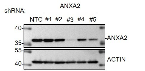
Immunocytochemistry/ Immunofluorescence: Annexin A2 Antibody (1G7) [H00000302-M02] - ANXA2 could regulate the activation of NF-kappa B through combined with the subunit p50. a, b. The change in localization of endogenous p50 was analyzed by IF-IC following treatment with or without TNF-alpha (100 ng/ml) for 1 h. Nuclei were stained with DAPI. The nuclear localization of endogenous p50 in each group were quantitated as a graph. The plotted error bars represent mean ± SEM. The nuclear localization of p50 was quantified according to data from 5 random figures in each group. c. Interaction between ANXA2 & p50 in SK-N-BE(2) cells was validated through Co-IP & analyzed by western blotting with the indicated antibodies **: P < 0.01. All results are from at least three independent experiments Image collected & cropped by CiteAb from the following publication (http://jeccr.biomedcentral.com/articles/10.1186/s13046-017-0581-6), licensed under a CC-BY license. Not internally tested by Novus Biologicals.Applications for Annexin A2 Antibody (1G7) - Azide and BSA Free
Immunocytochemistry/ Immunofluorescence
Western Blot
Reviewed Applications
Read 1 review rated 5 using H00000302-M02 in the following applications:
Flow Cytometry Panel Builder
Bio-Techne Knows Flow Cytometry
Save time and reduce costly mistakes by quickly finding compatible reagents using the Panel Builder Tool.
Advanced Features
- Spectra Viewer - Custom analysis of spectra from multiple fluorochromes
- Spillover Popups - Visualize the spectra of individual fluorochromes
- Antigen Density Selector - Match fluorochrome brightness with antigen density

Formulation, Preparation, and Storage
Purification
Formulation
Format
Preservative
Concentration
Shipping
Stability & Storage
Background: Annexin A2
Alternate Names
Entrez Gene IDs
Gene Symbol
OMIM
UniProt
Additional Annexin A2 Products
Product Documents for Annexin A2 Antibody (1G7) - Azide and BSA Free
Certificate of Analysis
To download a Certificate of Analysis, please enter a lot or batch number in the search box below.
Product Specific Notices for Annexin A2 Antibody (1G7) - Azide and BSA Free
This product is produced by and distributed for Abnova, a company based in Taiwan.
This product is for research use only and is not approved for use in humans or in clinical diagnosis. Primary Antibodies are guaranteed for 1 year from date of receipt.
Citations for Annexin A2 Antibody (1G7) - Azide and BSA Free
 Powered by Bioz
Customer Reviews for Annexin A2 Antibody (1G7) - Azide and BSA Free (1)
Have you used Annexin A2 Antibody (1G7) - Azide and BSA Free?
Submit a review and receive an Amazon gift card!
$25/€18/£15/$25CAN/¥2500 Yen for a review with an image
$10/€7/£6/$10CAN/¥1110 Yen for a review without an image
Submit a review
Customer Images
-
Application: Western BlotSample Tested: MDA-MB-231Species: HumanVerified Customer | Posted 09/12/2021Western Blot: MDA-MB-231 subclones whole cell lysates were loaded with 30 ug/lane. 10% SDS-PAGE. ANXA2 Antibody (H00000302-M02) was used for primary antibody: 1:2000, 4℃, overnight.

There are no reviews that match your criteria.
Protocols
Find general support by application which include: protocols, troubleshooting, illustrated assays, videos and webinars.
- 7-Amino Actinomycin D (7-AAD) Cell Viability Flow Cytometry Protocol
- Appropriate Fixation of IHC/ICC Samples
- Cellular Response to Hypoxia Protocols
- ClariTSA™ Fluorophore Kits
- Detection & Visualization of Antibody Binding
- ELISA Sample Preparation & Collection Guide
- ELISA Troubleshooting Guide
- Extracellular Membrane Flow Cytometry Protocol
- Flow Cytometry Protocol for Cell Surface Markers
- Flow Cytometry Protocol for Staining Membrane Associated Proteins
- Flow Cytometry Staining Protocols
- Flow Cytometry Troubleshooting Guide
- How to Run an R&D Systems DuoSet ELISA
- How to Run an R&D Systems Quantikine ELISA
- How to Run an R&D Systems Quantikine™ QuicKit™ ELISA
- ICC Cell Smear Protocol for Suspension Cells
- ICC Immunocytochemistry Protocol Videos
- ICC for Adherent Cells
- Immunocytochemistry (ICC) Protocol
- Immunocytochemistry Troubleshooting
- Immunofluorescence of Organoids Embedded in Cultrex Basement Membrane Extract
- Immunohistochemistry (IHC) and Immunocytochemistry (ICC) Protocols
- Immunoprecipitation Protocol
- Intracellular Flow Cytometry Protocol Using Alcohol (Methanol)
- Intracellular Flow Cytometry Protocol Using Detergents
- Intracellular Nuclear Staining Flow Cytometry Protocol Using Detergents
- Intracellular Staining Flow Cytometry Protocol Using Alcohol Permeabilization
- Intracellular Staining Flow Cytometry Protocol Using Detergents to Permeabilize Cells
- Preparing Samples for IHC/ICC Experiments
- Preventing Non-Specific Staining (Non-Specific Binding)
- Primary Antibody Selection & Optimization
- Propidium Iodide Cell Viability Flow Cytometry Protocol
- Protocol for Liperfluo
- Protocol for VisUCyte™ HRP Polymer Detection Reagent
- Protocol for the Characterization of Human Th22 Cells
- Protocol for the Characterization of Human Th9 Cells
- Protocol for the Fluorescent ICC Staining of Cell Smears - Graphic
- Protocol for the Fluorescent ICC Staining of Cultured Cells on Coverslips - Graphic
- Protocol for the Preparation and Fluorescent ICC Staining of Cells on Coverslips
- Protocol for the Preparation and Fluorescent ICC Staining of Non-adherent Cells
- Protocol for the Preparation and Fluorescent ICC Staining of Stem Cells on Coverslips
- Protocol for the Preparation of a Cell Smear for Non-adherent Cell ICC - Graphic
- Protocol: Annexin V and PI Staining by Flow Cytometry
- Protocol: Annexin V and PI Staining for Apoptosis by Flow Cytometry
- Quantikine HS ELISA Kit Assay Principle, Alkaline Phosphatase
- Quantikine HS ELISA Kit Principle, Streptavidin-HRP Polymer
- R&D Systems Quality Control Western Blot Protocol
- Sandwich ELISA (Colorimetric) – Biotin/Streptavidin Detection Protocol
- Sandwich ELISA (Colorimetric) – Direct Detection Protocol
- TUNEL and Active Caspase-3 Detection by IHC/ICC Protocol
- The Importance of IHC/ICC Controls
- Troubleshooting Guide: ELISA
- Troubleshooting Guide: Fluorokine Flow Cytometry Kits
- Troubleshooting Guide: Western Blot Figures
- Western Blot Conditions
- Western Blot Protocol
- Western Blot Protocol for Cell Lysates
- Western Blot Troubleshooting
- Western Blot Troubleshooting Guide
- View all Protocols, Troubleshooting, Illustrated assays and Webinars