APP Antibody (6E10) - Chimeric - Azide and BSA Free
Novus Biologicals | Catalog # NBP2-62566
Recombinant Monoclonal Antibody
 - Chimeric [NBP2-62566] -")
![Western Blot: APP Antibody (6E10)ChimericAzide and BSA Free [NBP2-62566]](https://resources.rndsystems.com/images/products/APP-Antibody-6E10-Chimeric-Western-Blot-NBP2-62566-img0002.jpg "Western Blot: APP Antibody (6E10)ChimericAzide and BSA Free [NBP2-62566]")
Loading...
Key Product Details
Validated by
Biological Validation
Species Reactivity
Validated:
Human, Mouse (Negative)
Cited:
Human, Mouse
Applications
Validated:
Immunohistochemistry, Immunohistochemistry-Paraffin, Immunohistochemistry-Frozen, Western Blot, ELISA, Immunoprecipitation, Electron Microscopy
Cited:
Immunohistochemistry, Immunohistochemistry-Paraffin, Immunohistochemistry-Frozen, Western Blot, ELISA, Immunocytochemistry/ Immunofluorescence, IF/IHC
Label
Unconjugated
Antibody Source
Monoclonal Rabbit IgG Kappa Clone # 6E10
Format
Azide and BSA Free
Loading...
Product Specifications
Immunogen
Amyloid-beta peptide
Specificity
This antibody binds to amino acid residues 1-16 of beta amyloid.
Clonality
Monoclonal
Host
Rabbit
Isotype
IgG Kappa
Scientific Data Images for APP Antibody (6E10) - Chimeric - Azide and BSA Free
Western Blot: APP Antibody (6E10)ChimericAzide and BSA Free [NBP2-62566]
Western Blot: APP Antibody (6E10) - Chimeric [NBP2-62566] - Anti-Amyloid beta antibody 6E10. Human brain cerebral cortex (A), frontal cortex (B) and cerebellum (C) lysate samples (35ug protein in RIPA buffer) were resolved on a 10% SDS PAGE gel and blots probed with the chimeric rabbit version of 6E10 (NBP2-62566) at 0.3 ug/ml before detection using an anti-rabbit secondary antibody. A primary incubation of 1h was used and protein was detected by chemiluminescence. The expected band size for Amyloid beta is ~35kDa, though this protein is known to form SDS-resistant low-molecular weight oligomers as seen in this blot (i.e. dimers, c.f. Kumar et al. PMID: 21527912). NBP2-62566 successfully detected aggregate human Amyloid beta in human brain cerebral cortex, frontal cortex and cerebellum lysate samples.![Immunohistochemistry-Paraffin: APP Antibody (6E10) - Chimeric - Azide and BSA Free [NBP2-62566]](https://resources.rndsystems.com/images/products/APP-Antibody-6E10-Chimeric-Immunohistochemistry-Paraffin-NBP2-62566-img0001.jpg "Immunohistochemistry-Paraffin: APP Antibody (6E10) - Chimeric - Azide and BSA Free [NBP2-62566]")
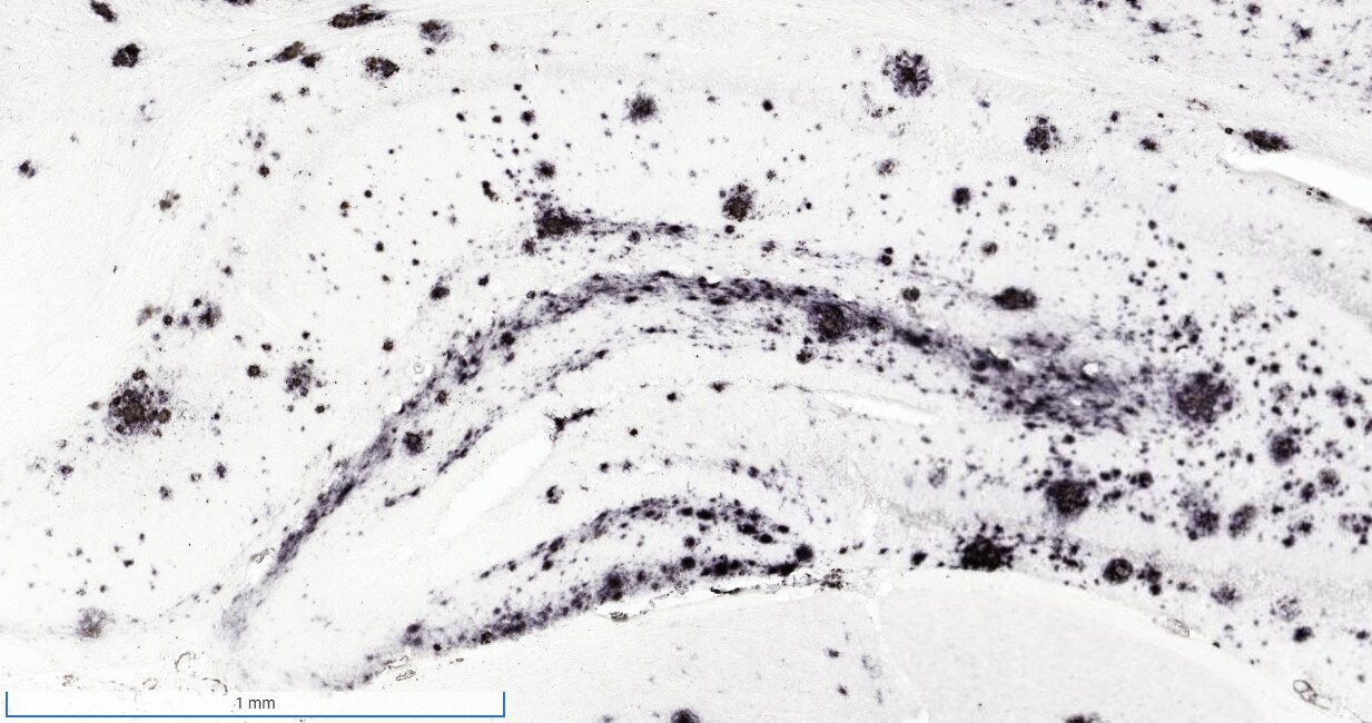
Immunohistochemistry-Paraffin: APP Antibody (6E10) - Chimeric - Azide and BSA Free [NBP2-62566]
Immunohistochemistry-Paraffin: APP Antibody (6E10) - Chimeric [NBP2-62566] - Staining of human Alzheimer's disease hippocampus tissue using anti-Amyloid Beta antibody. 6E10 Anti-Amyloid Beta staining of paraffin embedded human hippocampus affected by Alzheimer's disease using the rabbit-chimeric version of 6E10 (NBP2-62566). Antigen retreival was acheived by microwaving in citrate buffer (pH6), followed by blocking with protein block serum-free buffer. Primary antibody incubation with NBP2-62566 was carried out at 4 ug/ml for 30 minutes. Samples were then incubated with an anti-rabbit IgG HRP secondary antibody for 20 mins followed by DAB (3,3'-diaminobenzidine), and counter-staining with haemotoxylin. Staining of amyloid plaques in the parahippocampal gyrus may be observed. Recommended concentration, 2-4 ug/ml. - Chimeric [NBP2-62566] -")
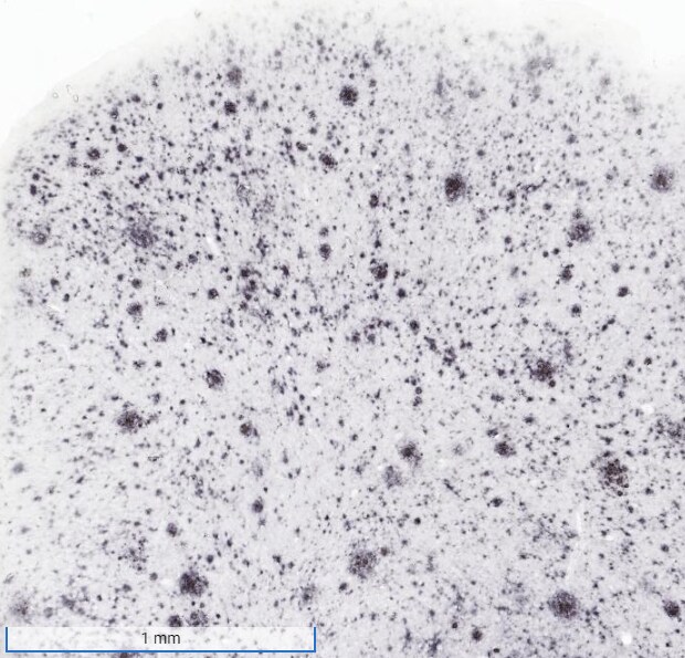
Immunohistochemistry-Frozen: APP Antibody (6E10) - Chimeric [NBP2-62566] -
Immunohistochemistry-Frozen: APP Antibody (6E10) - Chimeric [NBP2-62566] - 6E10 staining reveals amyloid plaque in frontal cortex of AD mouse model. Image from verified customer review.Applications for APP Antibody (6E10) - Chimeric - Azide and BSA Free
Application
Recommended Usage
ELISA
1:100 - 1:2000
Electron Microscopy
1:10 - 1:500
Immunohistochemistry
1:10 - 1:500
Immunoprecipitation
1:10 - 1:500
Western Blot
1:100 - 1:2000
Application Notes
This chimeric rabbit antibody was made using the variable domain sequences of the original Mouse IgG1 format, for improved compatibility with existing reagents, assays and techniques. APP Antibody (6E10) validated for Immunohistochemistry-Frozen from a verified customer review.
Reviewed Applications
Read 2 reviews rated 5 using NBP2-62566 in the following applications:
Formulation, Preparation, and Storage
Purification
Protein A purified
Formulation
PBS
Format
Azide and BSA Free
Preservative
0.02% Proclin 300
Concentration
1 mg/ml
Shipping
The product is shipped with polar packs. Upon receipt, store it immediately at the temperature recommended below.
Stability & Storage
Store at 4C for up to 3 months. For longer storage, aliquot and store at -20C.
Background: APP
Long Name
Amyloid Precursor Protein
Alternate Names
Amyloid beta, beta Amyloid, Protease Nexin II
Gene Symbol
APP
Additional APP Products
Product Documents for APP Antibody (6E10) - Chimeric - Azide and BSA Free
Certificate of Analysis
To download a Certificate of Analysis, please enter a lot or batch number in the search box below.
Product Specific Notices for APP Antibody (6E10) - Chimeric - Azide and BSA Free
This product is for research use only and is not approved for use in humans or in clinical diagnosis. Primary Antibodies are guaranteed for 1 year from date of receipt.
Related Research Areas
Citations for APP Antibody (6E10) - Chimeric - Azide and BSA Free
 Powered by Bioz
Customer Reviews for APP Antibody (6E10) - Chimeric - Azide and BSA Free (2)
5 out of 5
2 Customer Ratings
Have you used APP Antibody (6E10) - Chimeric - Azide and BSA Free?
Submit a review and receive an Amazon gift card!
$25/€18/£15/$25CAN/¥2500 Yen for a review with an image
$10/€7/£6/$10CAN/¥1110 Yen for a review without an image
Submit a review
Customer Images
Showing
1
-
2 of
2 reviews
Showing All
Filter By:
-
Application: Immunohistochemistry-FrozenSample Tested: Mouse brainSpecies: MouseVerified Customer | Posted 06/20/2023Plaque staining in AD Mouse model (Hippocampus)-without formic acid pretreatment

-
Application: Immunohistochemistry-FrozenSample Tested: Mouse brainSpecies: MouseVerified Customer | Posted 04/24/20236E10 staining reveals amyloid plaque in frontal cortex of AD mouse model.Free-floating IHC. Staining at a dilution of 1:500,000. Antibody worked phenomenally!
Bio-Techne ResponseThis review was submitted through the legacy Novus Innovators Program, reflecting a new species or application tested on a primary antibody.
There are no reviews that match your criteria.
Protocols
Find general support by application which include: protocols, troubleshooting, illustrated assays, videos and webinars.
- Antigen Retrieval Protocol (PIER)
- Antigen Retrieval for Frozen Sections Protocol
- Appropriate Fixation of IHC/ICC Samples
- Cellular Response to Hypoxia Protocols
- Chromogenic IHC Staining of Formalin-Fixed Paraffin-Embedded (FFPE) Tissue Protocol
- Chromogenic Immunohistochemistry Staining of Frozen Tissue
- ClariTSA™ Fluorophore Kits
- Detection & Visualization of Antibody Binding
- ELISA Sample Preparation & Collection Guide
- ELISA Troubleshooting Guide
- Fluorescent IHC Staining of Frozen Tissue Protocol
- Graphic Protocol for Heat-induced Epitope Retrieval
- Graphic Protocol for the Preparation and Fluorescent IHC Staining of Frozen Tissue Sections
- Graphic Protocol for the Preparation and Fluorescent IHC Staining of Paraffin-embedded Tissue Sections
- Graphic Protocol for the Preparation of Gelatin-coated Slides for Histological Tissue Sections
- How to Run an R&D Systems DuoSet ELISA
- How to Run an R&D Systems Quantikine ELISA
- How to Run an R&D Systems Quantikine™ QuicKit™ ELISA
- IHC Sample Preparation (Frozen sections vs Paraffin)
- Immunofluorescent IHC Staining of Formalin-Fixed Paraffin-Embedded (FFPE) Tissue Protocol
- Immunohistochemistry (IHC) and Immunocytochemistry (ICC) Protocols
- Immunohistochemistry Frozen Troubleshooting
- Immunohistochemistry Paraffin Troubleshooting
- Immunoprecipitation Protocol
- Preparing Samples for IHC/ICC Experiments
- Preventing Non-Specific Staining (Non-Specific Binding)
- Primary Antibody Selection & Optimization
- Protocol for Heat-Induced Epitope Retrieval (HIER)
- Protocol for Making a 4% Formaldehyde Solution in PBS
- Protocol for VisUCyte™ HRP Polymer Detection Reagent
- Protocol for the Preparation & Fixation of Cells on Coverslips
- Protocol for the Preparation and Chromogenic IHC Staining of Frozen Tissue Sections
- Protocol for the Preparation and Chromogenic IHC Staining of Frozen Tissue Sections - Graphic
- Protocol for the Preparation and Chromogenic IHC Staining of Paraffin-embedded Tissue Sections
- Protocol for the Preparation and Chromogenic IHC Staining of Paraffin-embedded Tissue Sections - Graphic
- Protocol for the Preparation and Fluorescent IHC Staining of Frozen Tissue Sections
- Protocol for the Preparation and Fluorescent IHC Staining of Paraffin-embedded Tissue Sections
- Protocol for the Preparation of Gelatin-coated Slides for Histological Tissue Sections
- Quantikine HS ELISA Kit Assay Principle, Alkaline Phosphatase
- Quantikine HS ELISA Kit Principle, Streptavidin-HRP Polymer
- R&D Systems Quality Control Western Blot Protocol
- Sandwich ELISA (Colorimetric) – Biotin/Streptavidin Detection Protocol
- Sandwich ELISA (Colorimetric) – Direct Detection Protocol
- TUNEL and Active Caspase-3 Detection by IHC/ICC Protocol
- The Importance of IHC/ICC Controls
- Troubleshooting Guide: ELISA
- Troubleshooting Guide: Immunohistochemistry
- Troubleshooting Guide: Western Blot Figures
- Western Blot Conditions
- Western Blot Protocol
- Western Blot Protocol for Cell Lysates
- Western Blot Troubleshooting
- Western Blot Troubleshooting Guide
- View all Protocols, Troubleshooting, Illustrated assays and Webinars


