ASC/TMS1 Antibody - BSA Free
Novus Biologicals | Catalog # NBP1-78977
![Simple Western: ASC/TMS1 AntibodyBSA Free [NBP1-78977]](https://resources.rndsystems.com/images/products/ASC-TMS1-Antibody-Simple-Western-NBP1-78977-img0005.jpg "Simple Western: ASC/TMS1 AntibodyBSA Free [NBP1-78977]")
Key Product Details
Validated by
Biological Validation
Species Reactivity
Validated:
Human, Mouse, Rat
Cited:
Human, Mouse, Rat
Applications
Validated:
Immunohistochemistry, Immunohistochemistry-Paraffin, Immunomicroscopy, Western Blot, Flow Cytometry, Flow (Intracellular), Immunocytochemistry/ Immunofluorescence, Simple Western, Immunoprecipitation
Cited:
Immunohistochemistry, Immunohistochemistry-Paraffin, Immunomicroscopy, Western Blot, Flow Cytometry, Immunocytochemistry/ Immunofluorescence, Immunoprecipitation, Flow Cytometry Control, IF/IHC
Label
Unconjugated
Antibody Source
Polyclonal Rabbit IgG
Format
BSA Free
Loading...
Product Specifications
Immunogen
This ASC/TMS1 Antibody was developed against a synthetic peptide made to an N-terminal portion of the human ASC/TMS1 protein (between residues 1-50) [Uniprot: Q9ULZ3]
Reactivity Notes
Reactivity with Rat reported in PMID 24464748
Localization
Cytoplasm. Note: Upstream of caspase activation, a redistribution from the cytoplasm to the aggregates occurs. These appear as hollow, perinuclear spherical, ball-like structures.
Clonality
Polyclonal
Host
Rabbit
Isotype
IgG
Scientific Data Images for ASC/TMS1 Antibody - BSA Free
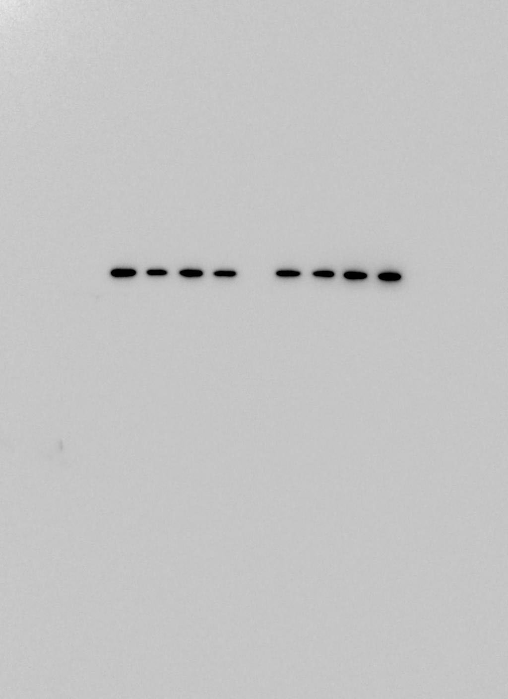
Simple Western: ASC/TMS1 AntibodyBSA Free [NBP1-78977]
Simple Western: ASC/TMS1 Antibody [NBP1-78977] - Lane view shows a specific band for ASC TMS1 in 0.5 mg/ml of MCF-7 lysate. This experiment was performed under reducing conditions using the 12-230 kDa separation system.![Immunocytochemistry/ Immunofluorescence: ASC/TMS1 Antibody - BSA Free [NBP1-78977]](https://resources.rndsystems.com/images/products/ASC-TMS1-Antibody-Immunocytochemistry-Immunofluorescence-NBP1-78977-img0008.jpg "Immunocytochemistry/ Immunofluorescence: ASC/TMS1 Antibody - BSA Free [NBP1-78977]")
Immunocytochemistry/ Immunofluorescence: ASC/TMS1 Antibody - BSA Free [NBP1-78977]
Immunocytochemistry/Immunofluorescence: ASC/TMS1 Antibody [NBP1-78977] - NLRP3 activation was evaluated by visualizing the co-localization of NLRP3 and apoptosis-associated speck-like protein (ASC) using immunocytochemistry (A). PLoS One. 2020 Jun 17;15(6):e0234039. doi: 10.1371/journal.pone.0234039. (Catalog # NBP1-78977AF647)![Immunohistochemistry: ASC/TMS1 Antibody - BSA Free [NBP1-78977]](https://resources.rndsystems.com/images/products/ASC-TMS1-Antibody-Immunohistochemistry-NBP1-78977-img0009.jpg "Immunohistochemistry: ASC/TMS1 Antibody - BSA Free [NBP1-78977]")
Immunohistochemistry: ASC/TMS1 Antibody - BSA Free [NBP1-78977]
Immunohistochemistry: ASC/TMS1 Antibody [NBP1-78977] - Rats were infected by S. pneumoniae or given PBS sham control. 21.5 umoles/kg Mito-Vit-E (MVE) or vehicle was administered orally 30 minutes post-inoculation, and heart tissues were harvested 24 hours later. Heart sections were co-stained with anti-ASC (brown) and haematoxylin (blue). Negative control was stained with secondary antibody alone. Images are representative of a random selection of at least 3 sections of N = 6. PLoS One. 2015 Oct 8;10(10):e0139416. doi: 10.1371/journal.pone.0139416.![Western Blot: ASC/TMS1 AntibodyBSA Free [NBP1-78977]](https://resources.rndsystems.com/images/products/ASC-TMS1-Antibody-Western-Blot-NBP1-78977-img0007.jpg "Western Blot: ASC/TMS1 AntibodyBSA Free [NBP1-78977]")
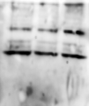
Western Blot: ASC/TMS1 AntibodyBSA Free [NBP1-78977]
Western Blot: ASC/TMS1 Antibody [NBP1-78977] - Whole cell protein from (1) THP1 and (2) HL60 was separated on a 4-15% gel by SDS-PAGE, transferred to 0.2 um PVDF membrane and blocked in 5% non-fat milk in TBST. The membrane was probed with 4.0 ug/ml anti-ACS/TMS1 in 1% milk, and detected with an anti-rabbit HRP secondary antibody using chemiluminescence.![Flow Cytometry: ASC/TMS1 Antibody - BSA Free [NBP1-78977]](https://resources.rndsystems.com/images/products/ASC-TMS1-Antibody---BSA-Free-Flow-Cytometry-NBP1-78977-img0010.jpg "Flow Cytometry: ASC/TMS1 Antibody - BSA Free [NBP1-78977]")
Flow Cytometry: ASC/TMS1 Antibody - BSA Free [NBP1-78977]
Flow Cytometry: ASC/TMS1 Antibody - BSA Free [NBP1-78977] - An intracellular stain was performed on MCF7 cells with ASC/TMS1 NBP1-78977 (blue) and a matched isotype control NBP2-24891 (orange). Cells were fixed with 4% PFA and then permeabilized with 0.1% saponin. Cells were incubated in an antibody dilution of 1 ug/mL for 30 minutes at room temperature, followed by Rabbit IgG (H+L) Cross-Adsorbed Secondary Antibody, Dylight 550 (SA5-10033, Thermo Fisher).![Immunocytochemistry/ Immunofluorescence: ASC/TMS1 Antibody - BSA Free [NBP1-78977]](https://resources.rndsystems.com/images/products/ASC-TMS1-Antibody-Immunocytochemistry-Immunofluorescence-NBP1-78977-img0003.jpg "Immunocytochemistry/ Immunofluorescence: ASC/TMS1 Antibody - BSA Free [NBP1-78977]")
Immunocytochemistry/ Immunofluorescence: ASC/TMS1 Antibody - BSA Free [NBP1-78977]
Immunocytochemistry/Immunofluorescence: ASC/TMS1 Antibody [NBP1-78977] - Tested in MCF-7 cells with FITC (green). Nuclei and alpha-tubulin were counterstained with DAPI (blue) and DyLight 550 (red).![Immunohistochemistry: ASC/TMS1 Antibody - BSA Free [NBP1-78977]](https://resources.rndsystems.com/images/products/ASC-TMS1-Antibody-Immunohistochemistry-NBP1-78977-img0004.jpg "Immunohistochemistry: ASC/TMS1 Antibody - BSA Free [NBP1-78977]")
Immunohistochemistry: ASC/TMS1 Antibody - BSA Free [NBP1-78977]
Immunohistochemistry: ASC/TMS1 Antibody [NBP1-78977] - Tested in mouse intestine at a 1:400 dilution.
Western Blot: ASC/TMS1 Antibody - BSA Free [NBP1-78977] -
Nicorandil alleviates cardiac apoptosis in type 2 diabetic rat. A: TUNEL staining and TUNEL‐positive cells rate. B: Western blot analysis of Bax/Bcl‐2 and cleaved caspase‐3. C: Level of nitric oxide and ADMA in serum. D: Western blot analysis of p‐eNOS. DM: Diabetic mellitus, N7.5: nicorandil, 7.5 mg/kg·day; N15: nicorandil, 15 mg/kg·day. *P < 0.05 compared with control; #P < 0.05 compared with DM; #P < 0.05 compared with HG + N, Data are means +/- SD Image collected and cropped by CiteAb from the following open publication (https://pubmed.ncbi.nlm.nih.gov/31131539), licensed under a CC-BY license. Not internally tested by Novus Biologicals.
Western Blot: ASC/TMS1 Antibody - BSA Free [NBP1-78977] -
PI3K/AKT pathway inhibition blocked the protection of nicorandil on H9c2 cardiomyocyte treated with high glucose. A: Western blot analysis of Bax/Bcl‐2 and cleaved caspase‐3 in high glucose‐induced H9c2 cardiomyocyte after nicorandil treatment or both nicorandil treatment and PI3K/mTOR inhibitors. B: TUNEL assay of apoptosis rate of high glucose‐induced H9c2 cardiomyocyte after nicorandil treatment or both nicorandil treatment and PI3K/mTOR inhibitors (scale bar: 20 um). C: Western blot analysis of p‐eNOS in high glucose‐induced H9c2 cardiomyocyte after nicorandil treatment or both nicorandil treatment and PI3K/mTOR inhibitors. N: Nicorandil (100 umol); MTF: miltefosine (100 umol); Rapa: rapamycin (100 umol) NG: normal glucose (5.5 mmol/L); HG: high glucose (25 mmol/L). *P < 0.05 compared with NG; #P < 0.05 compared with HG; &P < 0.05 compared with HG + N, Data are means +/- SD Image collected and cropped by CiteAb from the following open publication (https://pubmed.ncbi.nlm.nih.gov/31131539), licensed under a CC-BY license. Not internally tested by Novus Biologicals.
Western Blot: ASC/TMS1 Antibody - BSA Free [NBP1-78977] -
Nicorandil protects H9C2 cells from apoptosis through PI3K/AKT pathway. A: Western blot analysis of Bax/Bcl‐2 and cleaved caspase‐3 in high glucose‐induced H9c2 cardiomyocyte after nicorandil treatment or both nicorandil treatment and 5‐HD which is a inhibitor of nicorandil. B: TUNEL assay of apoptosis rate of high glucose‐induced H9c2 cardiomyocyte after nicorandil treatment or both nicorandil treatment and nicorandil inhibitor(5‐HD, 500 umol) (scale bar: 20 um). I:NG, II:HG, III:HG + N, IV:HG + N+5‐HD; C: Western blot analysis of phosphorylation level of PI3K, AKT, eNOS and mTOR in high glucose‐induced H9c2 cardiomyocyte after nicorandil treatment or both nicorandil treatment and nicorandil inhibitor(5‐HD). N: Nicorandil (100 umol); NG: normal glucose (5.5 mmol/L); HG: high glucose (25 mmol/L). *P < 0.05 compared with NG; #P < 0.05 compared with HG; & P < 0.05 compared with HG + N, Data are means +/- SD Image collected and cropped by CiteAb from the following open publication (https://pubmed.ncbi.nlm.nih.gov/31131539), licensed under a CC-BY license. Not internally tested by Novus Biologicals.
Western Blot: ASC/TMS1 Antibody - BSA Free [NBP1-78977] -
Effects of abrocitinib on NF kappa B-related inflammation and pyroptosis pathways. On the 1st day after TBI, NF kappa B-related inflammation and the activation of pyroptosis pathways were dramatically increased. After abrocitinib treatment, the indicators of NF kappa B-related inflammation and pyroptosis pathways were significantly decreased, as can be seen from the WB (n = 5–9 one-way ANOVA with Tukey’s post-hoc test) and GSDMD IHC staining (A–C) (n = 6 Kruskal-Wallis test). From the ELISA results, the changes in inflammatory cytokines (IL-1 beta and IL-18) after brain injury and the effects of abrocitinib were precisely revealed (D,E) (n = 5–6 one-way ANOVA with Tukey’s post-hoc test). All data are shown as mean +/- SD. * p < 0.05, ** p < 0.01, *** p < 0.001, **** p < 0.0001. Image collected and cropped by CiteAb from the following open publication (https://pubmed.ncbi.nlm.nih.gov/36429017), licensed under a CC-BY license. Not internally tested by Novus Biologicals.
Western Blot: ASC/TMS1 Antibody - BSA Free [NBP1-78977] -
Apoptosis level reduced after nicorandil treatment in high glucose‐induced H9c2 cardiomyocyte. A: Western blot analysis of bax and bcl‐2 in high glucose‐induced H9c2 cardiomyocyte after nicorandil treatment with different concentrations for 24 h. B: Western bolt analysis of cleaved caspase‐3 in high glucose‐induced H9c2 cardiomyocyte after nicorandil treatment. HG (33.3 mmol/L), NG (5.5 mmol/L), n1: nicorandil (10 umol); n2: nicorandil (50 umol); n3: nicorandil (100 umol). #P < 0.05 compared with NG; *P < 0.05 compared with HG, Data are means +/- SD Image collected and cropped by CiteAb from the following open publication (https://pubmed.ncbi.nlm.nih.gov/31131539), licensed under a CC-BY license. Not internally tested by Novus Biologicals.
Western Blot: ASC/TMS1 Antibody - BSA Free [NBP1-78977] -
Effects of Mito-Vit-E and TLR9 inhibitor OND-I in LPS-challenged cardiomyocytes.Cultured neonatal cardiomyocytes from rats were treated with +/-LPS (100 ng/ml), +/-Mito-Vit-E (MVE) (1μM), or +/-ODN-I (0.5 μM) 4 hours prior to harvesting. A. Mitochondrial superoxide was labeled with MitoSox Red and quantified by flow cytometry. B. Mitochondrial biogenesis was quantified in live cells using MitoBiogenesis In-Cell ELISA assay. C. Levels of mtDNA in cell medium and in cytoplasm were measured by real-time PCR. D. Cells apoptosis was evaluated by TUNEL assay (green). Cell nucleuses were identified by DAPI staining (blue). E. Expression of MyD88, RAGE, ASC and activated form of caspase 1 were determined in cell lysates by western blot using GAPDH as a loading control, and results were quantified by densitometry. F. Cellular production of IL–1 beta was measured by ELISA. All the measurements were normalized by cell numbers and obtained in triplicate. All values are means +/-SE. Significant differences are shown as * between control and LPS and delta between vehicle and drug-treated groups (p<0.02 for A-C and p<0.01 for E-F, n = 4). Image collected and cropped by CiteAb from the following open publication (https://pubmed.ncbi.nlm.nih.gov/26448624), licensed under a CC-BY license. Not internally tested by Novus Biologicals.
Western Blot: ASC/TMS1 Antibody - BSA Free [NBP1-78977] -
Analysis of NLRP3, NLRC4, NAIP, AIM2, ASC, Caspase-4, Caspase-1, beta -actin and GAPDH protein expression by immunoblotting. (A) Represents the immunoblotting results of NLRP3, NLRC4, NAIP, Caspase-1, GAPDH, AIM2, ASC, Caspase-4 and beta -actinin UPEC infected UTI patients and controls group. Scattered plots showing individual densitometric values (IDV) of NLRP3 (B), NLRC4 (C), NAIP (D), AIM2 (E), ASC (F), Caspase-4 (G), and Caspase-1 (H). Results were expressed as average densitometric ratio in patients and controls group +/- SD. P-value is ∗∗∗p = 0.0001 and n.s, non-significant. Image collected and cropped by CiteAb from the following open publication (https://pubmed.ncbi.nlm.nih.gov/31551961), licensed under a CC-BY license. Not internally tested by Novus Biologicals.
Western Blot: ASC/TMS1 Antibody - BSA Free [NBP1-78977] -
QWZK inhibited NLRP3 inflammasome activation in ALI rats induced by LPS. (A) Western blot assay of NLRP3, pro-caspase-1, cleaved caspase-1, and ASC in different groups. (B–D) The protein expression was analyzed by gray scale. Data were presented as the mean +/- SEM, n = 8. #p < 0.05 vs. control group, ##p < 0.01 vs. control group, ###p < 0.001 vs. control group. *p < 0.05 vs. LPS group, **p < 0.01 vs. LPS group, ***p < 0.001 vs. LPS group. Image collected and cropped by CiteAb from the following open publication (https://www.frontiersin.org/articles/10.3389/fphar.2021.790072/full), licensed under a CC-BY license. Not internally tested by Novus Biologicals.
Western Blot: ASC/TMS1 Antibody - BSA Free [NBP1-78977] -
Inhibition of FOXO1 or autophagy compromised the antioxidative and anti‐inflammatory actions of AVE in mice. (a, b) The selective FOXO1 inhibitor, AS, and the autophagy inhibitor, CQ, abrogated the immune‐regulatory effect of AVE on microglial polarization. (c) AS and CQ both inhibited AVE‐induced alleviation of microglial activation (IBA‐1 staining) and ROS generation (DHE staining) following LPS exposure. (d, e) Representative western blots (d) and statistical graphs (e) of the major components of NLRP3 inflammasomes. Scale bar = 50 μm. Data are means +/- SD (n = 6–7). *p < 0.05, **p < 0.01 compared to control group. +p < 0.05, ++p < 0.01 compared to LPS group. #p < 0.05, ##p < 0.01 compared to LPS + AVE group Image collected and cropped by CiteAb from the following open publication (https://pubmed.ncbi.nlm.nih.gov/34529881), licensed under a CC-BY license. Not internally tested by Novus Biologicals.
Western Blot: ASC/TMS1 Antibody - BSA Free [NBP1-78977] -
The measurement of pyroptosis-related markers after intervention in the mice. (A) IF staining (×200) (left) and the IOD of NLRP3 in GAS of db/db mice (n = 6). The protein expression levels of (B) NLRP3; (C) ASC; (D) Caspase-1 and (E) GSDMD were detected by western blot in the GAS of db/db mice (n = 6). Serum inflammatory markers (F) IL-1 beta and (G) IL-18 by ELISA (n = 8). (#) Significant difference compared with D group; (&) significant difference compared with DI group; ($) significant difference compared with DE group (p < 0.05). Image collected and cropped by CiteAb from the following open publication (https://www.mdpi.com/1420-3049/29/3/712), licensed under a CC-BY license. Not internally tested by Novus Biologicals.Applications for ASC/TMS1 Antibody - BSA Free
Application
Recommended Usage
Flow (Intracellular)
1 - 2 ug/ml. Use reported in scientific literature (PMID 35095880)
Flow Cytometry
1 - 2 ug/ml. Use reported in scientific literature (PMID 31214205)
Immunocytochemistry/ Immunofluorescence
1:40-1:100
Immunohistochemistry
1:200
Immunohistochemistry-Paraffin
1:200
Immunomicroscopy
reported in scientific literature (PMID 31054188)
Immunoprecipitation
reported in scientific literature (PMID 31551961)
Simple Western
1:1000
Western Blot
2.0 - 4.0 ug/ml
Application Notes
Prior to immunostaining paraffin tissues, antigen retrieval with sodium citrate buffer (pH 6.0) is recommended. In Simple Western only 10 - 15 uL of the recommended dilution is used per data point.
See Simple Western Antibody Database for Simple Western validation: Tested in MCF-7 lysate 0.5 mg/mL, separated by Size, antibody dilution of 1:1000, apparent MW was 27 kDa. Separated by Size-Wes, Sally Sue/Peggy Sue.
See Simple Western Antibody Database for Simple Western validation: Tested in MCF-7 lysate 0.5 mg/mL, separated by Size, antibody dilution of 1:1000, apparent MW was 27 kDa. Separated by Size-Wes, Sally Sue/Peggy Sue.
Reviewed Applications
Read 2 reviews rated 5 using NBP1-78977 in the following applications:
Flow Cytometry Panel Builder
Bio-Techne Knows Flow Cytometry
Save time and reduce costly mistakes by quickly finding compatible reagents using the Panel Builder Tool.
Advanced Features
- Spectra Viewer - Custom analysis of spectra from multiple fluorochromes
- Spillover Popups - Visualize the spectra of individual fluorochromes
- Antigen Density Selector - Match fluorochrome brightness with antigen density

Formulation, Preparation, and Storage
Purification
Immunogen affinity purified
Formulation
PBS
Format
BSA Free
Preservative
0.05% Sodium Azide
Concentration
1.0 mg/ml
Shipping
The product is shipped with polar packs. Upon receipt, store it immediately at the temperature recommended below.
Stability & Storage
Store at 4C short term. Aliquot and store at -20C long term. Avoid freeze-thaw cycles.
Background: ASC
In regard to immune and inflammatory response, ASC/TMS1 is involved in inflammasome function (3-4). The inflammasome is a multiprotein complex that responds to cellular stress or pathogens and activates inflammatory responses. Specifically, ASC/TMS1 helps assemble the NLRP3 inflammasome complex which then activates caspase-1, followed by stimulation of proinflammatory cytokines including IL-1b and IL-18 (3-4). In terms of the role in regulating apoptosis, multiple studies have revealed that the ASC/TMS1 gene is hypermethylated in many cancers including breast, lung, glioblastomas, and melanomas (2-5). The increased methylation results in decreased gene expression, or silencing, allowing those cancer cells to escape apoptosis (2-5).
References
1. Masumoto, J., Taniguchi, S., Ayukawa, K., Sarvotham, H., Kishino, T., Niikawa, N., Hidaka, E., Katsuyama, T., Higuchi, T., & Sagara, J. (1999). ASC, a novel 22-kDa protein, aggregates during apoptosis of human promyelocytic leukemia HL-60 cells. The Journal of biological chemistry, 274(48), 33835-33838. https://doi.org/10.1074/jbc.274.48.33835
2. McConnell, B. B., & Vertino, P. M. (2004). TMS1/ASC: the cancer connection. Apoptosis: an international journal on programmed cell death. https://doi.org/10.1023/B:APPT.0000012117.32430.0c
3. Salminen, A., Kauppinen, A., Hiltunen, M., & Kaarniranta, K. (2014). Epigenetic regulation of ASC/TMS1 expression: potential role in apoptosis and inflammasome function. Cellular and molecular life sciences : CMLS. https://doi.org/10.1007/s00018-013-1524-9
4. Protti, M. P., & De Monte, L. (2020). Dual Role of Inflammasome Adaptor ASC in Cancer. Frontiers in cell and developmental biology. https://doi.org/10.3389/fcell.2020.00040
5. Parsons, M. J., & Vertino, P. M. (2006). Dual role of TMS1/ASC in death receptor signaling. Oncogene. https://doi.org/10.1038/sj.onc.1209684
Long Name
Apoptosis-Associated Speck-Like Protein Containing A CARD
Alternate Names
CARD5, PYCARD, TMS1
Entrez Gene IDs
29108 (Human)
Gene Symbol
PYCARD
UniProt
Additional ASC Products
Product Documents for ASC/TMS1 Antibody - BSA Free
Certificate of Analysis
To download a Certificate of Analysis, please enter a lot or batch number in the search box below.
Product Specific Notices for ASC/TMS1 Antibody - BSA Free
This product is for research use only and is not approved for use in humans or in clinical diagnosis. Primary Antibodies are guaranteed for 1 year from date of receipt.
Related Research Areas
Citations for ASC/TMS1 Antibody - BSA Free
 Powered by Bioz
Customer Reviews for ASC/TMS1 Antibody - BSA Free (2)
5 out of 5
2 Customer Ratings
Have you used ASC/TMS1 Antibody - BSA Free?
Submit a review and receive an Amazon gift card!
$25/€18/£15/$25CAN/¥2500 Yen for a review with an image
$10/€7/£6/$10CAN/¥1110 Yen for a review without an image
Submit a review
Customer Images
Showing
1
-
2 of
2 reviews
Showing All
Filter By:
-
Application: Western BlotSample Tested: H9c2 whole cell lysateSpecies: RatVerified Customer | Posted 12/20/2017

-
Application: Western BlotSample Tested: Mouse macrophagesSpecies: MouseVerified Customer | Posted 05/08/2016ASC antibody
There are no reviews that match your criteria.
Protocols
View specific protocols for ASC/TMS1 Antibody - BSA Free (NBP1-78977):
Immunocytochemistry Protocol
Culture cells to appropriate density in 35 mm culture dishes or 6-well plates.
1. Remove culture medium and add 10% formalin to the dish. Fix at room temperature for 30 minutes.
2. Remove the formalin and add ice cold methanol. Incubate for 5-10 minutes.
3. Remove methanol and add washing solution (i.e. PBS). Be sure to not let the specimen dry out. Wash three times for 10 minutes.
4. To block nonspecific antibody binding incubate in 10% normal goat serum from 1 hour to overnight at room temperature.
5. Add primary antibody at appropriate dilution and incubate at room temperature from 2 hours to overnight at room temperature.
6. Remove primary antibody and replace with washing solution. Wash three times for 10 minutes.
7. Add secondary antibody at appropriate dilution. Incubate for 1 hour at room temperature.
8. Remove antibody and replace with wash solution, then wash for 10 minutes. Add Hoechst 33258 to wash solution at 1:25,0000 and incubate for 10 minutes. Wash a third time for 10 minutes.
9. Cells can be viewed directly after washing. The plates can also be stored in PBS containing Azide covered in Parafilm (TM). Cells can also be cover-slipped using Fluoromount, with appropriate sealing.
*The above information is only intended as a guide. The researcher should determine what protocol best meets their needs. Please follow safe laboratory procedures.
Immunohistochemistry-Paraffin Embedded Sections
Antigen Unmasking:
Bring slides to a boil in 10 mM sodium citrate buffer (pH 6.0) then maintain at a sub-boiling temperature for 10 minutes. Cool slides on bench-top for 30 minutes.
Staining:
1. Wash sections in deionized water three times for 5 minutes each.
2. Wash sections in wash buffer for 5 minutes.
3. Block each section with 100-400 ul blocking solution for 1 hour at room temperature.
4. Remove blocking solution and add 100-400 ul diluted primary antibody. Incubate overnight at 4C.
5. Remove antibody solution and wash sections in wash buffer three times for 5 minutes each.
6. Add 100-400 ul biotinylated diluted secondary antibody. Incubate 30 minutes at room temperature.
7. Remove secondary antibody solution and wash sections three times with wash buffer for 5 minutes each.
8. Add 100-400 ul Streptavidin-HRP reagent to each section and incubate for 30 minutes at room temperature.
9. Wash sections three times in wash buffer for 5 minutes each.
10. Add 100-400 ul DAB substrate to each section and monitor staining closely.
11. As soon as the sections develop, immerse slides in deionized water.
12. Counterstain sections in hematoxylin.
13. Wash sections in deionized water two times for 5 minutes each.
14. Dehydrate sections.
15. Mount coverslips.
*The above information is only intended as a guide. The researcher should determine what protocol best meets their needs. Please follow safe laboratory procedures.
Western Blot Protocol
1. Perform SDS-PAGE on samples to be analyzed, loading 10-25 ug of total protein per lane.
2. Transfer proteins to PVDF membrane according to the instructions provided by the manufacturer of the membrane and transfer apparatus.
3. Stain the membrane with Ponceau S (or similar product) to assess transfer success, and mark molecular weight standards where appropriate.
4. Rinse the blot TBS -0.05% Tween 20 (TBST).
5. Block the membrane in 5% Non-fat milk in TBST (blocking buffer) for at least 1 hour.
6. Wash the membrane in TBST three times for 10 minutes each.
7. Dilute primary antibody in blocking buffer and incubate overnight at 4C with gentle rocking.
8. Wash the membrane in TBST three times for 10 minutes each.
9. Incubate the membrane in diluted HRP conjugated secondary antibody in blocking buffer (as per manufacturer's instructions) for 1 hour at room temperature.
10. Wash the blot in TBST three times for 10 minutes each (this step can be repeated as required to reduce background).
11. Apply the detection reagent of choice in accordance with the manufacturer's instructions.
Find general support by application which include: protocols, troubleshooting, illustrated assays, videos and webinars.
- 7-Amino Actinomycin D (7-AAD) Cell Viability Flow Cytometry Protocol
- Antigen Retrieval Protocol (PIER)
- Antigen Retrieval for Frozen Sections Protocol
- Appropriate Fixation of IHC/ICC Samples
- Cellular Response to Hypoxia Protocols
- Chromogenic IHC Staining of Formalin-Fixed Paraffin-Embedded (FFPE) Tissue Protocol
- Chromogenic Immunohistochemistry Staining of Frozen Tissue
- ClariTSA™ Fluorophore Kits
- Detection & Visualization of Antibody Binding
- Extracellular Membrane Flow Cytometry Protocol
- Flow Cytometry Protocol for Cell Surface Markers
- Flow Cytometry Protocol for Staining Membrane Associated Proteins
- Flow Cytometry Staining Protocols
- Flow Cytometry Troubleshooting Guide
- Fluorescent IHC Staining of Frozen Tissue Protocol
- Graphic Protocol for Heat-induced Epitope Retrieval
- Graphic Protocol for the Preparation and Fluorescent IHC Staining of Frozen Tissue Sections
- Graphic Protocol for the Preparation and Fluorescent IHC Staining of Paraffin-embedded Tissue Sections
- Graphic Protocol for the Preparation of Gelatin-coated Slides for Histological Tissue Sections
- ICC Cell Smear Protocol for Suspension Cells
- ICC Immunocytochemistry Protocol Videos
- ICC for Adherent Cells
- IHC Sample Preparation (Frozen sections vs Paraffin)
- Immunocytochemistry (ICC) Protocol
- Immunocytochemistry Troubleshooting
- Immunofluorescence of Organoids Embedded in Cultrex Basement Membrane Extract
- Immunofluorescent IHC Staining of Formalin-Fixed Paraffin-Embedded (FFPE) Tissue Protocol
- Immunohistochemistry (IHC) and Immunocytochemistry (ICC) Protocols
- Immunohistochemistry Frozen Troubleshooting
- Immunohistochemistry Paraffin Troubleshooting
- Immunoprecipitation Protocol
- Intracellular Flow Cytometry Protocol Using Alcohol (Methanol)
- Intracellular Flow Cytometry Protocol Using Detergents
- Intracellular Nuclear Staining Flow Cytometry Protocol Using Detergents
- Intracellular Staining Flow Cytometry Protocol Using Alcohol Permeabilization
- Intracellular Staining Flow Cytometry Protocol Using Detergents to Permeabilize Cells
- Preparing Samples for IHC/ICC Experiments
- Preventing Non-Specific Staining (Non-Specific Binding)
- Primary Antibody Selection & Optimization
- Propidium Iodide Cell Viability Flow Cytometry Protocol
- Protocol for Heat-Induced Epitope Retrieval (HIER)
- Protocol for Liperfluo
- Protocol for Making a 4% Formaldehyde Solution in PBS
- Protocol for VisUCyte™ HRP Polymer Detection Reagent
- Protocol for the Characterization of Human Th22 Cells
- Protocol for the Characterization of Human Th9 Cells
- Protocol for the Fluorescent ICC Staining of Cell Smears - Graphic
- Protocol for the Fluorescent ICC Staining of Cultured Cells on Coverslips - Graphic
- Protocol for the Preparation & Fixation of Cells on Coverslips
- Protocol for the Preparation and Chromogenic IHC Staining of Frozen Tissue Sections
- Protocol for the Preparation and Chromogenic IHC Staining of Frozen Tissue Sections - Graphic
- Protocol for the Preparation and Chromogenic IHC Staining of Paraffin-embedded Tissue Sections
- Protocol for the Preparation and Chromogenic IHC Staining of Paraffin-embedded Tissue Sections - Graphic
- Protocol for the Preparation and Fluorescent ICC Staining of Cells on Coverslips
- Protocol for the Preparation and Fluorescent ICC Staining of Non-adherent Cells
- Protocol for the Preparation and Fluorescent ICC Staining of Stem Cells on Coverslips
- Protocol for the Preparation and Fluorescent IHC Staining of Frozen Tissue Sections
- Protocol for the Preparation and Fluorescent IHC Staining of Paraffin-embedded Tissue Sections
- Protocol for the Preparation of Gelatin-coated Slides for Histological Tissue Sections
- Protocol for the Preparation of a Cell Smear for Non-adherent Cell ICC - Graphic
- Protocol: Annexin V and PI Staining by Flow Cytometry
- Protocol: Annexin V and PI Staining for Apoptosis by Flow Cytometry
- R&D Systems Quality Control Western Blot Protocol
- TUNEL and Active Caspase-3 Detection by IHC/ICC Protocol
- The Importance of IHC/ICC Controls
- Troubleshooting Guide: Fluorokine Flow Cytometry Kits
- Troubleshooting Guide: Immunohistochemistry
- Troubleshooting Guide: Western Blot Figures
- Western Blot Conditions
- Western Blot Protocol
- Western Blot Protocol for Cell Lysates
- Western Blot Troubleshooting
- Western Blot Troubleshooting Guide
- View all Protocols, Troubleshooting, Illustrated assays and Webinars
Loading...
Associated Pathways

