ATM Antibody - BSA Free
Novus Biologicals | Catalog # NB100-104
![Western Blot: ATM Antibody [NB100-104]](https://resources.rndsystems.com/images/products/ATM-Antibody-Western-Blot-NB100-104-img0009.jpg "Western Blot: ATM Antibody [NB100-104]")
Key Product Details
Validated by
Biological Validation
Species Reactivity
Validated:
Human, Mouse, Rat, Canine, Kangaroo
Cited:
Human, Mouse, Canine
Applications
Validated:
Multiplex Immunofluorescence, Immunohistochemistry, Immunohistochemistry-Paraffin, Western Blot, Immunoblotting, ELISA, Flow Cytometry, Immunocytochemistry/ Immunofluorescence, Immunoprecipitation, COMET
Cited:
Western Blot, Immunoblotting, ELISA, Flow Cytometry, Immunocytochemistry/ Immunofluorescence, Immunoprecipitation, IF/IHC
Label
Unconjugated
Antibody Source
Polyclonal Rabbit IgG
Format
BSA Free
Loading...
Product Specifications
Immunogen
ATM Antibody was made to a fragment of the human ATM protein corresponding to the C-terminus (within the last third of the protein sequence). [Uniprot: Q13315]
Reactivity Notes
Canine reactivity reported in scientific literature (PMID: 31648115).
Localization
Nuclear
Clonality
Polyclonal
Host
Rabbit
Isotype
IgG
Theoretical MW
351 kDa.
Disclaimer note: The observed molecular weight of the protein may vary from the listed predicted molecular weight due to post translational modifications, post translation cleavages, relative charges, and other experimental factors.
Disclaimer note: The observed molecular weight of the protein may vary from the listed predicted molecular weight due to post translational modifications, post translation cleavages, relative charges, and other experimental factors.
Scientific Data Images for ATM Antibody - BSA Free
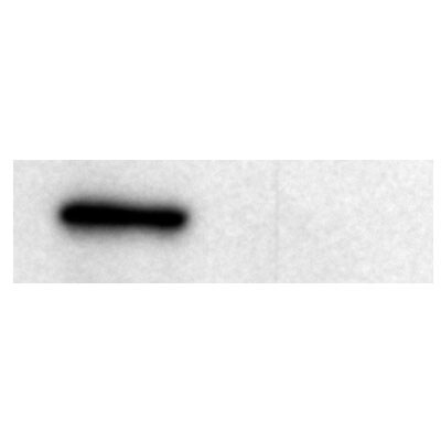
Western Blot: ATM Antibody [NB100-104]
Western Blot: ATM Antibody [NB100-104] - Detection of ATM in HeLa nuclear extract using ATM antibody [NB100-104]. Theoretical molecular weight 351 kDa.![Immunohistochemistry-Paraffin: ATM Antibody [NB100-104]](https://resources.rndsystems.com/images/products/ATM-Antibody-Immunohistochemistry-Paraffin-NB100-104-img0014.jpg "Immunohistochemistry-Paraffin: ATM Antibody [NB100-104]")
Immunohistochemistry-Paraffin: ATM Antibody [NB100-104]
Immunohistochemistry-Paraffin: ATM Antibody [NB100-104] - Staining of human tonsil, germinal center and mantle zone with ATM Antibody [NB100-104].![Immunocytochemistry/ Immunofluorescence: ATM Antibody [NB100-104]](https://resources.rndsystems.com/images/products/ATM-Antibody-Immunocytochemistry-Immunofluorescence-NB100-104-img0018.jpg "Immunocytochemistry/ Immunofluorescence: ATM Antibody [NB100-104]")
![Flow Cytometry: ATM Antibody [NB100-104]](https://resources.rndsystems.com/images/products/ATM-Antibody-Flow-Cytometry-NB100-104-img0016.jpg "Flow Cytometry: ATM Antibody [NB100-104]")
Flow Cytometry: ATM Antibody [NB100-104]
Flow Cytometry: ATM Antibody [NB100-104] - An intracellular stain was performed on HeLa cells with Dylight 550-conjugated ATM Antibody [NB100-104R] (blue) and a matched isotype control (orange). Cells were fixed with 4% PFA and then permeabilized with 0.1% saponin. Cells were incubated in an antibody dilution of 5 ug/mL for 30 minutes at room temperature. Both antibodies were conjugated to DyLight 550.![Western Blot: ATM Antibody [NB100-104]](https://resources.rndsystems.com/images/products/ATM-Antibody-Western-Blot-NB100-104-img0017.jpg "Western Blot: ATM Antibody [NB100-104]")
![Western Blot: ATM Antibody [NB100-104]](https://resources.rndsystems.com/images/products/ATM-Antibody-Western-Blot-NB100-104-img0001.jpg "Western Blot: ATM Antibody [NB100-104]")
Western Blot: ATM Antibody [NB100-104]
Western Blot: ATM Antibody [NB100-104] - Detection of ATM in Raji whole cell lysate using [NB100-104]. Observed molecular weight ~300 kDa. Theoretical molecular weight 351 kDa.![Immunocytochemistry/ Immunofluorescence: ATM Antibody [NB100-104]](https://resources.rndsystems.com/images/products/ATM-Antibody-Immunocytochemistry-Immunofluorescence-NB100-104-img0011.jpg "Immunocytochemistry/ Immunofluorescence: ATM Antibody [NB100-104]")
Immunocytochemistry/ Immunofluorescence: ATM Antibody [NB100-104]
Immunocytochemistry/Immunofluorescence: ATM Antibody [NB100-104] - HeLa cells were fixed for 10 minutes using 10% formalin and then permeabilized for 5 minutes using 1X TBS + 0.05% Triton X-100. Then cells were incubated with [NB100-104] at a 1:100 dilution overnight at 4C and detected with an anti-rabbit DyLight 488 (Green) at a 1:500 dilution. Alpha tubulin was used as a co-stain at a 1:1000 dilution and detected with an anti-mouse DyLight 550 (Red) at a 1:500 dilution. Nuclei were counterstained with DAPI (Blue). Cells were imaged using a 40X objective.![Flow Cytometry: ATM Antibody [NB100-104]](https://resources.rndsystems.com/images/products/ATM-Antibody-Flow-Cytometry-NB100-104-img0012.jpg "Flow Cytometry: ATM Antibody [NB100-104]")
Flow Cytometry: ATM Antibody [NB100-104]
Flow Cytometry: ATM Antibody [NB100-104] - Analysis of PE conjugate of ATM Antibody [NB100-104PE]. An intracellular stain was performed on HeLa cells with ATM antibody [NB100-104PE] (blue) and a matched isotype control [NBP2-24893PE] (orange). Cells were fixed with 4% PFA and then permeablized with 0.1% saponin.![Flow Cytometry: ATM Antibody [NB100-104]](https://resources.rndsystems.com/images/products/ATM-Antibody-Flow-Cytometry-NB100-104-img0015.jpg "Flow Cytometry: ATM Antibody [NB100-104]")
Flow Cytometry: ATM Antibody [NB100-104]
Flow Cytometry: ATM Antibody [NB100-104] - An intracellular stain was performed on HepG2 cells with PE-conjugated ATM antibody [NB100-104PE] (blue) and a matched isotype control (orange). Cells were fixed with 4% PFA and then permeablized with 0.1% saponin. Cells were incubated in an antibody dilution of 5 ug/mL for 30 minutes at room temperature. Both antibodies were conjugated to Phycoerythrin.
Immunocytochemistry/ Immunofluorescence: ATM Antibody [NB100-104] -
Immunocytochemistry/ Immunofluorescence: ATM Antibody [NB100-104] - ATM is activated by spermidine in GM00637 cells.GM00637 cells with or without KU55933 pretreatment (a,b), & GM05849 cells (c) were exposed to 50 μM spermidine or CCCP, followed by immunofluorescence analyses of total & p-ATM on Ser-1981. The scale bar is 10 μm. Ratios of cells expressing p-ATM Ser-1981 to cells expressing total ATM were presented (d). 20–45 cells/condition from three experiments were collected. Values are mean ± SD, *p < 0.05 vs. control. Image collected & cropped by CiteAb from the following publication (https://www.nature.com/articles/srep24700), licensed under a CC-BY license. Not internally tested by Novus Biologicals.
Western Blot: ATM Antibody [NB100-104] -
Western Blot: ATM Antibody [NB100-104] - Depletion of SMARCAL1 induces DNA damage & checkpoint activation in iSML1 iPSCs. (A,B) The iSML1 iPSCs were cultured for 7 & 14 days in the presence of doxycycline (DOX) to induce SMARCAL1 downregulation & then immunostained. The graphs (top) show quantification of the number of gamma -H2AX-positive cells (A) or ATM-pSer1981-positive cells (B). Representative images from triplicate experiments are shown (bottom). (C) Immunoblot detection of the indicated DDR proteins in iSML1 iPSCs after 14 days of continuous treatment with DOX. Lamin B1 was used as the loading control. Data are mean±s.e.m. from three independent experiments. *P≤0.05, **P≤0.01, ***P≤0.001 (two-way ANOVA test). ns, not significant. Scale bars: 10 µm. Image collected & cropped by CiteAb from the following publication (https://pubmed.ncbi.nlm.nih.gov/31515241), licensed under a CC-BY license. Not internally tested by Novus Biologicals.
Western Blot: ATM Antibody [NB100-104] -
Western Blot: ATM Antibody [NB100-104] - Analysis of double strand break repair in MCF-7 monolayer & mammosphere populations. (a) Cells were exposed to 4 Gy 60Co gamma -radiation & the relative degree of double-strand breakage (DSB) was determined by the comet assay under neutral conditions immediately after exposure & at the times indicated after exposure. (b) The 'comets' (n of about 100) were categorized according to the NIH LISTSERV (Comet Assay Interest Group web site) in which type 1 comets display the least DNA damage & type 5 the most. The error bars represent the mean ± standard error of the mean in both panels. The comets of the unirradiated cells are labeled Cont. (c) Expression of proteins involved in the NHEJ pathway of DSB repair in response to increasing doses of ionizing radiation. Lysates were prepared from unirradiated cells & from cells harvested one hour after exposure to 1 or 10-Gy 60Co gamma -radiation & analyzed by immunoblotting with antibodies against several DSB repair proteins. Phospho-ATM & phospho-DNA-PKcs refer to phosphorylation of these proteins at Ser1981 & Ser2056, respectively. Actin served as a loading control. Image collected & cropped by CiteAb from the following publication (http://breast-cancer-research.biomedcentral.com/articles/10.1186/bcr2583), licensed under a CC-BY license. Not internally tested by Novus Biologicals.Applications for ATM Antibody - BSA Free
Application
Recommended Usage
Immunoblotting
reported in scientific literature (PMID 28512243)
Immunocytochemistry/ Immunofluorescence
1 - 2 ug/ml
Immunohistochemistry
1:100 - 1:200
Immunohistochemistry-Paraffin
1:100 - 1:200
Immunoprecipitation
1:10-1:500
Western Blot
1:500-1:1000
Application Notes
In Western blot, it detects a band at ~350 kDa, representing ATM.
Reviewed Applications
Read 2 reviews rated 4.5 using NB100-104 in the following applications:
Flow Cytometry Panel Builder
Bio-Techne Knows Flow Cytometry
Save time and reduce costly mistakes by quickly finding compatible reagents using the Panel Builder Tool.
Advanced Features
- Spectra Viewer - Custom analysis of spectra from multiple fluorochromes
- Spillover Popups - Visualize the spectra of individual fluorochromes
- Antigen Density Selector - Match fluorochrome brightness with antigen density

Formulation, Preparation, and Storage
Purification
Immunogen affinity purified
Formulation
PBS
Format
BSA Free
Preservative
0.05% Sodium Azide
Concentration
1.0 mg/ml
Shipping
The product is shipped with polar packs. Upon receipt, store it immediately at the temperature recommended below.
Stability & Storage
Aliquot and store at -20C or -80C. Avoid freeze-thaw cycles.
Background: ATM
The theoretical molecular weight of ATM is 350 kDa and it has 3 main domains: a FAT (focal adhesion targeting) domain (aa 1960-2566), a PI-3/PI-4 kinase catalytic domain (aa 2712-2962), and a C-terminal FAT domain (aa 3024-3056). ATM exists as a dimer or tetramer in its inactive state. Upon sensing DNA damage, the MRE11-RAD50-NBS1 (MRN) complex recruits ATM. The intricate process of ATM activation involves acetylation by KAT5/TIP60, autophosphorylation at Ser-1981, and dissociation into catalytically active monomers (5). Following activation, ATM phosphorylates multiple substrates such as p53/TP53 and Chk2 involved in DNA repair, checkpoint signaling, and the apoptosis pathway.
References
1. Paull TT. (2015) Mechanisms of ATM Activation. Annu Rev Biochem. 84:711-38. PMID: 25580527
2. Chaudhary MW and Al-Baradie RS. (2014) Ataxia-telangiectasia: future prospects. Appl Clin Genet. 7:159-167. PMID: 25258552
3. Stagni V, Cirotti C, and Barila D. (2018) Ataxia-Telangiectasia Mutated Kinase in the Control of Oxidative Stress, Mitochondria, and Autophagy in Cancer: A Maestro With a Large Orchestra. Front Oncol. 8:73. PMID: 29616191
4. Gumy-Pause F, Wacker P, and Sappino AP. (2004) ATM gene and lymphoid malignancies. Leukemia. 18(2):238-42. PMID: 14628072
5. Adamowicz M. (2018) Breaking up with ATM. J Immunol Sci. 2(1):26-31. PMID: 29652413
Long Name
Ataxia Telangiectasia-mutated
Alternate Names
TEL1, TELO1, TPLL
Entrez Gene IDs
472 (Human)
Gene Symbol
ATM
UniProt
Additional ATM Products
Product Documents for ATM Antibody - BSA Free
Certificate of Analysis
To download a Certificate of Analysis, please enter a lot or batch number in the search box below.
Product Specific Notices for ATM Antibody - BSA Free
This product is for research use only and is not approved for use in humans or in clinical diagnosis. Primary Antibodies are guaranteed for 1 year from date of receipt.
Related Research Areas
Citations for ATM Antibody - BSA Free
 Powered by Bioz
Customer Reviews for ATM Antibody - BSA Free (2)
4.5 out of 5
2 Customer Ratings
Have you used ATM Antibody - BSA Free?
Submit a review and receive an Amazon gift card!
$25/€18/£15/$25CAN/¥2500 Yen for a review with an image
$10/€7/£6/$10CAN/¥1110 Yen for a review without an image
Submit a review
Customer Images
Showing
1
-
2 of
2 reviews
Showing All
Filter By:
-
Application: ImmunofluorescenceSample Tested: cellSpecies: OtherVerified Customer | Posted 10/08/2014
-
Application: Western BlotSample Tested: Lymphoblastoid (LCL) nuclear lysateSpecies: HumanVerified Customer | Posted 08/15/2013

There are no reviews that match your criteria.
Protocols
View specific protocols for ATM Antibody - BSA Free (NB100-104):
Immunocytochemistry Protocol
Culture cells to appropriate density in 35 mm culture dishes or 6-well plates.
1. Remove culture medium and wash the cells briefly in PBS. Add 4% paraformaldehyde to the dish and fix at room temperature for 10 minutes.
2. Remove the paraformaldehyde and wash the cells in PBS.
3. Permeabilize the cells with 0.1% Triton X100 or other suitable detergent for 2 min.
4. Remove the permeabilization buffer and wash three times for 5 minutes each in PBS. Be sure to not let the specimen dry out.
5. To block nonspecific antibody binding, incubate in 10% normal goat serum from 1 hour to overnight at room temperature.
6. Add primary antibody at appropriate dilution and incubate overnight at 4C.
7. Remove primary antibody and replace with PBS. Wash three times for 5 minutes each.
8. Add secondary antibody at appropriate dilution. Incubate for 1 hour at room temperature.
9. Remove secondary antibody and replace with PBS. Wash three times for 5 minutes each.
10. Counter stain DNA with DAPI if required.
Immunohistochemistry-Paraffin Embedded Sections
Antigen Unmasking:
Bring slides to a boil in 10 mM sodium citrate buffer (pH 6.0) then maintain at a sub-boiling temperature for 10 minutes. Cool slides on bench-top for 30 minutes (keep slides in the sodium citrate buffer at all times).
Staining:
1. Wash sections in deionized water three times for 5 minutes each.
2. Wash sections in PBS for 5 minutes.
3. Block each section with 100-400 ul blocking solution (1% BSA in PBS) for 1 hour at room temperature.
4. Remove blocking solution and add 100-400 ul diluted primary antibody. Incubate overnight at 4 C.
5. Remove antibody solution and wash sections in wash buffer three times for 5 minutes each.
6. Add 100-400 ul HRP polymer conjugated secondary antibody. Incubate 30 minutes at room temperature.
7. Wash sections three times in wash buffer for 5 minutes each.
8. Add 100-400 ul DAB substrate to each section and monitor staining closely.
9. As soon as the sections develop, immerse slides in deionized water.
10. Counterstain sections in hematoxylin.
11. Wash sections in deionized water two times for 5 minutes each.
12. Dehydrate sections.
13. Mount coverslips.
Western Blot Protocol
1. Perform SDS-PAGE on samples to be analyzed, loading 10-25 ug of total protein per lane.
2. Transfer proteins to PVDF membrane according to the instructions provided by the manufacturer of the membrane and transfer apparatus.
3. Stain the membrane with Ponceau S (or similar product) to assess transfer success, and mark molecular weight standards where appropriate.
4. Rinse the blot TBS -0.05% Tween 20 (TBST).
5. Block the membrane in 5% Non-fat milk in TBST (blocking buffer) for at least 1 hour.
6. Wash the membrane in TBST three times for 10 minutes each.
7. Dilute primary antibody in blocking buffer and incubate overnight at 4C with gentle rocking.
8. Wash the membrane in TBST three times for 10 minutes each.
9. Incubate the membrane in diluted HRP conjugated secondary antibody in blocking buffer (as per manufacturer's instructions) for 1 hour at room temperature.
10. Wash the blot in TBST three times for 10 minutes each (this step can be repeated as required to reduce background).
11. Apply the detection reagent of choice in accordance with the manufacturer's instructions.
Find general support by application which include: protocols, troubleshooting, illustrated assays, videos and webinars.
- 7-Amino Actinomycin D (7-AAD) Cell Viability Flow Cytometry Protocol
- Antigen Retrieval Protocol (PIER)
- Antigen Retrieval for Frozen Sections Protocol
- Appropriate Fixation of IHC/ICC Samples
- Cellular Response to Hypoxia Protocols
- Chromogenic IHC Staining of Formalin-Fixed Paraffin-Embedded (FFPE) Tissue Protocol
- Chromogenic Immunohistochemistry Staining of Frozen Tissue
- ClariTSA™ Fluorophore Kits
- Detection & Visualization of Antibody Binding
- ELISA Sample Preparation & Collection Guide
- ELISA Troubleshooting Guide
- Extracellular Membrane Flow Cytometry Protocol
- Flow Cytometry Protocol for Cell Surface Markers
- Flow Cytometry Protocol for Staining Membrane Associated Proteins
- Flow Cytometry Staining Protocols
- Flow Cytometry Troubleshooting Guide
- Fluorescent IHC Staining of Frozen Tissue Protocol
- Graphic Protocol for Heat-induced Epitope Retrieval
- Graphic Protocol for the Preparation and Fluorescent IHC Staining of Frozen Tissue Sections
- Graphic Protocol for the Preparation and Fluorescent IHC Staining of Paraffin-embedded Tissue Sections
- Graphic Protocol for the Preparation of Gelatin-coated Slides for Histological Tissue Sections
- How to Run an R&D Systems DuoSet ELISA
- How to Run an R&D Systems Quantikine ELISA
- How to Run an R&D Systems Quantikine™ QuicKit™ ELISA
- ICC Cell Smear Protocol for Suspension Cells
- ICC Immunocytochemistry Protocol Videos
- ICC for Adherent Cells
- IHC Sample Preparation (Frozen sections vs Paraffin)
- Immunocytochemistry (ICC) Protocol
- Immunocytochemistry Troubleshooting
- Immunofluorescence of Organoids Embedded in Cultrex Basement Membrane Extract
- Immunofluorescent IHC Staining of Formalin-Fixed Paraffin-Embedded (FFPE) Tissue Protocol
- Immunohistochemistry (IHC) and Immunocytochemistry (ICC) Protocols
- Immunohistochemistry Frozen Troubleshooting
- Immunohistochemistry Paraffin Troubleshooting
- Immunoprecipitation Protocol
- Intracellular Flow Cytometry Protocol Using Alcohol (Methanol)
- Intracellular Flow Cytometry Protocol Using Detergents
- Intracellular Nuclear Staining Flow Cytometry Protocol Using Detergents
- Intracellular Staining Flow Cytometry Protocol Using Alcohol Permeabilization
- Intracellular Staining Flow Cytometry Protocol Using Detergents to Permeabilize Cells
- Preparing Samples for IHC/ICC Experiments
- Preventing Non-Specific Staining (Non-Specific Binding)
- Primary Antibody Selection & Optimization
- Propidium Iodide Cell Viability Flow Cytometry Protocol
- Protocol for Heat-Induced Epitope Retrieval (HIER)
- Protocol for Liperfluo
- Protocol for Making a 4% Formaldehyde Solution in PBS
- Protocol for VisUCyte™ HRP Polymer Detection Reagent
- Protocol for the Characterization of Human Th22 Cells
- Protocol for the Characterization of Human Th9 Cells
- Protocol for the Fluorescent ICC Staining of Cell Smears - Graphic
- Protocol for the Fluorescent ICC Staining of Cultured Cells on Coverslips - Graphic
- Protocol for the Preparation & Fixation of Cells on Coverslips
- Protocol for the Preparation and Chromogenic IHC Staining of Frozen Tissue Sections
- Protocol for the Preparation and Chromogenic IHC Staining of Frozen Tissue Sections - Graphic
- Protocol for the Preparation and Chromogenic IHC Staining of Paraffin-embedded Tissue Sections
- Protocol for the Preparation and Chromogenic IHC Staining of Paraffin-embedded Tissue Sections - Graphic
- Protocol for the Preparation and Fluorescent ICC Staining of Cells on Coverslips
- Protocol for the Preparation and Fluorescent ICC Staining of Non-adherent Cells
- Protocol for the Preparation and Fluorescent ICC Staining of Stem Cells on Coverslips
- Protocol for the Preparation and Fluorescent IHC Staining of Frozen Tissue Sections
- Protocol for the Preparation and Fluorescent IHC Staining of Paraffin-embedded Tissue Sections
- Protocol for the Preparation of Gelatin-coated Slides for Histological Tissue Sections
- Protocol for the Preparation of a Cell Smear for Non-adherent Cell ICC - Graphic
- Protocol: Annexin V and PI Staining by Flow Cytometry
- Protocol: Annexin V and PI Staining for Apoptosis by Flow Cytometry
- Quantikine HS ELISA Kit Assay Principle, Alkaline Phosphatase
- Quantikine HS ELISA Kit Principle, Streptavidin-HRP Polymer
- R&D Systems Quality Control Western Blot Protocol
- Sandwich ELISA (Colorimetric) – Biotin/Streptavidin Detection Protocol
- Sandwich ELISA (Colorimetric) – Direct Detection Protocol
- TUNEL and Active Caspase-3 Detection by IHC/ICC Protocol
- The Importance of IHC/ICC Controls
- Troubleshooting Guide: ELISA
- Troubleshooting Guide: Fluorokine Flow Cytometry Kits
- Troubleshooting Guide: Immunohistochemistry
- Troubleshooting Guide: Western Blot Figures
- Western Blot Conditions
- Western Blot Protocol
- Western Blot Protocol for Cell Lysates
- Western Blot Troubleshooting
- Western Blot Troubleshooting Guide
- View all Protocols, Troubleshooting, Illustrated assays and Webinars
FAQs for ATM Antibody - BSA Free
Showing
1
-
4 of
4 FAQs
Showing All
-
Q: What is the expiration date of this product?
A: We guarantee the stability of this product for 1 year from the date of receipt.
-
Q: What is the isotype of this antibody?
A: The isotype for this ATM Antibody is IgG.
-
Q: What is the theoretical molecular weight for your ATM antibodies?
A: The theoretical molecular weight for our ATM antibodies is 351 kDa.
-
Q: Which applications can I use this antibody in?
A: This antibody has been cited in multiple publications for use in ELISA, flow cytometry, immunoblotting, ICC/IF, immunohistochemistry, immunoprecipitation, and western blot. This antibody has also been tested in IHC-Paraffin; therefore all of these applications fall under our 100% guarantee. If you would like to test this antibody in an application or species not listed, please check out our Innovator's Reward Program.
-
Q: What is the expiration date of this product?
A: We guarantee the stability of this product for 1 year from the date of receipt.
-
Q: What is the isotype of this antibody?
A: The isotype for this ATM Antibody is IgG.
-
Q: What is the theoretical molecular weight for your ATM antibodies?
A: The theoretical molecular weight for our ATM antibodies is 351 kDa.
-
Q: Which applications can I use this antibody in?
A: This antibody has been cited in multiple publications for use in ELISA, flow cytometry, immunoblotting, ICC/IF, immunohistochemistry, immunoprecipitation, and western blot. This antibody has also been tested in IHC-Paraffin; therefore all of these applications fall under our 100% guarantee. If you would like to test this antibody in an application or species not listed, please check out our Innovator's Reward Program.
Loading...