
ATP6V0A1 Antibody - BSA Free
Novus Biologicals | Catalog # NBP1-89342

![Western Blot: ATP6V0A1 Antibody [NBP1-89342]](https://resources.rndsystems.com/images/products/ATP6V0A1-Antibody-Western-Blot-NBP1-89342-img0018.jpg "Western Blot: ATP6V0A1 Antibody [NBP1-89342]")
![Immunohistochemistry-Paraffin: ATP6V0A1 Antibody [NBP1-89342]](https://resources.rndsystems.com/images/products/ATP6V0A1-Antibody-Immunohistochemistry-Paraffin-NBP1-89342-img0015.jpg "Immunohistochemistry-Paraffin: ATP6V0A1 Antibody [NBP1-89342]")
Loading...
Key Product Details
Validated by
Orthogonal Validation, Biological Validation
Species Reactivity
Validated:
Human
Cited:
Human, Mouse
Predicted:
Mouse (100%), Rat (100%). Backed by our 100% Guarantee.
Applications
Validated:
Immunohistochemistry, Immunohistochemistry-Paraffin, Western Blot, Immunocytochemistry/ Immunofluorescence
Cited:
Immunohistochemistry, Immunohistochemistry-Paraffin, Western Blot, Immunocytochemistry/ Immunofluorescence, Immunoprecipitation
Label
Unconjugated
Antibody Source
Polyclonal Rabbit IgG
Format
BSA Free
Loading...
Product Specifications
Immunogen
This antibody was developed against Recombinant Protein corresponding to amino acids: VQFRDLNPDVNVFQRKFVNEVRRCEEMDRKLRFVEKEIRKANIPIMDTGENPEVPFPRDMIDLEANFEKIENELKEINTNQEALKRNFLELTELK
Clonality
Polyclonal
Host
Rabbit
Isotype
IgG
Scientific Data Images for ATP6V0A1 Antibody - BSA Free
![Western Blot: ATP6V0A1 Antibody [NBP1-89342]](https://resources.rndsystems.com/images/products/ATP6V0A1-Antibody-Western-Blot-NBP1-89342-img0017.jpg "Western Blot: ATP6V0A1 Antibody [NBP1-89342]")
Western Blot: ATP6V0A1 Antibody [NBP1-89342]
ATP6V0A1-Antibody-Western-Blot-NBP1-89342-img0017.jpg![Immunocytochemistry/ Immunofluorescence: ATP6V0A1 Antibody [NBP1-89342]](https://resources.rndsystems.com/images/products/ATP6V0A1-Antibody-Immunocytochemistry-Immunofluorescence-NBP1-89342-img0006.jpg "Immunocytochemistry/ Immunofluorescence: ATP6V0A1 Antibody [NBP1-89342]")
Immunocytochemistry/ Immunofluorescence: ATP6V0A1 Antibody [NBP1-89342]
Immunocytochemistry/Immunofluorescence: ATP6V0A1 Antibody [NBP1-89342] - Staining of human cell line A-431 shows localization to the Golgi apparatus & vesicles. Antibody staining is shown in green.![Immunohistochemistry-Paraffin: ATP6V0A1 Antibody [NBP1-89342]](https://resources.rndsystems.com/images/products/ATP6V0A1-Antibody-Immunohistochemistry-Paraffin-NBP1-89342-img0016.jpg "Immunohistochemistry-Paraffin: ATP6V0A1 Antibody [NBP1-89342]")
Immunohistochemistry-Paraffin: ATP6V0A1 Antibody [NBP1-89342]
Immunohistochemistry-Paraffin: ATP6V0A1 Antibody [NBP1-89342] - Staining of human cerebral cortex shows strong cytoplasmic positivity in neuropil.![Immunohistochemistry-Paraffin: ATP6V0A1 Antibody [NBP1-89342]](https://resources.rndsystems.com/images/products/ATP6V0A1-Antibody-Immunohistochemistry-Paraffin-NBP1-89342-img0011.jpg "Immunohistochemistry-Paraffin: ATP6V0A1 Antibody [NBP1-89342]")
Immunohistochemistry-Paraffin: ATP6V0A1 Antibody [NBP1-89342]
Immunohistochemistry-Paraffin: ATP6V0A1 Antibody [NBP1-89342] - Staining of human tonsil shows low expression as expected.![Immunohistochemistry-Paraffin: ATP6V0A1 Antibody [NBP1-89342]](https://resources.rndsystems.com/images/products/ATP6V0A1-Antibody-Immunohistochemistry-Paraffin-NBP1-89342-img0013.jpg "Immunohistochemistry-Paraffin: ATP6V0A1 Antibody [NBP1-89342]")
Immunohistochemistry-Paraffin: ATP6V0A1 Antibody [NBP1-89342]
Immunohistochemistry-Paraffin: ATP6V0A1 Antibody [NBP1-89342] - Staining of human kidney shows strong cytoplasmic postivity in cells in tubules.![Immunohistochemistry-Paraffin: ATP6V0A1 Antibody [NBP1-89342]](https://resources.rndsystems.com/images/products/ATP6V0A1-Antibody-Immunohistochemistry-Paraffin-NBP1-89342-img0014.jpg "Immunohistochemistry-Paraffin: ATP6V0A1 Antibody [NBP1-89342]")
Immunohistochemistry-Paraffin: ATP6V0A1 Antibody [NBP1-89342]
Immunohistochemistry-Paraffin: ATP6V0A1 Antibody [NBP1-89342] - Staining of human cerebellum shows strong cytoplasmic postivity in cells in granular layer.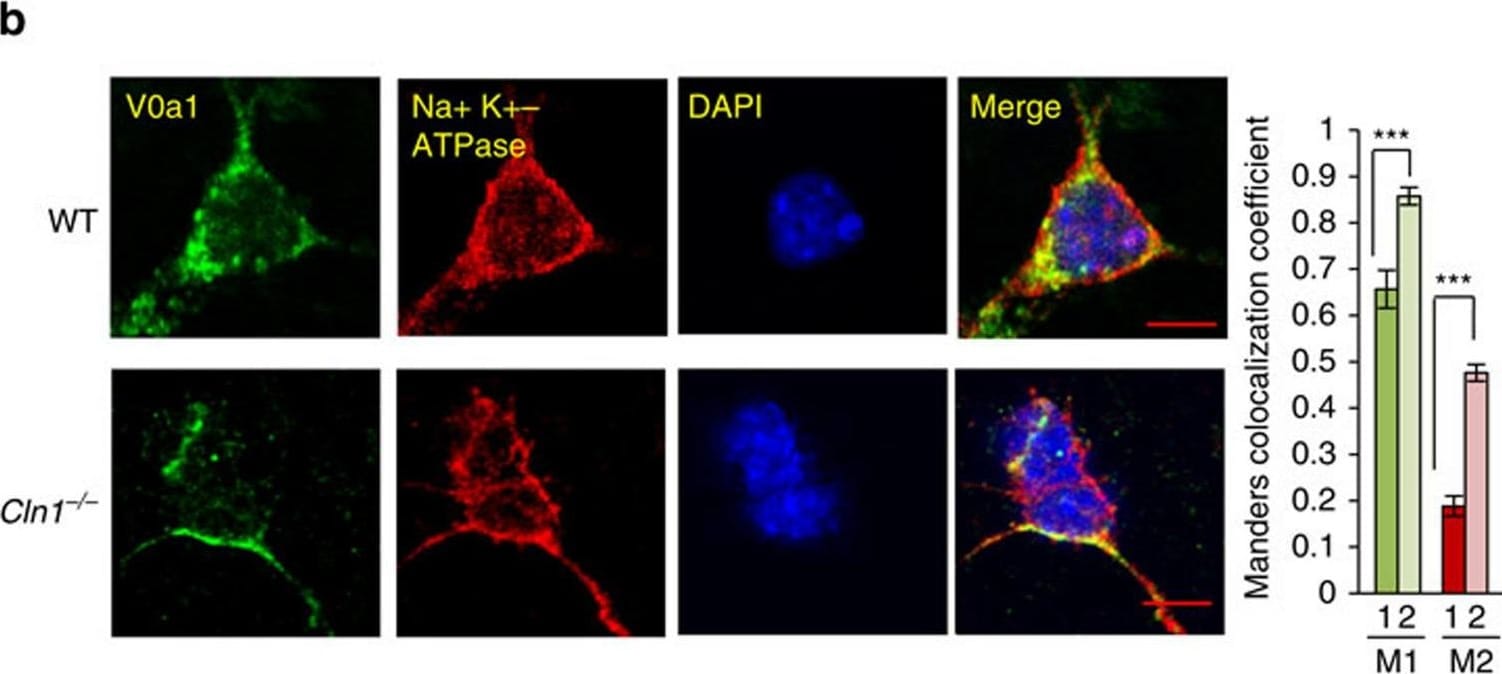
Immunocytochemistry/ Immunofluorescence: ATP6V0A1 Antibody [NBP1-89342] -
Immunocytochemistry/ Immunofluorescence: ATP6V0A1 Antibody [NBP1-89342] - In Cln1−/− mice V0a1 is misrouted to plasma membrane preventing its interaction with AP-3.(a) Western blot analysis & densitometric quantitation of V0a1 in isolated plasma membrane fraction from WT & Cln1−/− mouse brain (n=4, *P<0.05). (b) Localization of V0a1 in the plasma membrane in WT & Cln1−/− neurons using Na+, K+-ATPase as membrane marker. Colocalization between V0a1 & Na+, K+-ATPase was assessed using the Manders' colocalization coefficients M1 & M2 (n=18 for WT & n=22 for Cln1−/−, ***P<0.001; scale bars, 5 μm. (c) Pull-down assay with AP-3 antibody detects V0a1 in total brain lysates from WT & Cln1−/− mouse brain (n=4, *P<0.05). (d) Confocal imaging of PLA reaction showing V0a1 & AP-3 delta interaction in neurons isolated from WT & Cln1−/−mouse brain (n=188 for WT & n=158 for Cln1−/−, ***P<0.001); scale bars, 20 μm. (e) Pull-down assay with AP-3 antibody using total lysates from untreated (lane 1) & bromopalmitate-treated (lane 2) WT brain slices to detect V0a1 & its densitometric quantitation (n=4, *P<0.05). (f) HEK-293 cells were transfected with WT GFP-V0a1 & GFP-V0a1-Cys25Ser mutant construct, & pull-down experiments were conducted with AP-3 antibody to detect GFP-V0a1,*P<0.05(n=4). Image collected & cropped by CiteAb from the following publication (https://www.nature.com/articles/ncomms14612), licensed under a CC-BY license. Not internally tested by Novus Biologicals.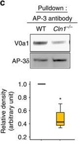
Western Blot: ATP6V0A1 Antibody [NBP1-89342] -
Western Blot: ATP6V0A1 Antibody [NBP1-89342] - In Cln1−/− mice V0a1 is misrouted to plasma membrane preventing its interaction with AP-3.(a) Western blot analysis & densitometric quantitation of V0a1 in isolated plasma membrane fraction from WT & Cln1−/− mouse brain (n=4, *P<0.05). (b) Localization of V0a1 in the plasma membrane in WT & Cln1−/− neurons using Na+, K+-ATPase as membrane marker. Colocalization between V0a1 & Na+, K+-ATPase was assessed using the Manders' colocalization coefficients M1 & M2 (n=18 for WT & n=22 for Cln1−/−, ***P<0.001; scale bars, 5 μm. (c) Pull-down assay with AP-3 antibody detects V0a1 in total brain lysates from WT & Cln1−/− mouse brain (n=4, *P<0.05). (d) Confocal imaging of PLA reaction showing V0a1 & AP-3 delta interaction in neurons isolated from WT & Cln1−/−mouse brain (n=188 for WT & n=158 for Cln1−/−, ***P<0.001); scale bars, 20 μm. (e) Pull-down assay with AP-3 antibody using total lysates from untreated (lane 1) & bromopalmitate-treated (lane 2) WT brain slices to detect V0a1 & its densitometric quantitation (n=4, *P<0.05). (f) HEK-293 cells were transfected with WT GFP-V0a1 & GFP-V0a1-Cys25Ser mutant construct, & pull-down experiments were conducted with AP-3 antibody to detect GFP-V0a1,*P<0.05(n=4). Image collected & cropped by CiteAb from the following publication (https://www.nature.com/articles/ncomms14612), licensed under a CC-BY license. Not internally tested by Novus Biologicals.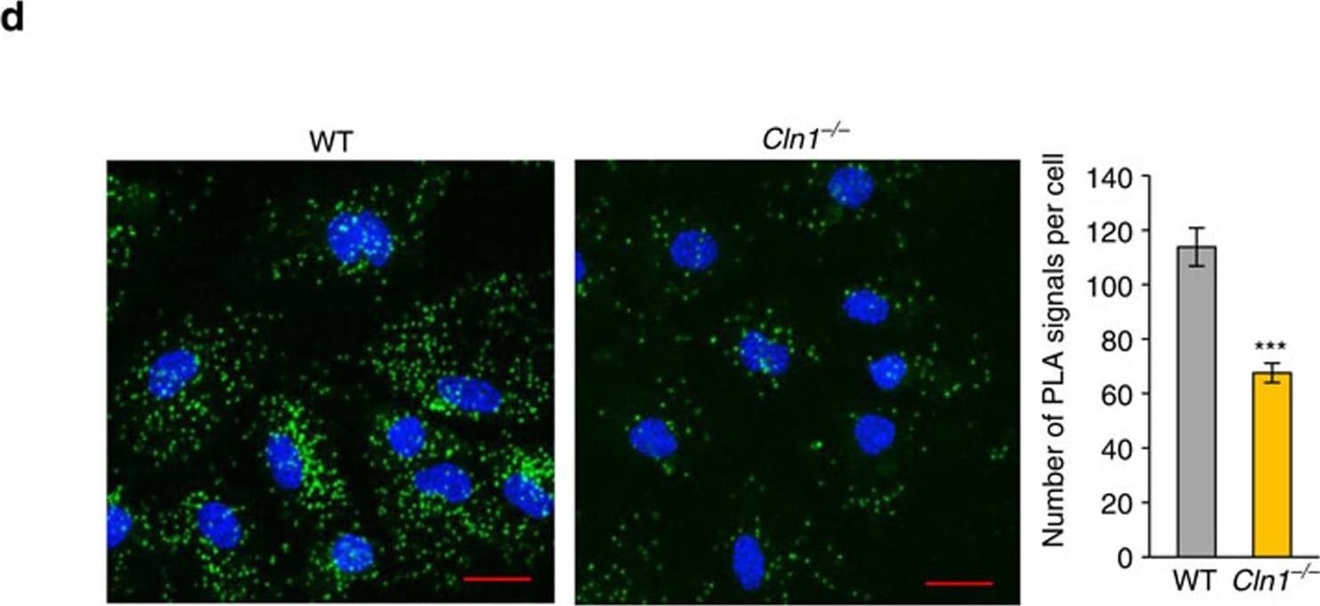
Immunocytochemistry/ Immunofluorescence: ATP6V0A1 Antibody [NBP1-89342] -
Immunocytochemistry/ Immunofluorescence: ATP6V0A1 Antibody [NBP1-89342] - In Cln1−/− mice V0a1 is misrouted to plasma membrane preventing its interaction with AP-3.(a) Western blot analysis & densitometric quantitation of V0a1 in isolated plasma membrane fraction from WT & Cln1−/− mouse brain (n=4, *P<0.05). (b) Localization of V0a1 in the plasma membrane in WT & Cln1−/− neurons using Na+, K+-ATPase as membrane marker. Colocalization between V0a1 & Na+, K+-ATPase was assessed using the Manders' colocalization coefficients M1 & M2 (n=18 for WT & n=22 for Cln1−/−, ***P<0.001; scale bars, 5 μm. (c) Pull-down assay with AP-3 antibody detects V0a1 in total brain lysates from WT & Cln1−/− mouse brain (n=4, *P<0.05). (d) Confocal imaging of PLA reaction showing V0a1 & AP-3 delta interaction in neurons isolated from WT & Cln1−/−mouse brain (n=188 for WT & n=158 for Cln1−/−, ***P<0.001); scale bars, 20 μm. (e) Pull-down assay with AP-3 antibody using total lysates from untreated (lane 1) & bromopalmitate-treated (lane 2) WT brain slices to detect V0a1 & its densitometric quantitation (n=4, *P<0.05). (f) HEK-293 cells were transfected with WT GFP-V0a1 & GFP-V0a1-Cys25Ser mutant construct, & pull-down experiments were conducted with AP-3 antibody to detect GFP-V0a1,*P<0.05(n=4). Image collected & cropped by CiteAb from the following publication (https://www.nature.com/articles/ncomms14612), licensed under a CC-BY license. Not internally tested by Novus Biologicals.Applications for ATP6V0A1 Antibody - BSA Free
Application
Recommended Usage
Immunocytochemistry/ Immunofluorescence
0.25 - 2 ug/mL
Immunohistochemistry
1:200 - 1:500
Immunohistochemistry-Paraffin
1:200 - 1:500
Western Blot
Image collected and cropped by CiteAb from the following publication (http://www.nature.com/doifinder/10.1038/ncomms14612), licensed under a CC-BY license.
Application Notes
IHC-Paraffin, HIER pH 6 retrieval is recommended. ICC/IF, Fixation Permeabilization: Use PFA/Triton X-100.
Reviewed Applications
Read 2 reviews rated 4.5 using NBP1-89342 in the following applications:
Formulation, Preparation, and Storage
Purification
Affinity purified
Formulation
PBS (pH 7.2) and 40% Glycerol
Format
BSA Free
Preservative
0.02% Sodium Azide
Concentration
Concentrations vary lot to lot. See vial label for concentration. If unlisted please contact technical services.
Shipping
The product is shipped with polar packs. Upon receipt, store it immediately at the temperature recommended below.
Stability & Storage
Store at 4C short term. Aliquot and store at -20C long term. Avoid freeze-thaw cycles.
Background: ATP6V0A1
Alternate Names
a1, ATP6N1, ATP6N1AATPase, H+ transporting, lysosomal non-catalytic accessory protein 1(110/116kD), ATP6V0, ATPase, H+ transporting, lysosomal (vacuolar proton pump) non-catalyticaccessory protein 1A (110/116kD), ATPase, H+ transporting, lysosomal V0 subunit a isoform 1, ATPase, H+ transporting, lysosomal V0 subunit a1, Clathrin-coated vesicle/synaptic vesicle proton pump 116 kDa subunit, DKFZp781J1951, Stv1, Vacuolar adenosine triphosphatase subunit Ac116, Vacuolar proton pump subunit 1, vacuolar proton pump, subunit 1, vacuolar proton translocating ATPase 116 kDa subunit A, Vacuolar proton translocating ATPase 116 kDa subunit a isoform 1, vacuolar-type H(+)-ATPase 115 kDa subunit, V-ATPase 116 kDa, V-ATPase 116 kDa isoform a1, Vph1, VPP1H(+)-transporting two-sector ATPase, 116 kDa accessory protein A1, V-type proton ATPase 116 kDa subunit a, V-type proton ATPase 116 kDa subunit a isoform 1
Gene Symbol
ATP6V0A1
Additional ATP6V0A1 Products
Product Documents for ATP6V0A1 Antibody - BSA Free
Certificate of Analysis
To download a Certificate of Analysis, please enter a lot or batch number in the search box below.
Product Specific Notices for ATP6V0A1 Antibody - BSA Free
This product is for research use only and is not approved for use in humans or in clinical diagnosis. Primary Antibodies are guaranteed for 1 year from date of receipt.
Citations for ATP6V0A1 Antibody - BSA Free
 Powered by Bioz
Customer Reviews for ATP6V0A1 Antibody - BSA Free (2)
4.5 out of 5
2 Customer Ratings
Have you used ATP6V0A1 Antibody - BSA Free?
Submit a review and receive an Amazon gift card!
$25/€18/£15/$25CAN/¥2500 for a review with an image
$10/€7/£6/$10CAN/¥1110 for a review without an image
Submit a review
Customer Images
Showing
1
-
2 of
2 reviews
Showing All
Filter By:
-
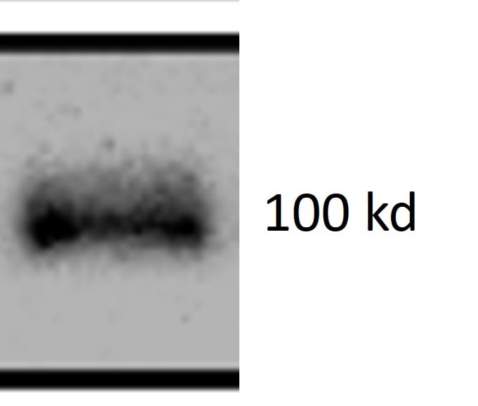
Application: Western BlotSample Tested: Brain (cortex) tissueSpecies: MouseVerified Customer | Posted 02/03/2025A nice ATP6V0A1 antibody1:2000 dilution at 4 degrees overnight to detect endogenous V0A1 from mouse (20 ug total protein).

-
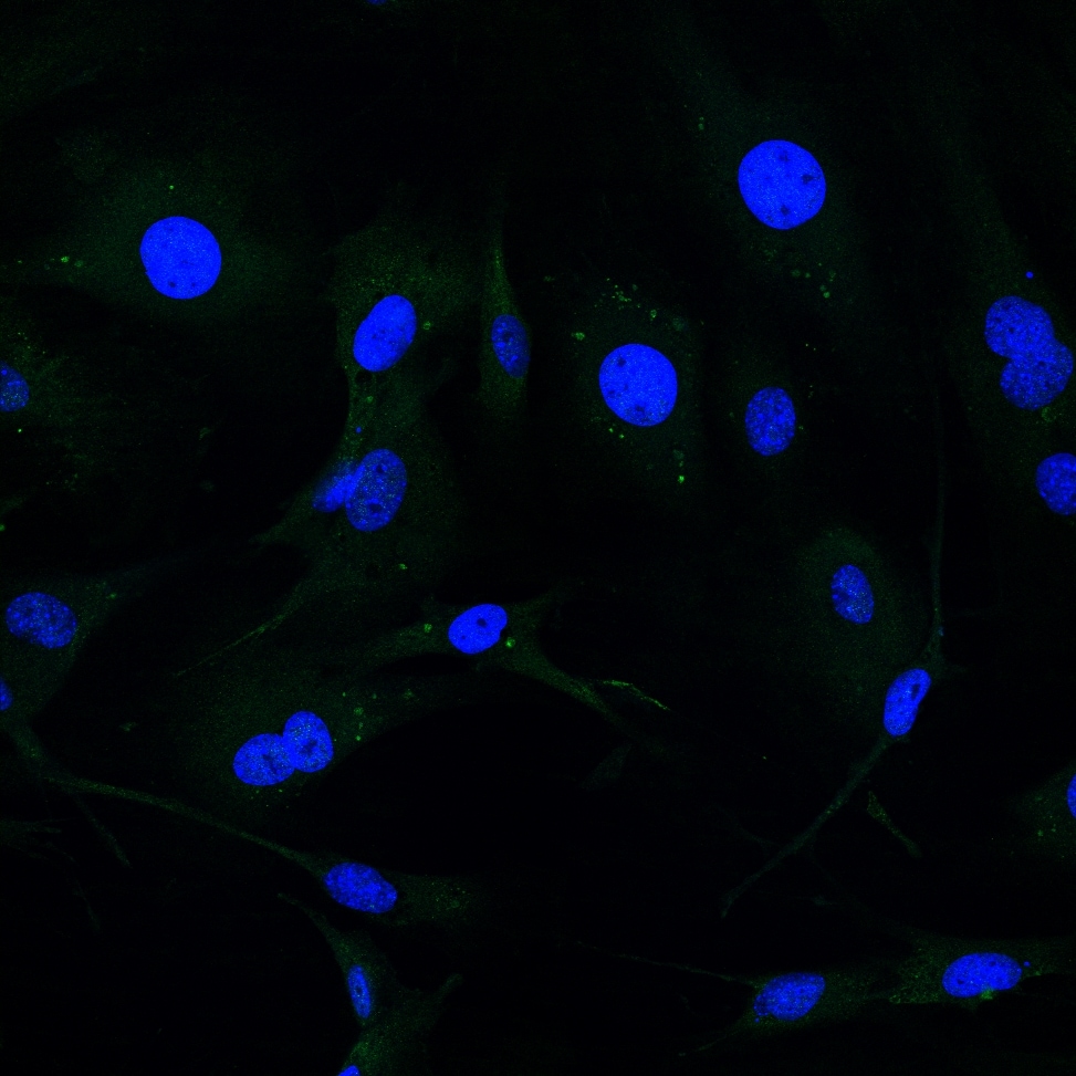
Application: ImmunocytochemistrySample Tested: Mouse embryonic fibroblastsSpecies: MouseVerified Customer | Posted 10/13/2020MEFs stained for ATP6v0A1 (green&#41 and nuclei (DAPI). Antibody diluted 1:200 and incubated for 1 h RT.
There are no reviews that match your criteria.
Protocols
Find general support by application which include: protocols, troubleshooting, illustrated assays, videos and webinars.
- Antigen Retrieval Protocol (PIER)
- Antigen Retrieval for Frozen Sections Protocol
- Appropriate Fixation of IHC/ICC Samples
- Cellular Response to Hypoxia Protocols
- Chromogenic IHC Staining of Formalin-Fixed Paraffin-Embedded (FFPE) Tissue Protocol
- Chromogenic Immunohistochemistry Staining of Frozen Tissue
- ClariTSA™ Fluorophore Kits
- Detection & Visualization of Antibody Binding
- Fluorescent IHC Staining of Frozen Tissue Protocol
- Graphic Protocol for Heat-induced Epitope Retrieval
- Graphic Protocol for the Preparation and Fluorescent IHC Staining of Frozen Tissue Sections
- Graphic Protocol for the Preparation and Fluorescent IHC Staining of Paraffin-embedded Tissue Sections
- Graphic Protocol for the Preparation of Gelatin-coated Slides for Histological Tissue Sections
- ICC Cell Smear Protocol for Suspension Cells
- ICC Immunocytochemistry Protocol Videos
- ICC for Adherent Cells
- IHC Sample Preparation (Frozen sections vs Paraffin)
- Immunocytochemistry (ICC) Protocol
- Immunocytochemistry Troubleshooting
- Immunofluorescence of Organoids Embedded in Cultrex Basement Membrane Extract
- Immunofluorescent IHC Staining of Formalin-Fixed Paraffin-Embedded (FFPE) Tissue Protocol
- Immunohistochemistry (IHC) and Immunocytochemistry (ICC) Protocols
- Immunohistochemistry Frozen Troubleshooting
- Immunohistochemistry Paraffin Troubleshooting
- Preparing Samples for IHC/ICC Experiments
- Preventing Non-Specific Staining (Non-Specific Binding)
- Primary Antibody Selection & Optimization
- Protocol for Heat-Induced Epitope Retrieval (HIER)
- Protocol for Making a 4% Formaldehyde Solution in PBS
- Protocol for VisUCyte™ HRP Polymer Detection Reagent
- Protocol for the Fluorescent ICC Staining of Cell Smears - Graphic
- Protocol for the Fluorescent ICC Staining of Cultured Cells on Coverslips - Graphic
- Protocol for the Preparation & Fixation of Cells on Coverslips
- Protocol for the Preparation and Chromogenic IHC Staining of Frozen Tissue Sections
- Protocol for the Preparation and Chromogenic IHC Staining of Frozen Tissue Sections - Graphic
- Protocol for the Preparation and Chromogenic IHC Staining of Paraffin-embedded Tissue Sections
- Protocol for the Preparation and Chromogenic IHC Staining of Paraffin-embedded Tissue Sections - Graphic
- Protocol for the Preparation and Fluorescent ICC Staining of Cells on Coverslips
- Protocol for the Preparation and Fluorescent ICC Staining of Non-adherent Cells
- Protocol for the Preparation and Fluorescent ICC Staining of Stem Cells on Coverslips
- Protocol for the Preparation and Fluorescent IHC Staining of Frozen Tissue Sections
- Protocol for the Preparation and Fluorescent IHC Staining of Paraffin-embedded Tissue Sections
- Protocol for the Preparation of Gelatin-coated Slides for Histological Tissue Sections
- Protocol for the Preparation of a Cell Smear for Non-adherent Cell ICC - Graphic
- R&D Systems Quality Control Western Blot Protocol
- TUNEL and Active Caspase-3 Detection by IHC/ICC Protocol
- The Importance of IHC/ICC Controls
- Troubleshooting Guide: Immunohistochemistry
- Troubleshooting Guide: Western Blot Figures
- Western Blot Conditions
- Western Blot Protocol
- Western Blot Protocol for Cell Lysates
- Western Blot Troubleshooting
- Western Blot Troubleshooting Guide
- View all Protocols, Troubleshooting, Illustrated assays and Webinars
Loading...