Bcl-2 Antibody - BSA Free
Novus Biologicals | Catalog # NB100-56098
![Western Blot: Bcl-2 Antibody [NB100-56098]](https://resources.rndsystems.com/images/products/Bcl-2-Antibody-Western-Blot-NB100-56098-img0012.jpg "Western Blot: Bcl-2 Antibody [NB100-56098]")
![Western Blot: Bcl-2 Antibody [NB100-56098]](https://resources.rndsystems.com/images/products/Bcl-2-Antibody-Western-Blot-NB100-56098-img0004.jpg "Western Blot: Bcl-2 Antibody [NB100-56098]")
Loading...
Key Product Details
Validated by
Knockout/Knockdown, Biological Validation
Species Reactivity
Validated:
Human, Mouse, Rat, Canine
Cited:
Human, Mouse, Rat, Canine
Predicted:
Bovine (93%), Feline (100%), Porcine (100%). Backed by our 100% Guarantee.
Applications
Validated:
Immunohistochemistry, Immunohistochemistry-Paraffin, Western Blot, Immunoprecipitation
Cited:
Immunohistochemistry, Immunohistochemistry-Paraffin, Western Blot, IF/IHC
Label
Unconjugated
Antibody Source
Polyclonal Rabbit IgG
Format
BSA Free
Loading...
Product Specifications
Immunogen
A synthetic peptide corresponding to amino acids 41-54 (GAAPAPGIFSSQPG) of human Bcl-2 was used as immunogen; GenBank no. NP_000648.2. This sequence is 100% conserved in human Bcl-2 alpha (239 aa) and beta (205 aa) isoforms.
Reactivity Notes
Use in Rat reported in scientific literature (PMID:34575863). Mouse, Canine, Rat reactivity reported in scientific literature (PMID: 15380478).
Clonality
Polyclonal
Host
Rabbit
Isotype
IgG
Scientific Data Images for Bcl-2 Antibody - BSA Free
![Immunohistochemistry-Paraffin: Bcl-2 Antibody [NB100-56098]](https://resources.rndsystems.com/images/products/Bcl-2-Antibody-Immunohistochemistry-Paraffin-NB100-56098-img0011.jpg "Immunohistochemistry-Paraffin: Bcl-2 Antibody [NB100-56098]")
Immunohistochemistry-Paraffin: Bcl-2 Antibody [NB100-56098]
Immunohistochemistry-Paraffin: Bcl-2 Antibody [NB100-56098] - Analysis of a FFPE human breast carcinoma tissue section using 1:1000 dilution of Bcl-2 antibody (NB100-56098) on a Bond Rx autostainer (Leica Biosystems). The assay involved 20 minutes of heat induced antigen retrieval (HIER) with 10mM sodium citrate buffer (pH 6.0) and endogenous peroxidase quenching using peroxide block. The sections were incubated with primary antibody for 30 minutes. Bond Polymer Refine Detection (Leica Biosystems) and DAB were used for signal detection which followed counterstaining with hematoxylin. Whole slide scanning and capturing of representative images (20X) were performed using Aperio AT2 (Leica Biosystems). This antibody generated a diffused cytoplasmic staining of BCL2 in the cancer cells with relatively intense signal in their nuclear membranes. Staining was performed by Histowiz.![Western Blot: Bcl-2 Antibody [NB100-56098]](https://resources.rndsystems.com/images/products/Bcl-2-Antibody-Western-Blot-NB100-56098-img0005.jpg "Western Blot: Bcl-2 Antibody [NB100-56098]")
Western Blot: Bcl-2 Antibody [NB100-56098]
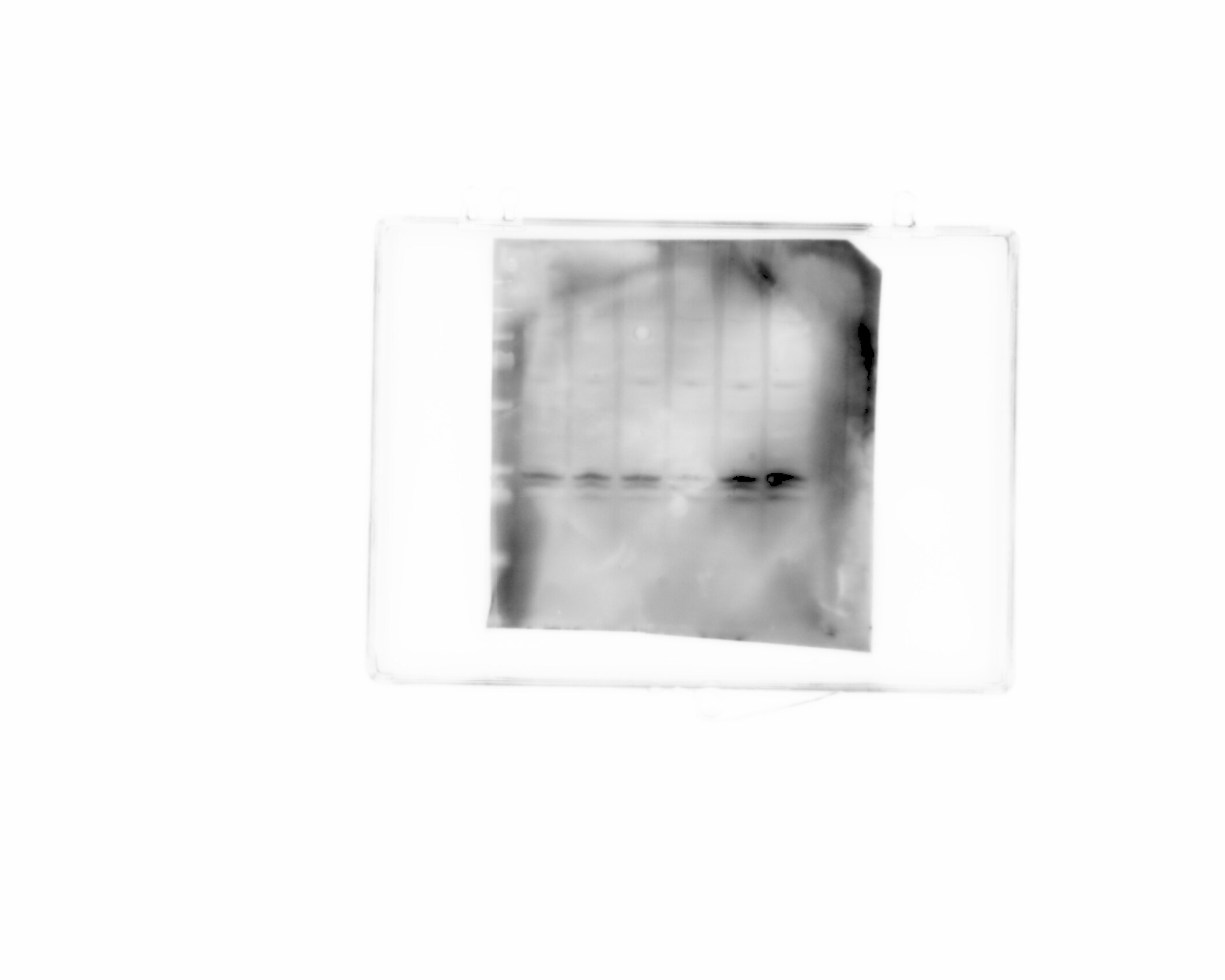
Western Blot: Bcl-2 Antibody [NB100-56098] - Analysis of Bcl-2 using this antibody at 1:2000, 20 ug/protein was loaded per lane. (1) Human Jurkat T cells. (2) Human RS11 lymphoma cells. (3-6) Human breast cancer cases. Patient sample in lane 5 lacked detectible Bcl-2 expression.![Western Blot: Bcl-2 Antibody [NB100-56098]](https://resources.rndsystems.com/images/products/Bcl-2-Antibody-Western-Blot-NB100-56098-img0006.jpg "Western Blot: Bcl-2 Antibody [NB100-56098]")
Western Blot: Bcl-2 Antibody [NB100-56098]
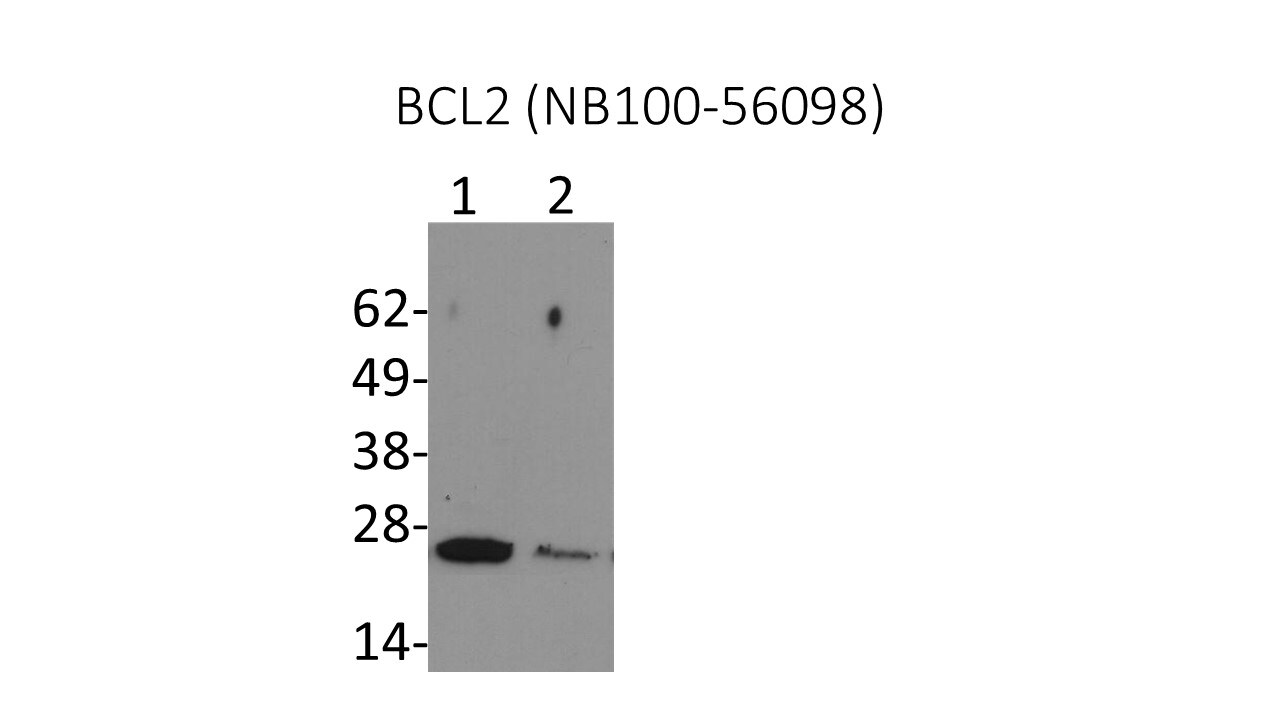
Western Blot: Bcl-2 Antibody [NB100-56098] - Analysis of Bcl-2 in 293T cell lysate (30ug) using anti-Bcl-2 antibody. Image from verified customer review.![Western Blot: Bcl-2 Antibody [NB100-56098]](https://resources.rndsystems.com/images/products/Bcl-2-Antibody-Western-Blot-NB100-56098-img0007.jpg "Western Blot: Bcl-2 Antibody [NB100-56098]")
Western Blot: Bcl-2 Antibody [NB100-56098]
Western Blot: Bcl-2 Antibody [NB100-56098] - Analysis of whole cell lysate (WCL) of Jurkat cells using anti-Bcl-2 antibody (NB100-56098) at 1:1000 dilution. HRP conjugated goat anti-rabbit IgG (H+L) cross adsorbed secondary antibody was used with ECL substrate for the detection of Bcl2 antibody bound to the blotted protein. This Bcl2 antibody detected a specific band at expected molecular weight marker position of Bcl2 protein (26kDa).![Immunohistochemistry-Paraffin: Bcl-2 Antibody [NB100-56098]](https://resources.rndsystems.com/images/products/Bcl-2-Antibody-Immunohistochemistry-Paraffin-NB100-56098-img0010.jpg "Immunohistochemistry-Paraffin: Bcl-2 Antibody [NB100-56098]")
Immunohistochemistry-Paraffin: Bcl-2 Antibody [NB100-56098]
Immunohistochemistry-Paraffin: Bcl-2 Antibody [NB100-56098] - Analysis of a FFPE human breast carcinoma tissue section using 1:1000 dilution of Bcl-2 antibody (NB100-56098) on a Bond Rx autostainer (Leica Biosystems). The assay involved 20 minutes of heat induced antigen retrieval (HIER) with 10mM sodium citrate buffer (pH 6.0) and endogenous peroxidase quenching using peroxide block. The sections were incubated with primary antibody for 30 minutes. Bond Polymer Refine Detection (Leica Biosystems) and DAB were used for signal detection which followed counterstaining with hematoxylin. Whole slide scanning and capturing of representative images (20X) were performed using Aperio AT2 (Leica Biosystems). This antibody generated a diffused cytoplasmic staining of BCL2 in the cancer cells with relatively intense signal in their nuclear membranes. The endothelial cells and the stromal cells showed a weak signal while the RBCs were negative for BCL2.Applications for Bcl-2 Antibody - BSA Free
Application
Recommended Usage
Immunohistochemistry-Paraffin
1:1000-1:5000
Immunoprecipitation
1:50-1:200
Western Blot
1:1000-1:2000
Reviewed Applications
Read 3 reviews rated 5 using NB100-56098 in the following applications:
Formulation, Preparation, and Storage
Purification
Unpurified
Formulation
Whole antisera
Format
BSA Free
Preservative
0.05% Sodium Azide
Concentration
This product is unpurified. The exact concentration of antibody is not quantifiable.
Shipping
The product is shipped with polar packs. Upon receipt, store it immediately at the temperature recommended below.
Stability & Storage
Store at -20C. Avoid freeze-thaw cycles.
Background: Bcl-2
Long Name
B Cell Lymphoma/Leukemia 2
Alternate Names
Bcl2
Entrez Gene IDs
596 (Human)
Gene Symbol
BCL2
Additional Bcl-2 Products
Product Documents for Bcl-2 Antibody - BSA Free
Certificate of Analysis
To download a Certificate of Analysis, please enter a lot or batch number in the search box below.
Product Specific Notices for Bcl-2 Antibody - BSA Free
This product is for research use only and is not approved for use in humans or in clinical diagnosis. Primary Antibodies are guaranteed for 1 year from date of receipt.
Related Research Areas
Citations for Bcl-2 Antibody - BSA Free
 Powered by Bioz
Customer Reviews for Bcl-2 Antibody - BSA Free (3)
5 out of 5
3 Customer Ratings
Have you used Bcl-2 Antibody - BSA Free?
Submit a review and receive an Amazon gift card!
$25/€18/£15/$25CAN/¥2500 Yen for a review with an image
$10/€7/£6/$10CAN/¥1110 Yen for a review without an image
Submit a review
Customer Images
-(005-ml)_NB100-56098_11346.jpg)
Showing
1
-
3 of
3 reviews
Showing All
Filter By:
-
Application: Western BlotSample Tested: Cancer cell lysatesSpecies: Mouse and HumanVerified Customer | Posted 04/19/2024Bcl2 wester blot after apoptosis induction

-
Application: Western BlotSample Tested: Whole cell lysate from Daoy cellsSpecies: HumanVerified Customer | Posted 07/13/2016Daoy cells transfected with BCL2 targeting (Lane 2) or scrambled control siRNAs (Lane 1).
-
Application: Western BlotSample Tested: Whole cell lysate from 293T cells, in lysis bufferSpecies: HumanVerified Customer | Posted 10/17/2014Bcl2 western in 293T cells
There are no reviews that match your criteria.
Protocols
Find general support by application which include: protocols, troubleshooting, illustrated assays, videos and webinars.
- Antigen Retrieval Protocol (PIER)
- Antigen Retrieval for Frozen Sections Protocol
- Appropriate Fixation of IHC/ICC Samples
- Cellular Response to Hypoxia Protocols
- Chromogenic IHC Staining of Formalin-Fixed Paraffin-Embedded (FFPE) Tissue Protocol
- Chromogenic Immunohistochemistry Staining of Frozen Tissue
- ClariTSA™ Fluorophore Kits
- Detection & Visualization of Antibody Binding
- Fluorescent IHC Staining of Frozen Tissue Protocol
- Graphic Protocol for Heat-induced Epitope Retrieval
- Graphic Protocol for the Preparation and Fluorescent IHC Staining of Frozen Tissue Sections
- Graphic Protocol for the Preparation and Fluorescent IHC Staining of Paraffin-embedded Tissue Sections
- Graphic Protocol for the Preparation of Gelatin-coated Slides for Histological Tissue Sections
- IHC Sample Preparation (Frozen sections vs Paraffin)
- Immunofluorescent IHC Staining of Formalin-Fixed Paraffin-Embedded (FFPE) Tissue Protocol
- Immunohistochemistry (IHC) and Immunocytochemistry (ICC) Protocols
- Immunohistochemistry Frozen Troubleshooting
- Immunohistochemistry Paraffin Troubleshooting
- Immunoprecipitation Protocol
- Preparing Samples for IHC/ICC Experiments
- Preventing Non-Specific Staining (Non-Specific Binding)
- Primary Antibody Selection & Optimization
- Protocol for Heat-Induced Epitope Retrieval (HIER)
- Protocol for Making a 4% Formaldehyde Solution in PBS
- Protocol for VisUCyte™ HRP Polymer Detection Reagent
- Protocol for the Preparation & Fixation of Cells on Coverslips
- Protocol for the Preparation and Chromogenic IHC Staining of Frozen Tissue Sections
- Protocol for the Preparation and Chromogenic IHC Staining of Frozen Tissue Sections - Graphic
- Protocol for the Preparation and Chromogenic IHC Staining of Paraffin-embedded Tissue Sections
- Protocol for the Preparation and Chromogenic IHC Staining of Paraffin-embedded Tissue Sections - Graphic
- Protocol for the Preparation and Fluorescent IHC Staining of Frozen Tissue Sections
- Protocol for the Preparation and Fluorescent IHC Staining of Paraffin-embedded Tissue Sections
- Protocol for the Preparation of Gelatin-coated Slides for Histological Tissue Sections
- R&D Systems Quality Control Western Blot Protocol
- TUNEL and Active Caspase-3 Detection by IHC/ICC Protocol
- The Importance of IHC/ICC Controls
- Troubleshooting Guide: Immunohistochemistry
- Troubleshooting Guide: Western Blot Figures
- Western Blot Conditions
- Western Blot Protocol
- Western Blot Protocol for Cell Lysates
- Western Blot Troubleshooting
- Western Blot Troubleshooting Guide
- View all Protocols, Troubleshooting, Illustrated assays and Webinars


