![Western Blot: BDNF Antibody [NBP1-46750]](https://resources.rndsystems.com/images/products/BDNF-Antibody-Western-Blot-NBP1-46750-img0002.jpg "Western Blot: BDNF Antibody [NBP1-46750]")
Loading...
Key Product Details
Species Reactivity
Validated:
Human, Mouse, Rat
Cited:
Mouse, Rat
Applications
Validated:
Immunohistochemistry, Immunohistochemistry-Paraffin, Western Blot
Cited:
Immunohistochemistry-Paraffin, Western Blot
Label
Unconjugated
Antibody Source
Polyclonal Rabbit IgG
Loading...
Product Specifications
Immunogen
A synthetic peptide from internal region of human mature BDNF conjugated to blue carrier protein was used as the antigen. The peptide is homologous in rat and mouse.
Specificity
Specific for mature BDNF.
Clonality
Polyclonal
Host
Rabbit
Isotype
IgG
Scientific Data Images for BDNF Antibody
Western Blot: BDNF Antibody [NBP1-46750]
Western Blot: BDNF Antibody [NBP1-46750] - Human platelet lysate. Blocking: 1% LFDM for 30 min at RT; primary antibody incubated at 4C overnight. Click on eLAB BOOK tab to see the details. Mature BDNF: (1:2000 dilution) ; proBDNF: (1:1000 dilution)![Immunohistochemistry-Paraffin: BDNF Antibody [NBP1-46750]](https://resources.rndsystems.com/images/products/BDNF-Antibody-Immunohistochemistry-Paraffin-NBP1-46750-img0008.jpg "Immunohistochemistry-Paraffin: BDNF Antibody [NBP1-46750]")
Immunohistochemistry-Paraffin: BDNF Antibody [NBP1-46750]
Immunohistochemistry-Paraffin: BDNF Antibody [NBP1-46750] - IHC-P on paraffin sections of rat hippocampus.The animal was perfused using Autoperfuser at a pressure of 130 mmHg with 300 ml 4% FA before being processed for paraffin embedding. HIER: Tris-EDTA, pH 9 for 20 min.Blocking: 0.2% LFDM in TBST filtered thru 0.2 um.Detection was done using HRP polymer following manufacturers instructions; DAB chromogen.Primary antibody: dilution 1: 1000, incubated 30 min at RT using Autostainer.Sections were counterstained with Harris Hematoxylin.Small neurons are stained and also some nuclear staining is observed.![Immunohistochemistry-Paraffin: BDNF Antibody [NBP1-46750]](https://resources.rndsystems.com/images/products/BDNF-Antibody-Immunohistochemistry-Paraffin-NBP1-46750-img0003.jpg "Immunohistochemistry-Paraffin: BDNF Antibody [NBP1-46750]")
Immunohistochemistry-Paraffin: BDNF Antibody [NBP1-46750]
Immunohistochemistry-Paraffin: BDNF Antibody [NBP1-46750] - Paraffin sections of rat spinal cord. Dilution 1: 1000.![Immunohistochemistry-Paraffin: BDNF Antibody [NBP1-46750]](https://resources.rndsystems.com/images/products/BDNF-Antibody-Immunohistochemistry-Paraffin-NBP1-46750-img0004.jpg "Immunohistochemistry-Paraffin: BDNF Antibody [NBP1-46750]")
Immunohistochemistry-Paraffin: BDNF Antibody [NBP1-46750]
Immunohistochemistry-Paraffin: BDNF Antibody [NBP1-46750] - Paraffin sections of rat hippocampus. Dilution 1: 1000.![Immunohistochemistry-Paraffin: BDNF Antibody [NBP1-46750]](https://resources.rndsystems.com/images/products/BDNF-Antibody-Immunohistochemistry-Paraffin-NBP1-46750-img0005.jpg "Immunohistochemistry-Paraffin: BDNF Antibody [NBP1-46750]")
Immunohistochemistry-Paraffin: BDNF Antibody [NBP1-46750]
Immunohistochemistry-Paraffin: BDNF Antibody [NBP1-46750] - Paraffin sections of mouse hippocampus. Dilution 1: 1000.![Immunohistochemistry-Paraffin: BDNF Antibody [NBP1-46750]](https://resources.rndsystems.com/images/products/BDNF-Antibody-Immunohistochemistry-Paraffin-NBP1-46750-img0007.jpg "Immunohistochemistry-Paraffin: BDNF Antibody [NBP1-46750]")
Immunohistochemistry-Paraffin: BDNF Antibody [NBP1-46750]
Immunohistochemistry-Paraffin: BDNF Antibody [NBP1-46750] - IHC-P on paraffin sections of rat hippocampus.The animal was perfused using Autoperfuser at a pressure of 130 mmHg with 300 ml 4% FA before being processed for paraffin embedding. HIER: Tris-EDTA, pH 9 for 20 min.Blocking: 0.2% LFDM in TBST filtered thru 0.2 um.Detection was done using HRP polymer following manufacturers instructions; DAB chromogen.Primary antibody: dilution 1: 1000, incubated 30 min at RT using Autostainer.Sections were counterstained with Harris Hematoxylin.Small neurons are stained and also some nuclear staining is observed.
Western Blot: BDNF Antibody [NBP1-46750] -
Aging reduces BDNF maturation in the brain. Brains of C57BL/6J mice at 6, 12 and 24 months of age were homogenized for quantitative Western blot of BDNF (A). Normal aging significantly reduced the protein levels of maturated BDNF, but not pro-BDNF (B and C; One-way ANOVA followed by Bonferroni post-hoc test; n = 5–11 per group). Aging did not decrease phosphorylated TrkB (D; One-way ANOVA; n = 4–5 per group), but significantly increased the protein level of total TrkB (E; One-way ANOVA followed by Bonferroni post-hoc test; n = 4–5 per group) Image collected and cropped by CiteAb from the following open publication (https://actaneurocomms.biomedcentral.com/articles/10.1186/s40478-025-02…), licensed under a CC-BY license. Not internally tested by Novus Biologicals.
Western Blot: BDNF Antibody [NBP1-46750] -
Deficiency of astrocyte BDNF reduces vasculature and pericytes in the brain. Brain homogenates from 10 month-old C57BL/6 mice with (ko) and without (wt) knockout of Bdnf gene in astrocytes for 3 months were detected for protein levels of BDNF (A). Knockout of Bdnf gene significantly reduced mature BDNF but not pro-BDNF (B and C; t-test, n = 9–11 per group). Brain sections were then stained for collagen type IV and quantified for the vasculature (D). Deficiency of astrocyte BDNF significantly reduced both the length and density of branch points of cerebral vessels (E and F; t-test, n = 8 per group). Additionally, microvessels were isolated from brains and detected with Western blot for pericyte markers and relevant signaling molecules (G and J). Deficiency of astrocyte BDNF decreased the protein level of PDGFR beta, but not CD13 (H and I; t-test, n = 11–12 per group), and reduced the phosphorylation of both Akt and Erk1/2 (K and L; t-test, n = 4–9 per group) Image collected and cropped by CiteAb from the following open publication (https://actaneurocomms.biomedcentral.com/articles/10.1186/s40478-025-02…), licensed under a CC-BY license. Not internally tested by Novus Biologicals.Applications for BDNF Antibody
Application
Recommended Usage
Immunohistochemistry
1:1000-1:2000
Immunohistochemistry-Paraffin
1:1000-1:2000
Western Blot
1:1000-1:2000
Reviewed Applications
Read 2 reviews rated 2 using NBP1-46750 in the following applications:
Formulation, Preparation, and Storage
Purification
Unpurified
Reconstitution
Reconstitute in 0.1 ml of sterile water. Centrifuge to remove any insoluble material. Glycerol may be added (1:1) for additional stability. Please note the sample size is provided in reconstituted format.
Formulation
Lyophilized from whole antisera
Preservative
No Preservative
Concentration
This product is unpurified. The exact concentration of antibody is not quantifiable.
Shipping
The product is shipped with polar packs. Upon receipt, store it immediately at the temperature recommended below.
Stability & Storage
Store at 4C short term. Aliquot and store at -20C long term. Avoid freeze-thaw cycles.
Calculators
Background: BDNF
Long Name
Brain-derived Neurotrophic Factor
Alternate Names
Abrineurin, ANON2, BULN2, Neurotrophin
Entrez Gene IDs
627 (Human)
Gene Symbol
BDNF
Additional BDNF Products
Product Documents for BDNF Antibody
Certificate of Analysis
To download a Certificate of Analysis, please enter a lot or batch number in the search box below.
Product Specific Notices for BDNF Antibody
This product is for research use only and is not approved for use in humans or in clinical diagnosis. Primary Antibodies are guaranteed for 1 year from date of receipt.
Related Research Areas
Citations for BDNF Antibody
 Powered by Bioz
Customer Reviews for BDNF Antibody (2)
2 out of 5
2 Customer Ratings
Have you used BDNF Antibody?
Submit a review and receive an Amazon gift card!
$25/€18/£15/$25CAN/¥2500 Yen for a review with an image
$10/€7/£6/$10CAN/¥1110 Yen for a review without an image
Submit a review
Customer Images

Showing
1
-
2 of
2 reviews
Showing All
Filter By:
-
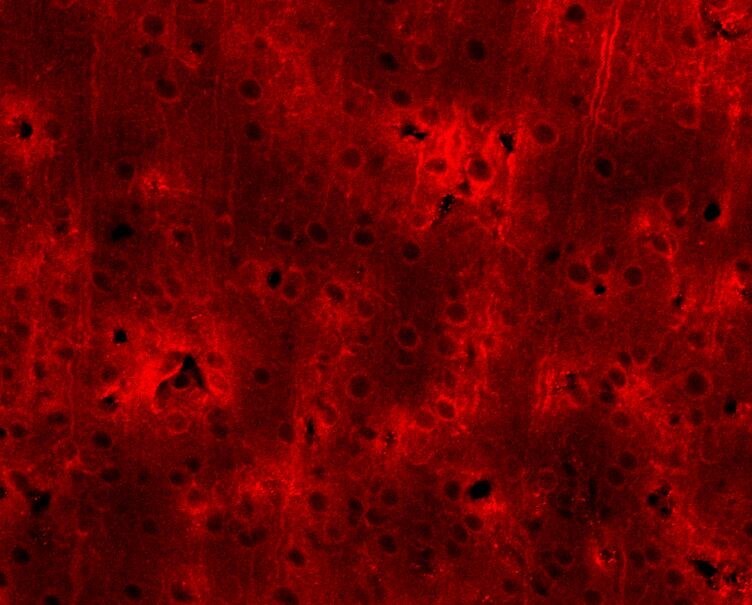
Application: Immunohistochemistry-FrozenSample Tested: rat brain sectionsSpecies: RatVerified Customer | Posted 10/16/2017Immunofluoro (Alexa 555)-labeled BDNF signals using NBP1-46750 (1:200). When we overlayed images with BDNF and NeuN signals, they showed colocalization, and we confirmed those signals were positive. This was IHC w/ frozen rat brain section.This figure represents immunofluoro (Alexa 555)-labeled BDNF signals using antibody NBP1-46750 (1:200). When we overlayed images with BDNF and NeuN signals, they showed colocalization, and we confirmed those signals were positive. However, the patterns are ring-shaped, different from what we expected. This was immunohistochemistry with frozen rat brain section.

-
Application: Simple WesternSample Tested: BDNF Overexpression Lysate (NBL1-07962)Species: HumanVerified Customer | Posted 11/24/2016
There are no reviews that match your criteria.
Protocols
Find general support by application which include: protocols, troubleshooting, illustrated assays, videos and webinars.
- Antigen Retrieval Protocol (PIER)
- Antigen Retrieval for Frozen Sections Protocol
- Appropriate Fixation of IHC/ICC Samples
- Cellular Response to Hypoxia Protocols
- Chromogenic IHC Staining of Formalin-Fixed Paraffin-Embedded (FFPE) Tissue Protocol
- Chromogenic Immunohistochemistry Staining of Frozen Tissue
- ClariTSA™ Fluorophore Kits
- Detection & Visualization of Antibody Binding
- Fluorescent IHC Staining of Frozen Tissue Protocol
- Graphic Protocol for Heat-induced Epitope Retrieval
- Graphic Protocol for the Preparation and Fluorescent IHC Staining of Frozen Tissue Sections
- Graphic Protocol for the Preparation and Fluorescent IHC Staining of Paraffin-embedded Tissue Sections
- Graphic Protocol for the Preparation of Gelatin-coated Slides for Histological Tissue Sections
- IHC Sample Preparation (Frozen sections vs Paraffin)
- Immunofluorescent IHC Staining of Formalin-Fixed Paraffin-Embedded (FFPE) Tissue Protocol
- Immunohistochemistry (IHC) and Immunocytochemistry (ICC) Protocols
- Immunohistochemistry Frozen Troubleshooting
- Immunohistochemistry Paraffin Troubleshooting
- Preparing Samples for IHC/ICC Experiments
- Preventing Non-Specific Staining (Non-Specific Binding)
- Primary Antibody Selection & Optimization
- Protocol for Heat-Induced Epitope Retrieval (HIER)
- Protocol for Making a 4% Formaldehyde Solution in PBS
- Protocol for VisUCyte™ HRP Polymer Detection Reagent
- Protocol for the Preparation & Fixation of Cells on Coverslips
- Protocol for the Preparation and Chromogenic IHC Staining of Frozen Tissue Sections
- Protocol for the Preparation and Chromogenic IHC Staining of Frozen Tissue Sections - Graphic
- Protocol for the Preparation and Chromogenic IHC Staining of Paraffin-embedded Tissue Sections
- Protocol for the Preparation and Chromogenic IHC Staining of Paraffin-embedded Tissue Sections - Graphic
- Protocol for the Preparation and Fluorescent IHC Staining of Frozen Tissue Sections
- Protocol for the Preparation and Fluorescent IHC Staining of Paraffin-embedded Tissue Sections
- Protocol for the Preparation of Gelatin-coated Slides for Histological Tissue Sections
- R&D Systems Quality Control Western Blot Protocol
- TUNEL and Active Caspase-3 Detection by IHC/ICC Protocol
- The Importance of IHC/ICC Controls
- Troubleshooting Guide: Immunohistochemistry
- Troubleshooting Guide: Western Blot Figures
- Western Blot Conditions
- Western Blot Protocol
- Western Blot Protocol for Cell Lysates
- Western Blot Troubleshooting
- Western Blot Troubleshooting Guide
- View all Protocols, Troubleshooting, Illustrated assays and Webinars

