Bestrophin 1 Antibody (E6-6) - Azide and BSA Free
Novus Biologicals | Catalog # NBP2-80593
Key Product Details
Species Reactivity
Human, Mouse, Porcine, Canine, Primate, Rat (Negative)
Applications
Immunohistochemistry, Immunohistochemistry-Paraffin, Immunohistochemistry-Frozen, Western Blot, Dual RNAscope ISH-IHC, Immunocytochemistry/ Immunofluorescence, Immunoprecipitation
Label
Unconjugated
Antibody Source
Monoclonal Mouse IgG1 kappa Clone # E6-6
Format
Azide and BSA Free
Loading...
Product Specifications
Immunogen
Synthetic peptide conjugated to KLH corresponding to the C-terminus of human Bestrophin 1 (KDHMDPYWALENRDEAHS) [Uniprot: O76090]
Reactivity Notes
Human, Primate, Porcine reactivity reported in scientific literature (PMID: 11050159).
Clonality
Monoclonal
Host
Mouse
Isotype
IgG1 kappa
Scientific Data Images for Bestrophin 1 Antibody (E6-6) - Azide and BSA Free
![Western Blot: Bestrophin 1 Antibody (E6-6)Azide and BSA Free [NBP2-80593]](https://resources.rndsystems.com/images/products/Bestrophin-1-Antibody-E6-6-Azide-and-BSA-Free-Western-Blot-NBP2-80593-img0002.jpg "Western Blot: Bestrophin 1 Antibody (E6-6)Azide and BSA Free [NBP2-80593]")
Western Blot: Bestrophin 1 Antibody (E6-6)Azide and BSA Free [NBP2-80593]
Western Blot: Bestrophin 1 Antibody (E6-6) - Azide and BSA Free [NBP2-80593] - Western blot analysis of the normal and mutant human Best1 protein in transiently transfected MDCK cells. Best1 proteins are detectable as a 68 kDa band in all transfected cells, but not in non-transfected controls (MDCK lane). Actin bands are shown to indicate equal loading of cell lysates. Image collected and cropped by CiteAb from the following publication (https://www.mdpi.com/1422-0067/14/7/15121), licensed under a CC-BY license. Image from the standard format of this antibody.![Immunocytochemistry: Bestrophin 1 Antibody (E6-6) - Azide and BSA Free [NBP2-80593]](https://resources.rndsystems.com/images/products/Bestrophin-1-Antibody-E6-6-Azide-and-BSA-Free-Immunocytochemistry-NBP2-80593-img0005.jpg "Immunocytochemistry: Bestrophin 1 Antibody (E6-6) - Azide and BSA Free [NBP2-80593]")
Immunocytochemistry: Bestrophin 1 Antibody (E6-6) - Azide and BSA Free [NBP2-80593]
Immunocytochemistry: Bestrophin 1 Antibody (E6-6) - Azide and BSA Free [NBP2-80593] - Bestrophin 1 was detected in immersion fixed paraffin-embedded sections of human brain using Mouse Anti-Human Bestrophin 1 (E6-6) Monoclonal Antibody (Catalog # NB300-164) at 1:300 for 1 hour at room temperature followed by incubation with the Anti-Mouse![Immunohistochemistry: Bestrophin 1 Antibody (E6-6) - Azide and BSA Free [NBP2-80593]](https://resources.rndsystems.com/images/products/Bestrophin-1-Antibody-E6-6-Azide-and-BSA-Free-Immunohistochemistry-NBP2-80593-img0004.jpg "Immunohistochemistry: Bestrophin 1 Antibody (E6-6) - Azide and BSA Free [NBP2-80593]")
Immunohistochemistry: Bestrophin 1 Antibody (E6-6) - Azide and BSA Free [NBP2-80593]
Immunohistochemistry: Bestrophin 1 Antibody (E6-6) - Azide and BSA Free [NBP2-80593] - Bestrophin 1 was detected in immersion fixed paraffin sections of human small intestine using t Mouse Anti-Human Bestrophin 1 Monoclonal Antibody (Catalog # NB300-164) at 5 ug/mL for 1 hour at room temperature followed by incubation with the Anti-Mouse Ig![Western Blot: Bestrophin 1 Antibody (E6-6)Azide and BSA Free [NBP2-80593]](https://resources.rndsystems.com/images/products/Bestrophin-1-Antibody-E6-6-Azide-and-BSA-Free-Western-Blot-NBP2-80593-img0001.jpg "Western Blot: Bestrophin 1 Antibody (E6-6)Azide and BSA Free [NBP2-80593]")
Western Blot: Bestrophin 1 Antibody (E6-6)Azide and BSA Free [NBP2-80593]
Western Blot: Bestrophin 1 Antibody (E6-6) - Azide and BSA Free [NBP2-80593] - Detection of Bestrophin (68 kDa) from human RPE cell lysate. Image from the standard format of this antibody.![Immunocytochemistry/ Immunofluorescence: Bestrophin 1 Antibody (E6-6) - Azide and BSA Free [NBP2-80593]](https://resources.rndsystems.com/images/products/Bestrophin-1-Antibody-E6-6-Azide-and-BSA-Free-Immunocytochemistry-Immunofluorescence-NBP2-80593-img0003.jpg "Immunocytochemistry/ Immunofluorescence: Bestrophin 1 Antibody (E6-6) - Azide and BSA Free [NBP2-80593]")
Immunocytochemistry/ Immunofluorescence: Bestrophin 1 Antibody (E6-6) - Azide and BSA Free [NBP2-80593]
Immunocytochemistry/Immunofluorescence: Bestrophin 1 Antibody (E6-6) - Azide and BSA Free [NBP2-80593] - X-Z confocal single image scan of transiently transfected cells with different BEST1 cDNA constructs showing mislocalization of mutants Y85H, Q96R, L100R and Y227N. Cells were stained for Best1 (green), beta-catenin (red) and nuclei (blue). Scale bar = 10 um. Image collected and cropped by CiteAb from the following publication (https://www.mdpi.com/1422-0067/14/7/15121), licensed under a CC-BY license. Image from the standard format of this antibody.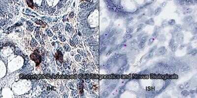
Dual RNAscope ISH-IHC: Bestrophin 1 Antibody (E6-6) - Azide and BSA Free [NBP2-80593] - Formalin-fixed paraffin-embedded tissue sections of human duodenum were probed for Bestrophin 1 mRNA (ACD RNAScope Probe, catalog # 433181; Fast Red chromogen, ACD catalog # 322360). Adjacent tissue section was processed for immunohistochemistry using mouse anti-human (Novus Biologicals catalog # NB300-164) at 0.3ug/mL with overnight incubation at 4 degrees Celsius followed by incubation with anti-mouse IgG VisUCyte HRP Polymer Antibody (Catalog # VC001) and DAB chromogen (yellow-brown). Tissue was counterstained with hematoxylin (blue). IHC signal is confined to cytoplasm. Image from the standard format of this antibody.
Applications for Bestrophin 1 Antibody (E6-6) - Azide and BSA Free
Application
Recommended Usage
Immunohistochemistry
reported in scientific literature (PMID 30048622)
Immunohistochemistry-Paraffin
reported in scientific literature (PMID 24345323)
Western Blot
1:1000
Application Notes
In Western blot, this antibody recognizes a band at ~68 kDa representing Bestrophin. Please see protocol for treatment of cell extracts. The observed molecular weight of the protein may vary from the listed predicted molecular weight due to post translational modifications, post translation cleavages, relative charges, and other experimental factors.
Formulation, Preparation, and Storage
Purification
Protein A or G purified
Formulation
PBS
Format
Azide and BSA Free
Preservative
No Preservative
Concentration
1 mg/ml
Shipping
The product is shipped with polar packs. Upon receipt, store it immediately at the temperature recommended below.
Stability & Storage
Aliquot and store at -20C or -80C. Avoid freeze-thaw cycles.
Background: Bestrophin 1
Alternate Names
ARB, BEST, Best disease, bestrophin 1, bestrophin-1, BMD, TU15B, vitelliform macular dystrophy 2, Vitelliform macular dystrophy protein 2, VMD2RP50
Gene Symbol
BEST1
Additional Bestrophin 1 Products
Product Documents for Bestrophin 1 Antibody (E6-6) - Azide and BSA Free
Certificate of Analysis
To download a Certificate of Analysis, please enter a lot or batch number in the search box below.
Product Specific Notices for Bestrophin 1 Antibody (E6-6) - Azide and BSA Free
This product is for research use only and is not approved for use in humans or in clinical diagnosis. Primary Antibodies are guaranteed for 1 year from date of receipt.
Customer Reviews for Bestrophin 1 Antibody (E6-6) - Azide and BSA Free
There are currently no reviews for this product. Be the first to review Bestrophin 1 Antibody (E6-6) - Azide and BSA Free and earn rewards!
Have you used Bestrophin 1 Antibody (E6-6) - Azide and BSA Free?
Submit a review and receive an Amazon gift card!
$25/€18/£15/$25CAN/¥2500 Yen for a review with an image
$10/€7/£6/$10CAN/¥1110 Yen for a review without an image
Submit a review
Protocols
Find general support by application which include: protocols, troubleshooting, illustrated assays, videos and webinars.
- Antigen Retrieval Protocol (PIER)
- Antigen Retrieval for Frozen Sections Protocol
- Appropriate Fixation of IHC/ICC Samples
- Cellular Response to Hypoxia Protocols
- Chromogenic IHC Staining of Formalin-Fixed Paraffin-Embedded (FFPE) Tissue Protocol
- Chromogenic Immunohistochemistry Staining of Frozen Tissue
- ClariTSA™ Fluorophore Kits
- Detection & Visualization of Antibody Binding
- Fluorescent IHC Staining of Frozen Tissue Protocol
- Graphic Protocol for Heat-induced Epitope Retrieval
- Graphic Protocol for the Preparation and Fluorescent IHC Staining of Frozen Tissue Sections
- Graphic Protocol for the Preparation and Fluorescent IHC Staining of Paraffin-embedded Tissue Sections
- Graphic Protocol for the Preparation of Gelatin-coated Slides for Histological Tissue Sections
- ICC Cell Smear Protocol for Suspension Cells
- ICC Immunocytochemistry Protocol Videos
- ICC for Adherent Cells
- IHC Sample Preparation (Frozen sections vs Paraffin)
- ISH-IHC Protocol for Chromogenic Detection on Formalin Fixed Paraffin Embedded (FFPE) Tissue
- Immunocytochemistry (ICC) Protocol
- Immunocytochemistry Troubleshooting
- Immunofluorescence of Organoids Embedded in Cultrex Basement Membrane Extract
- Immunofluorescent IHC Staining of Formalin-Fixed Paraffin-Embedded (FFPE) Tissue Protocol
- Immunohistochemistry (IHC) and Immunocytochemistry (ICC) Protocols
- Immunohistochemistry Frozen Troubleshooting
- Immunohistochemistry Paraffin Troubleshooting
- Immunoprecipitation Protocol
- Preparing Samples for IHC/ICC Experiments
- Preventing Non-Specific Staining (Non-Specific Binding)
- Primary Antibody Selection & Optimization
- Protocol for Heat-Induced Epitope Retrieval (HIER)
- Protocol for Making a 4% Formaldehyde Solution in PBS
- Protocol for VisUCyte™ HRP Polymer Detection Reagent
- Protocol for the Fluorescent ICC Staining of Cell Smears - Graphic
- Protocol for the Fluorescent ICC Staining of Cultured Cells on Coverslips - Graphic
- Protocol for the Preparation & Fixation of Cells on Coverslips
- Protocol for the Preparation and Chromogenic IHC Staining of Frozen Tissue Sections
- Protocol for the Preparation and Chromogenic IHC Staining of Frozen Tissue Sections - Graphic
- Protocol for the Preparation and Chromogenic IHC Staining of Paraffin-embedded Tissue Sections
- Protocol for the Preparation and Chromogenic IHC Staining of Paraffin-embedded Tissue Sections - Graphic
- Protocol for the Preparation and Fluorescent ICC Staining of Cells on Coverslips
- Protocol for the Preparation and Fluorescent ICC Staining of Non-adherent Cells
- Protocol for the Preparation and Fluorescent ICC Staining of Stem Cells on Coverslips
- Protocol for the Preparation and Fluorescent IHC Staining of Frozen Tissue Sections
- Protocol for the Preparation and Fluorescent IHC Staining of Paraffin-embedded Tissue Sections
- Protocol for the Preparation of Gelatin-coated Slides for Histological Tissue Sections
- Protocol for the Preparation of a Cell Smear for Non-adherent Cell ICC - Graphic
- R&D Systems Quality Control Western Blot Protocol
- TUNEL and Active Caspase-3 Detection by IHC/ICC Protocol
- The Importance of IHC/ICC Controls
- Troubleshooting Guide: Immunohistochemistry
- Troubleshooting Guide: Western Blot Figures
- Western Blot Conditions
- Western Blot Protocol
- Western Blot Protocol for Cell Lysates
- Western Blot Troubleshooting
- Western Blot Troubleshooting Guide
- View all Protocols, Troubleshooting, Illustrated assays and Webinars
Loading...