Bestrophin 1 Antibody (E6-6) - BSA Free
Novus Biologicals | Catalog # NB300-164
Key Product Details
Validated by
Species Reactivity
Validated:
Cited:
Applications
Validated:
Cited:
Label
Antibody Source
Format
Product Specifications
Immunogen
Reactivity Notes
Clonality
Host
Isotype
Scientific Data Images for Bestrophin 1 Antibody (E6-6) - BSA Free
![Western Blot: Bestrophin 1 Antibody (E6-6)BSA Free [NB300-164]](https://resources.rndsystems.com/images/products/Bestrophin-1-Antibody-E6-6-BSA-Free-Western-Blot-NB300-164-img0007.jpg "Western Blot: Bestrophin 1 Antibody (E6-6)BSA Free [NB300-164]")
Western Blot: Bestrophin 1 Antibody (E6-6)BSA Free [NB300-164]
Bestrophin-1-Antibody-E6-6-BSA-Free-Western-Blot-NB300-164-img0007.jpg![Immunocytochemistry/ Immunofluorescence: Bestrophin 1 Antibody (E6-6) - BSA Free [NB300-164]](https://resources.rndsystems.com/images/products/Bestrophin-1-Antibody-E6-6-BSA-Free-Immunocytochemistry-Immunofluorescence-NB300-164-img0008.jpg "Immunocytochemistry/ Immunofluorescence: Bestrophin 1 Antibody (E6-6) - BSA Free [NB300-164]")
Immunocytochemistry/ Immunofluorescence: Bestrophin 1 Antibody (E6-6) - BSA Free [NB300-164]
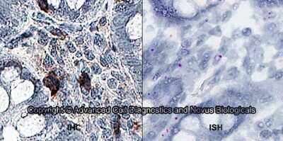
Bestrophin-1-Antibody-E6-6-BSA-Free-Immunocytochemistry-Immunofluorescence-NB300-164-img0008.jpg![Immunohistochemistry-Paraffin: Bestrophin 1 Antibody (E6-6) - BSA Free [NB300-164]](https://resources.rndsystems.com/images/products/Bestrophin-1-Antibody-E6-6-BSA-Free-Immunohistochemistry-Paraffin-NB300-164-img0005.jpg "Immunohistochemistry-Paraffin: Bestrophin 1 Antibody (E6-6) - BSA Free [NB300-164]")
Immunohistochemistry-Paraffin: Bestrophin 1 Antibody (E6-6) - BSA Free [NB300-164]
Immunohistochemistry-Paraffin: Bestrophin 1 Antibody (E6-6) - BSA Free [NB300-164] - Bestrophin 1 was detected in immersion fixed paraffin sections of human small intestine using t Mouse Anti-Human Bestrophin 1 Monoclonal Antibody (Catalog # NB300-164) at 5 ug/mL for 1 hour at room temperature followed by incubation with the Anti-Mouse IgG VisUCyte™ HRP Polymer Antibody (Catalog # VC001). Tissue was stained using DAB (brown) and counterstained with hematoxylin (blue). Specific staining was localized to the cell surface and extracellular.![Western Blot: Bestrophin 1 Antibody (E6-6)BSA Free [NB300-164]](https://resources.rndsystems.com/images/products/Bestrophin-1-Antibody-E6-6-BSA-Free-Western-Blot-NB300-164-img0003.jpg "Western Blot: Bestrophin 1 Antibody (E6-6)BSA Free [NB300-164]")
Western Blot: Bestrophin 1 Antibody (E6-6)BSA Free [NB300-164]
Western Blot: Bestrophin 1 Antibody (E6-6) - BSA Free [NB300-164] - Detection of Bestrophin (68 kDa) from human RPE cell lysate.![Immunohistochemistry-Paraffin: Bestrophin 1 Antibody (E6-6) - BSA Free [NB300-164]](https://resources.rndsystems.com/images/products/Bestrophin-1-Antibody-E6-6-BSA-Free-Immunohistochemistry-Paraffin-NB300-164-img0004.jpg "Immunohistochemistry-Paraffin: Bestrophin 1 Antibody (E6-6) - BSA Free [NB300-164]")
Immunohistochemistry-Paraffin: Bestrophin 1 Antibody (E6-6) - BSA Free [NB300-164]
Immunohistochemistry-Paraffin: Bestrophin 1 Antibody (E6-6) - BSA Free [NB300-164] - Bestrophin 1 was detected in immersion fixed paraffin-embedded sections of human brain using Mouse Anti-Human Bestrophin 1 (E6-6) Monoclonal Antibody (Catalog # NB300-164) at 1:300 for 1 hour at room temperature followed by incubation with the Anti-Mouse IgG VisUCyte™ HRP Polymer Antibody (Catalog # VC001). Tissue was stained using DAB (brown) and counterstained with hematoxylin (blue). Specific staining was localized to the cytoplasm in neurons.![Knockout Validated: Bestrophin 1 Antibody (E6-6) - BSA Free [NB300-164]](https://resources.rndsystems.com/images/products/Bestrophin-1-Antibody-E6-6-BSA-Free-Knockout-Validated-NB300-164-img0009.jpg "Western Blot: Bestrophin 1 Antibody (E6-6) - BSA Free [NB300-164]")
 - BSA Free [NB300-164] -")
Western Blot: Bestrophin 1 Antibody (E6-6) - BSA Free [NB300-164] -
Expression of RPE-specific marker proteins in hPSC-RPE & iPSC-RPE cells.(a–b) Immunoblotting showing the expression of RPE-specific proteins BEST1, RPE65, CRALBP, & the loading control beta -Actin in hPSC-RPE (a) & iPSC-RPE (b) cells. Two gels/blots in the same panel were prepared from the same cell lysate of each PSC-RPE to detect BEST1 + beta -Actin, & RPE65 + CRALBP, respectively. Image collected & cropped by CiteAb from the following publication (https://pubmed.ncbi.nlm.nih.gov/34061021), licensed under a CC-BY license. Not internally tested by Novus Biologicals. - BSA Free [NB300-164] -")
Western Blot: Bestrophin 1 Antibody (E6-6) - BSA Free [NB300-164] -
Western Blot: Bestrophin 1 Antibody (E6-6) - BSA Free [NB300-164] - (a) Schematic drawing showing localization of the different mutations (black diamonds) tested in our study (modified from [12]); (b) Western blot analysis of the normal & mutant human Best1 protein in transiently transfected MDCK cells. Best1 proteins are detectable as a 68 kDa band in all transfected cells, but not in non-transfected controls (MDCK lane). Actin bands are shown to indicate equal loading of cell lysates. Image collected & cropped by CiteAb from the following publication (https://pubmed.ncbi.nlm.nih.gov/23880862), licensed under a CC-BY license. Not internally tested by Novus Biologicals. - BSA Free [NB300-164] -")
Immunocytochemistry/ Immunofluorescence: Bestrophin 1 Antibody (E6-6) - BSA Free [NB300-164] -
Immunocytochemistry/ Immunofluorescence: Bestrophin 1 Antibody (E6-6) - BSA Free [NB300-164] - Subcellular localization of WT & mutant BEST1 in iPSC-RPEs. Confocal images showing the co-staining of BEST1, Collagen IV & Hoechst in iPSC-RPEs derived from a WT donor or patients. Image collected & cropped by CiteAb from the following publication (https://pubmed.ncbi.nlm.nih.gov/31836750), licensed under a CC-BY license. Not internally tested by Novus Biologicals. - BSA Free [NB300-164] -")
Western Blot: Bestrophin 1 Antibody (E6-6) - BSA Free [NB300-164] -
Western Blot: Bestrophin 1 Antibody (E6-6) - BSA Free [NB300-164] - (a) HPRT & BEST1 mRNAs are expressed in MDCK & RPE-J cells. M–100 bp ladder, N–negative control; (b) Quantification of BEST1 expression levels between MDCK & RPE-J cells using quantitative Real-Time PCR. Fold change variation in BEST1 expression levels is reported as 2^-delta delta Ct value, the reference mRNA being HPRT (mean ± SEM., n = 2); (c) Western blot analysis—Best1 protein is not synthesized by RPE-J or MDCK cells. After transfection, MDCK produce human Best1 at 68 kDa. Image collected & cropped by CiteAb from the following publication (https://pubmed.ncbi.nlm.nih.gov/23880862), licensed under a CC-BY license. Not internally tested by Novus Biologicals. - BSA Free [NB300-164] -")
Immunocytochemistry/ Immunofluorescence: Bestrophin 1 Antibody (E6-6) - BSA Free [NB300-164] -
Immunocytochemistry/ Immunofluorescence: Bestrophin 1 Antibody (E6-6) - BSA Free [NB300-164] - Characterization of WT iPSC & iPSC-RPE.(A) Phase picture of established WT iPSC line before differentiation. Scale bar, 400 μm. (B) Immunocytofluorescence images of pluripotency markers in established iPSC. Scale bar, 200 μm. (C) Confocal images showing plasma membrane localization of BEST1. Scale bar, 10 μm. (D) Comparison of current amplitudes in iPSC-RPEs from two BEST1 WT donors. Bar chart showing the steady-state current amplitudes at 0 [Ca2+]i, 1.2 μM [Ca2+]i, & 1.2 μM [Ca2+]i + 100 μM NFA in RPEs from two distinct BEST1 WT human donors; n = 5–6. ∗$p<0.05 compared to current amplitudes at 1.2 μM [Ca2+]i from donor #1 & #2, respectively, using two-tailed unpaired Student t test. Image collected & cropped by CiteAb from the following publication (https://pubmed.ncbi.nlm.nih.gov/29063836), licensed under a CC-BY license. Not internally tested by Novus Biologicals. - BSA Free [NB300-164] -")
Western Blot: Bestrophin 1 Antibody (E6-6) - BSA Free [NB300-164] -
Western Blot: Bestrophin 1 Antibody (E6-6) - BSA Free [NB300-164] - CRISPR/Cas9-mediated gene silencing in combination with augmentation.(a) Augmented BEST1-GFP & endogenous BEST1 were detected by immunoblotting in hPSC-RPE cells. (b) Schematic of the baculovirus-based silencing (BVSi) vector. (c) Immunoblotting showing the knockdown of endogenous BEST1 expression with BVSi vectors & augmentation of wobble BEST1-mCherry in WT hPSC-RPE cells. (d) Immunoblotting showing the knockdown of endogenous BEST1 expression with BVSi 3–8 & augmentation of wobble BEST1-mCherry in hPSC-RPE cells carrying BEST1 gain-of-function mutations. Image collected & cropped by CiteAb from the following publication (https://pubmed.ncbi.nlm.nih.gov/34061021), licensed under a CC-BY license. Not internally tested by Novus Biologicals. - BSA Free [NB300-164] -")
Western Blot: Bestrophin 1 Antibody (E6-6) - BSA Free [NB300-164] -
Western Blot: Bestrophin 1 Antibody (E6-6) - BSA Free [NB300-164] - Expression of RPE-specific marker proteins in hPSC-RPE & iPSC-RPE cells.(a–b) Immunoblotting showing the expression of RPE-specific proteins BEST1, RPE65, CRALBP, & the loading control beta -Actin in hPSC-RPE (a) & iPSC-RPE (b) cells. Two gels/blots in the same panel were prepared from the same cell lysate of each PSC-RPE to detect BEST1 + beta -Actin, & RPE65 + CRALBP, respectively. Image collected & cropped by CiteAb from the following publication (https://pubmed.ncbi.nlm.nih.gov/34061021), licensed under a CC-BY license. Not internally tested by Novus Biologicals. - BSA Free [NB300-164] -")
Immunocytochemistry/ Immunofluorescence: Bestrophin 1 Antibody (E6-6) - BSA Free [NB300-164] -
Immunocytochemistry/ Immunofluorescence: Bestrophin 1 Antibody (E6-6) - BSA Free [NB300-164] - Impact of BEST1 pathogenic variants in bestrophin-1 RPE localization. Cryosections obtained from the BD donors & an 88-year-old control were labeled with antibodies specific to bestrophin-1 (green), while cell nuclei have been labeled with TO-PRO-3 (blue). Bruch’s membrane is indicated by the hashed white line. Arrow = mislocalized apical RPE distribution of bestrophin-1; arrowheads = basolateral RPE distribution of bestrophin-1; double arrowheads = intracellular bestrophin-1. Scale bar = 40 μm (all images). Image collected & cropped by CiteAb from the following publication (https://pubmed.ncbi.nlm.nih.gov/33154968), licensed under a CC-BY license. Not internally tested by Novus Biologicals. - BSA Free [NB300-164] -")
Immunocytochemistry/ Immunofluorescence: Bestrophin 1 Antibody (E6-6) - BSA Free [NB300-164] -
Immunocytochemistry/ Immunofluorescence: Bestrophin 1 Antibody (E6-6) - BSA Free [NB300-164] - Subcellular localization of WT & mutant BEST1 in iPSC-RPEs. Confocal images showing the co-staining of BEST1, Collagen IV & Hoechst in iPSC-RPEs derived from a WT donor or patients. Image collected & cropped by CiteAb from the following publication (https://pubmed.ncbi.nlm.nih.gov/31836750), licensed under a CC-BY license. Not internally tested by Novus Biologicals. - BSA Free [NB300-164] -")
Immunocytochemistry/ Immunofluorescence: Bestrophin 1 Antibody (E6-6) - BSA Free [NB300-164] -
Immunocytochemistry/ Immunofluorescence: Bestrophin 1 Antibody (E6-6) - BSA Free [NB300-164] - (a) X-Z confocal single image scan of transiently transfected cells with different BEST1 cDNA constructs showing mislocalization of mutants Y85H, Q96R, L100R & Y227N. Cells were stained for Best1 (green), beta -catenin (red) & nuclei (blue). Scale bar = 10 μm; (b) Z-series confocal stack signals corresponding to each labeling were quantified. Curves indicate the pixel intensity of each section along the Z-axis for each cell (Best1, green; beta -catenin, red; nuclei, blue). The black vertical line indicates the Z-focal plane chosen as threshold for apical & basolateral domains separation. Basolateral & apical sides are as indicated. Horizontal axis represents μm distance & vertical axis shows pixel intensities; (c) Bar graph illustrating quantification of Best1 mutants distribution in the basolateral & apical domains of the cells compared with normal protein (mean ± SEM., n = 10, *p < 0.01, ***p < 0.0001). Image collected & cropped by CiteAb from the following publication (https://pubmed.ncbi.nlm.nih.gov/23880862), licensed under a CC-BY license. Not internally tested by Novus Biologicals.Applications for Bestrophin 1 Antibody (E6-6) - BSA Free
Immunohistochemistry
Immunohistochemistry-Paraffin
Proximity Ligation Assay
Western Blot
Reviewed Applications
Read 1 review rated 5 using NB300-164 in the following applications:
Formulation, Preparation, and Storage
Purification
Formulation
Format
Preservative
Concentration
Shipping
Stability & Storage
Background: Bestrophin 1
Alternate Names
Gene Symbol
UniProt
Additional Bestrophin 1 Products
Product Documents for Bestrophin 1 Antibody (E6-6) - BSA Free
Certificate of Analysis
To download a Certificate of Analysis, please enter a lot or batch number in the search box below.
Product Specific Notices for Bestrophin 1 Antibody (E6-6) - BSA Free
This product is for research use only and is not approved for use in humans or in clinical diagnosis. Primary Antibodies are guaranteed for 1 year from date of receipt.
Citations for Bestrophin 1 Antibody (E6-6) - BSA Free
 Powered by Bioz
Customer Reviews for Bestrophin 1 Antibody (E6-6) - BSA Free (1)
Have you used Bestrophin 1 Antibody (E6-6) - BSA Free?
Submit a review and receive an Amazon gift card!
$25/€18/£15/$25CAN/¥2500 Yen for a review with an image
$10/€7/£6/$10CAN/¥1110 Yen for a review without an image
Submit a review
Customer Images
-
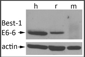
Application: Western BlotSample Tested: RPE whole cell lysateSpecies: Human and RatVerified Customer | Posted 12/29/2016Western blot was performed to detect Bestrophin-1 in human, rat, and mouse RPE tissue (upper panel). b-actin re-probing confirmed sample load (lower panel). Bestrophin-1 was detected in human and rat RPE. The antibody did not recognize mouse Bestrophin-1.E6-6 used at 1:2000 over night. next day secondary antibody donkey anti-mouse peroxidase was applied followed by detection using enhanced chemiluminescence.

There are no reviews that match your criteria.
Protocols
View specific protocols for Bestrophin 1 Antibody (E6-6) - BSA Free (NB300-164):
Immunofluorescence
1. Paraffin slides. Deparaffinize as follows:
a. 2x 5 min in Xylene
b. 2x 5 min in 100% ethanol
c. 2x 5 min in 95% ethanol
d. 1x 5 min in 70% ethanol
e. 1x 5 min or more in PBS
2. Cryosections:
a. air dry for >30 min
b. rehydrate in PBS-CM (PBS + 0.1mM CaCl2 and 1mM MgCl2) + 3% BSA
3. Use pap pen to draw circles around sections
4. Block in PBS-CM + BSA for 30 min at RT
5. Dilute anti-Bestrophin [cat# NB 300-164] in PBS-CM + BSA and incubate at RT for 1 hour or overnight at 4C.
6. Wash the slides with PBS-CM + BSA 5x 5 min
7. Dilute the secondary antibody in PBS-CM + BSA and incubate at RT for >1 hour (if staining nuclei with propidium iodide add saponin to 0.1% and RNAse A at 1:500)
8. Wash 3x 8 min with PBS-CM + BSA and then 1x 5 min with PBS-CM
a. If staining nuclei with DAPI or propidium iodide, dilute into PBS-CM at 1:1000
b. Wash 3x with PBS-CM, if using propidium iodide
c. Proceed directly to step 9, if using DAPI
9. Mount in Flourmount.
**NOTE: Immunofluorescence Considerations
1. Aldehyde fixatives (ie: PFA and formalin) will not work in immunofluorescence with this antibody.
A) Transfected cells on coverslips can be fixed in acetone or methanol, as can tissue.
B) Paraformaldehyde for paraffin sections can be used if the tissue is subject to heat and pressure mediated antigen retrieval [see specific reference 1 on datasheet]
2. To date, endogenous protein in human or pig eyes cannot be detected, even in methanol/acetone fixed sections directly.
3. Immunohistochemistry, using this antibody, has been done using the vector ABC kit, which includes a signal amplification step.
Procedure Guide for NB 300-164 Monoclonal Anti-Bestrophin
Western Blot Procedure
1. Run cell lysates** on an SDS-PAGE gel.
2. Transfer the proteins to PVDF.
3. Block the membrane in 1% Carnation instant milk in PBS + 0.1% Tween 20 (with 0.1mM CaCl2 and 1mM MgCl2) for 1 hour at RT.
4. Dilute the anti-Bestrophin [NB 300-164] to 1:1,000 in 10 ml of fresh blocking buffer and incubate for 1 hour at RT.
5. Wash the membrane with blocking buffer, 3x 5-10 minutes.
6. Dilute the secondary antibody in fresh blocking buffer, as recommended by the secondary vendor and incubate for 1hour at RT.
7. Wash the membrane with blocking buffer, 5x 8 minutes and rinse 1x with PBS (containing 0.1mM CaCl2 and 1mM MgCl2).
8. Detect the protein-antibody complex with alkaline phosphatase, if using NBT/BCIP or with HRP, if using ECL.
**Cell Lysate Preparation
A. Lysates were prepared in lysis buffer [50mM Tris-HCl, pH 8 / 120mM NaCl / 0.5% Nonidet P-40 / 10 ug/ml aprotinin / 10 ug/ml leupeptin / 1mM phenylmethylsulfonyl fluoride / 1mM sodium orthovanadate].
B. Total protein content was determined by bicinchoninic acid assay (Pierce).
Find general support by application which include: protocols, troubleshooting, illustrated assays, videos and webinars.
- Antigen Retrieval Protocol (PIER)
- Antigen Retrieval for Frozen Sections Protocol
- Appropriate Fixation of IHC/ICC Samples
- Cellular Response to Hypoxia Protocols
- Chromogenic IHC Staining of Formalin-Fixed Paraffin-Embedded (FFPE) Tissue Protocol
- Chromogenic Immunohistochemistry Staining of Frozen Tissue
- ClariTSA™ Fluorophore Kits
- Detection & Visualization of Antibody Binding
- Fluorescent IHC Staining of Frozen Tissue Protocol
- Graphic Protocol for Heat-induced Epitope Retrieval
- Graphic Protocol for the Preparation and Fluorescent IHC Staining of Frozen Tissue Sections
- Graphic Protocol for the Preparation and Fluorescent IHC Staining of Paraffin-embedded Tissue Sections
- Graphic Protocol for the Preparation of Gelatin-coated Slides for Histological Tissue Sections
- ICC Cell Smear Protocol for Suspension Cells
- ICC Immunocytochemistry Protocol Videos
- ICC for Adherent Cells
- IHC Sample Preparation (Frozen sections vs Paraffin)
- ISH-IHC Protocol for Chromogenic Detection on Formalin Fixed Paraffin Embedded (FFPE) Tissue
- Immunocytochemistry (ICC) Protocol
- Immunocytochemistry Troubleshooting
- Immunofluorescence of Organoids Embedded in Cultrex Basement Membrane Extract
- Immunofluorescent IHC Staining of Formalin-Fixed Paraffin-Embedded (FFPE) Tissue Protocol
- Immunohistochemistry (IHC) and Immunocytochemistry (ICC) Protocols
- Immunohistochemistry Frozen Troubleshooting
- Immunohistochemistry Paraffin Troubleshooting
- Immunoprecipitation Protocol
- Preparing Samples for IHC/ICC Experiments
- Preventing Non-Specific Staining (Non-Specific Binding)
- Primary Antibody Selection & Optimization
- Protocol for Heat-Induced Epitope Retrieval (HIER)
- Protocol for Making a 4% Formaldehyde Solution in PBS
- Protocol for VisUCyte™ HRP Polymer Detection Reagent
- Protocol for the Fluorescent ICC Staining of Cell Smears - Graphic
- Protocol for the Fluorescent ICC Staining of Cultured Cells on Coverslips - Graphic
- Protocol for the Preparation & Fixation of Cells on Coverslips
- Protocol for the Preparation and Chromogenic IHC Staining of Frozen Tissue Sections
- Protocol for the Preparation and Chromogenic IHC Staining of Frozen Tissue Sections - Graphic
- Protocol for the Preparation and Chromogenic IHC Staining of Paraffin-embedded Tissue Sections
- Protocol for the Preparation and Chromogenic IHC Staining of Paraffin-embedded Tissue Sections - Graphic
- Protocol for the Preparation and Fluorescent ICC Staining of Cells on Coverslips
- Protocol for the Preparation and Fluorescent ICC Staining of Non-adherent Cells
- Protocol for the Preparation and Fluorescent ICC Staining of Stem Cells on Coverslips
- Protocol for the Preparation and Fluorescent IHC Staining of Frozen Tissue Sections
- Protocol for the Preparation and Fluorescent IHC Staining of Paraffin-embedded Tissue Sections
- Protocol for the Preparation of Gelatin-coated Slides for Histological Tissue Sections
- Protocol for the Preparation of a Cell Smear for Non-adherent Cell ICC - Graphic
- R&D Systems Quality Control Western Blot Protocol
- TUNEL and Active Caspase-3 Detection by IHC/ICC Protocol
- The Importance of IHC/ICC Controls
- Troubleshooting Guide: Immunohistochemistry
- Troubleshooting Guide: Western Blot Figures
- Western Blot Conditions
- Western Blot Protocol
- Western Blot Protocol for Cell Lysates
- Western Blot Troubleshooting
- Western Blot Troubleshooting Guide
- View all Protocols, Troubleshooting, Illustrated assays and Webinars
FAQs for Bestrophin 1 Antibody (E6-6) - BSA Free
-
Q: According to the data sheet, for Immunohistochemistry, it references (PMID: 21249129). I see a 1:25 dilution in the journal article referenced under immunocytochemistry. Is this the same dilution recommended for IHC? If not do you have a recommended dilution? Antigen retrieval: Do you have any information on AR? Citrate buffer or higher pH (EDTA)? Temperature? Time? Incubation: Is 1 hour at room temperature or overnight at 4C recommended?
A: This antibody was validated in IHC in the reference PMID 11050159. Generally we recommend using antigen retrieval with citrate buffer and incubating for 1 hour at RT. Some antigens may be tricky and require overnight incubation. I imagine the specific protocol can be found in the reference, however.
-
Q: Can I get the recipe for RIPA buffer and the lysate preparation protocol for membrane bound proteins using NB300-164?
A: Here is both the recipe we use for RIPA and a good protocol for tissue lysate prep. RIPA buffer: 150 mM sodium chloride 1.0% NP-40 or Triton X-100 0.5% sodium deoxycholate 0.1% SDS (sodium dodecyl sulphate) 50 mM Tris, pH 8.0 RIPA buffer is also useful for whole cell extracts and membrane-bound proteins, and may be preferable to NP-40 or Triton X100-only buffers for extracting nuclear proteins. It will disrupt protein-protein interactions and may therefore be problematic for immunoprecipitations/pull down assays. Preparation of lysate from tissues Dissect the tissue of interest with clean tools, on ice preferably, and as quickly as possible to prevent degradation by proteases. Place the tissue in round-bottom microfuge tubes or Eppendorf tubes and immerse in liquid nitrogen to snap freeze. Store samples at -80C for later use or keep on ice for immediate homogenization. For a approximately 5 mg piece of tissue, add approximately 300 ul lysis buffer rapidly to the tube, homogenize with an electric homogenizer, rinse the blade twice with another 2x300 ul lysis buffer, then maintain constant agitation for 2 hours at 4C (e.g place on an orbital shaker in the fridge). Volumes of lysis buffer must be determined in relation to the amount of tissue present (protein extract should not be too diluted to avoid loss of protein and large volumes of samples to be loaded onto gels. The minimum concentration is 0.1 mg/ml, optimal concentration is 1-5 mg/ml). Centrifuge for 20 min at 12000 rpm at 4C in a microcentrifuge. Gently remove the tubes from the centrifuge and place on ice, aspirate the supernatant and place in a fresh tube kept on ice; discard the pellet.
-
Q: I want to know the concentration of Bestrophin 1 antibody [NB300-164].
A: For our product NB300-164, this antibody is provided as unpurified ascites. Therefore, the antibody concentration has not been determined.
-
Q: We are interested in your item, NB300-164 and would you please let us know the antibody concentration of it?
A: The total protein concentration of the ascites (Lot# B-3) is 11.7 mg/ml.
-
Q: According to the data sheet, for Immunohistochemistry, it references (PMID: 21249129). I see a 1:25 dilution in the journal article referenced under immunocytochemistry. Is this the same dilution recommended for IHC? If not do you have a recommended dilution? Antigen retrieval: Do you have any information on AR? Citrate buffer or higher pH (EDTA)? Temperature? Time? Incubation: Is 1 hour at room temperature or overnight at 4C recommended?
A: This antibody was validated in IHC in the reference PMID 11050159. Generally we recommend using antigen retrieval with citrate buffer and incubating for 1 hour at RT. Some antigens may be tricky and require overnight incubation. I imagine the specific protocol can be found in the reference, however.
-
Q: Can I get the recipe for RIPA buffer and the lysate preparation protocol for membrane bound proteins using NB300-164?
A: Here is both the recipe we use for RIPA and a good protocol for tissue lysate prep. RIPA buffer: 150 mM sodium chloride 1.0% NP-40 or Triton X-100 0.5% sodium deoxycholate 0.1% SDS (sodium dodecyl sulphate) 50 mM Tris, pH 8.0 RIPA buffer is also useful for whole cell extracts and membrane-bound proteins, and may be preferable to NP-40 or Triton X100-only buffers for extracting nuclear proteins. It will disrupt protein-protein interactions and may therefore be problematic for immunoprecipitations/pull down assays. Preparation of lysate from tissues Dissect the tissue of interest with clean tools, on ice preferably, and as quickly as possible to prevent degradation by proteases. Place the tissue in round-bottom microfuge tubes or Eppendorf tubes and immerse in liquid nitrogen to snap freeze. Store samples at -80C for later use or keep on ice for immediate homogenization. For a approximately 5 mg piece of tissue, add approximately 300 ul lysis buffer rapidly to the tube, homogenize with an electric homogenizer, rinse the blade twice with another 2x300 ul lysis buffer, then maintain constant agitation for 2 hours at 4C (e.g place on an orbital shaker in the fridge). Volumes of lysis buffer must be determined in relation to the amount of tissue present (protein extract should not be too diluted to avoid loss of protein and large volumes of samples to be loaded onto gels. The minimum concentration is 0.1 mg/ml, optimal concentration is 1-5 mg/ml). Centrifuge for 20 min at 12000 rpm at 4C in a microcentrifuge. Gently remove the tubes from the centrifuge and place on ice, aspirate the supernatant and place in a fresh tube kept on ice; discard the pellet.
-
Q: I want to know the concentration of Bestrophin 1 antibody [NB300-164].
A: For our product NB300-164, this antibody is provided as unpurified ascites. Therefore, the antibody concentration has not been determined.
-
Q: We are interested in your item, NB300-164 and would you please let us know the antibody concentration of it?
A: The total protein concentration of the ascites (Lot# B-3) is 11.7 mg/ml.