beta-Actin Antibody - BSA Free
Novus Biologicals | Catalog # NB100-56874
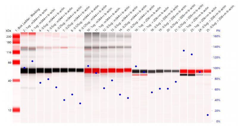
![Simple Western: beta-Actin Antibody [NB100-56874]](https://resources.rndsystems.com/images/products/beta-Actin-Antibody-Simple-Western-NB100-56874-img0007.jpg "Simple Western: beta-Actin Antibody [NB100-56874]")
Loading...
Key Product Details
Validated by
Biological Validation
Species Reactivity
Validated:
Human, Mouse, Rat, Porcine, Bovine, Chinese Hamster, Invertebrate
Cited:
Human, Mouse, Rat, Porcine, Bovine, Hamster - Cricetulus (Chinese Hamster), Invertebrate, Nematode - Caenorhabditis elegans
Applications
Validated:
Immunohistochemistry, Immunohistochemistry-Paraffin, Western Blot, Immunocytochemistry/ Immunofluorescence, Simple Western, Knockdown Validated
Cited:
Western Blot, IF/IHC, Knockdown Validated
Label
Unconjugated
Antibody Source
Polyclonal Rabbit IgG
Format
BSA Free
Loading...
Product Specifications
Immunogen
Amino acids 2-16 (CDDDIAALVIDNGSG) of actin protein were used as the immunogen sequence for this beta-Actin Antibody.
Reactivity Notes
Use in Rat reported in scientific literature (PMID:34443654). Invertebrate, Porcine, Bovine, and Chinese Hamster reactivity reported in scientific literature (PMID: 25240497, 26593451, 18650284, and 19291423 respectively)..
Clonality
Polyclonal
Host
Rabbit
Isotype
IgG
Theoretical MW
42 kDa.
Disclaimer note: The observed molecular weight of the protein may vary from the listed predicted molecular weight due to post translational modifications, post translation cleavages, relative charges, and other experimental factors.
Disclaimer note: The observed molecular weight of the protein may vary from the listed predicted molecular weight due to post translational modifications, post translation cleavages, relative charges, and other experimental factors.
Scientific Data Images for beta-Actin Antibody - BSA Free
Simple Western: beta-Actin Antibody [NB100-56874]
Simple Western: beta-Actin Antibody [NB100-56874] - Image shows a specific band for Beta Actin in 1.0 mg/mL of HeLa lysate. This experiment was performed under reducing conditions using the 12-230 kDa separation system.![Western Blot: beta-Actin Antibody [NB100-56874]](https://resources.rndsystems.com/images/products/beta-Actin-Antibody-Western-Blot-NB100-56874-img0004.jpg "Western Blot: beta-Actin Antibody [NB100-56874]")
Western Blot: beta-Actin Antibody [NB100-56874]
Western Blot: beta-Actin Antibody [NB100-56874] - Specific bands are seen in various cell lysates at the same molecular weight. A: human brain, B: mouse brain, C: rat brain, D: human lung, and E: human spleen tissue probed using beta actin antibody at 0.25 ug/mL.![Immunocytochemistry/ Immunofluorescence: beta-Actin Antibody [NB100-56874]](https://resources.rndsystems.com/images/products/beta-Actin-Antibody-Immunocytochemistry-Immunofluorescence-NB100-56874-img0009.jpg "Immunocytochemistry/ Immunofluorescence: beta-Actin Antibody [NB100-56874]")
Immunocytochemistry/ Immunofluorescence: beta-Actin Antibody [NB100-56874]
Immunocytochemistry/Immunofluorescence: beta-Actin Antibody [NB100-56874] - Actin was detected in NIH-3T3 cells fixed with methanol using rabbit anti-mouse Actin antibody (NB100-56874) at 1:100 dilution, overnight at 4C. Cells were stained using Northern Lights 557 conjugated anti-rabbit IgG secondary antibodies (NL004) and counterstained with DAPI.![Western Blot: beta-Actin Antibody [NB100-56874]](https://resources.rndsystems.com/images/products/beta-Actin-Antibody-Western-Blot-NB100-56874-img0010.jpg "Western Blot: beta-Actin Antibody [NB100-56874]")
![Immunohistochemistry-Paraffin: beta-Actin Antibody [NB100-56874]](https://resources.rndsystems.com/images/products/beta-Actin-Antibody-Immunohistochemistry-Paraffin-NB100-56874-img0005.jpg "Immunohistochemistry-Paraffin: beta-Actin Antibody [NB100-56874]")
Immunohistochemistry-Paraffin: beta-Actin Antibody [NB100-56874]
Immunohistochemistry-Paraffin: beta-Actin Antibody [NB100-56874] - Analysis of human spleen using antibody at 10 ug/mL.![Western Blot: beta-Actin Antibody [NB100-56874]](https://resources.rndsystems.com/images/products/beta-Actin-Antibody-Western-Blot-NB100-56874-img0003.jpg "Western Blot: beta-Actin Antibody [NB100-56874]")
Western Blot: beta-Actin Antibody [NB100-56874]
Western Blot: beta-Actin Antibody [NB100-56874] - Decreasing amounts of human ovary tissue lysate were probed using beta actin antibody at 0.25 ug/mL: A) 40 ug, B) 30 ug, C) 20 ug, D) 10 ug per lane.
Western Blot: beta-Actin Antibody [NB100-56874] -
Western Blot: beta-Actin Antibody [NB100-56874] - Simultaneous downregulation of multiple genes more effectively inhibits HCV replication: Huh-7.5 cells in a similar experimental setup as mentioned in Figure 1 were transfected with following combinations of siRNAs: La autoantigen + NS5B, La autoantigen + hVAP-A, La autoantigen + PSMA7, NS5B + hVAP-A, NS5B + PSMA7, & PSMA7 + hVAP-A. Downregulation of viral replication were determined by RT-PCR as shown in (a). (b) represents densitometry analysis of HCV NS5B downregulation. The data represent mean ± standard deviation. ∗P value < 0.05 versus control was considered statistically significant. Image collected & cropped by CiteAb from the following publication (https://pubmed.ncbi.nlm.nih.gov/27446609), licensed under a CC-BY license. Not internally tested by Novus Biologicals.
Western Blot: beta-Actin Antibody [NB100-56874] -
Western Blot: beta-Actin Antibody [NB100-56874] - Downregulation of La autoantigen, hVAP-A, PSMA7, & NS5B leads to reduction in viral replication: overnight grown Huh-7.5 cells were transfected with La autoantigen, hVAP-A, & PSMA7 siRNAs for 48 h. Cells were then infected with the virus & again transfected with same siRNAs. After incubation total RNA was isolated & subjected to RT-PCR. NS5B siRNA was transfected once only after cells were infected. (a), (c), (e), & (g) represent RT-PCR results for target genes & NS5B gene upon treatment with La autoantigen, hVAP-A, PSMA7, & NS5B siRNAs, respectively. Densitometric analysis of (a), (c), (e), & (g) images is expressed in percentage in (b), (d), (f), & (h), respectively. The data represent mean ± standard deviation. ∗P value < 0.05 versus control considered statistically significant. Image collected & cropped by CiteAb from the following publication (https://pubmed.ncbi.nlm.nih.gov/27446609), licensed under a CC-BY license. Not internally tested by Novus Biologicals.
Western Blot: beta-Actin Antibody [NB100-56874] -
Western Blot: beta-Actin Antibody [NB100-56874] - Downregulation of La autoantigen, hVAP-A, PSMA7, & NS5B leads to reduction in viral replication: overnight grown Huh-7.5 cells were transfected with La autoantigen, hVAP-A, & PSMA7 siRNAs for 48 h. Cells were then infected with the virus & again transfected with same siRNAs. After incubation total RNA was isolated & subjected to RT-PCR. NS5B siRNA was transfected once only after cells were infected. (a), (c), (e), & (g) represent RT-PCR results for target genes & NS5B gene upon treatment with La autoantigen, hVAP-A, PSMA7, & NS5B siRNAs, respectively. Densitometric analysis of (a), (c), (e), & (g) images is expressed in percentage in (b), (d), (f), & (h), respectively. The data represent mean ± standard deviation. ∗P value < 0.05 versus control considered statistically significant. Image collected & cropped by CiteAb from the following publication (https://pubmed.ncbi.nlm.nih.gov/27446609), licensed under a CC-BY license. Not internally tested by Novus Biologicals.Applications for beta-Actin Antibody - BSA Free
Application
Recommended Usage
Immunocytochemistry/ Immunofluorescence
1:100
Immunohistochemistry
reported in scientific literature (PMID 27371029)
Immunohistochemistry-Paraffin
10 ug/mL
Knockdown Validated
reported in scientific literature (PMID 31835509)
Simple Western
1:12.5
Western Blot
0.25-1 ug/ml
Application Notes
This antibody is not suitable for testing heart or muscle lysate. In Simple Western only 10 - 15 uL of the recommended dilution is used per data point.
See Simple Western Antibody Database for Simple Western validation: Tested in HeLa lysate 1.0 mg/mL, separated by Size, antibody dilution of 1:12.5, apparent MW was 49 kDa. Separated by Size-Wes, Sally Sue/Peggy Sue.
See Simple Western Antibody Database for Simple Western validation: Tested in HeLa lysate 1.0 mg/mL, separated by Size, antibody dilution of 1:12.5, apparent MW was 49 kDa. Separated by Size-Wes, Sally Sue/Peggy Sue.
Reviewed Applications
Read 4 reviews rated 4 using NB100-56874 in the following applications:
Formulation, Preparation, and Storage
Purification
Protein G purified
Formulation
PBS
Format
BSA Free
Preservative
0.05% Sodium Azide
Concentration
1.0 mg/ml
Shipping
The product is shipped with polar packs. Upon receipt, store it immediately at the temperature recommended below.
Stability & Storage
Store at 4C short term. Aliquot and store at -20C long term. Avoid freeze-thaw cycles.
Background: beta-Actin
References
1. Vandekerckhove J, Weber K. 1978. At least six different actins are expressed in a higher mammal: an analysis based on the amino acid sequence of the amino-terminal tryptic peptide. J Mol Biol. 126(4):783-802. PMID: 745245
2. Gimona M, Vandekerckhove J, Goethals M, Herzog M, Lando Z, Small JV. (1994) Beta-actin specific monoclonal antibody. Cell Motil Cytoskeleton. 27(2):108-16. PMID: 8162619
3. Holden VI, Lenio S, Kuick R, Ramakrishnan SK, Shah YM, Bachman MA. (2014) Bacterial siderophores that evade or overwhelm Lipocalin 2 induce HIF-1a and pro-inflammatory cytokine secretion in cultured respiratory epithelial cells Infect Immun. 82(9):3826-36 PMID: 24980968
4. Tsai WL, Yeh PH, Tsai CY, Ting CT, Chiu YH, Tao MH, Li WC, Hung SC. (2016) Efficient Programming of Human Mesenchymal Stem Cell Derived Hepatocytes by Epigenetic Regulations. J. Gastroenterol. Hepatol. 32(1):261-269. PMID: 27218433
Alternate Names
ACTB, betaActin
Gene Symbol
ACTB
Additional beta-Actin Products
Product Documents for beta-Actin Antibody - BSA Free
Certificate of Analysis
To download a Certificate of Analysis, please enter a lot or batch number in the search box below.
Product Specific Notices for beta-Actin Antibody - BSA Free
This product is for research use only and is not approved for use in humans or in clinical diagnosis. Primary Antibodies are guaranteed for 1 year from date of receipt.
Related Research Areas
Citations for beta-Actin Antibody - BSA Free
 Powered by Bioz
Customer Reviews for beta-Actin Antibody - BSA Free (4)
4 out of 5
4 Customer Ratings
Have you used beta-Actin Antibody - BSA Free?
Submit a review and receive an Amazon gift card!
$25/€18/£15/$25CAN/¥2500 Yen for a review with an image
$10/€7/£6/$10CAN/¥1110 Yen for a review without an image
Submit a review
Customer Images

-(01-mg)_NB100-56874_9446.jpg)
Showing
1
-
4 of
4 reviews
Showing All
Filter By:
-
Application: Simple WesternSample Tested: liverSpecies: HumanVerified Customer | Posted 03/20/2020lane 2-15, rabbit anti-b-actin antibody (N100-56874). lane 2-9, detected by anti-rabbit-NIR, lane 10-15, detected by anti-rabbit-HRP

-
Application: Western BlotSample Tested: Kidney tissue homogenatesSpecies: FelineVerified Customer | Posted 05/18/2017LiCOR Odyssey CLx imaging System. A specific single band is shown as green.
-
Application: Western BlotVerified Customer | Posted 09/15/2014
-
Application: Western BlotSample Tested: Lysate from 293T cellsSpecies: HumanVerified Customer | Posted 08/19/2014WB for beta-actin in 293T cells as control
There are no reviews that match your criteria.
Protocols
Find general support by application which include: protocols, troubleshooting, illustrated assays, videos and webinars.
- Antigen Retrieval Protocol (PIER)
- Antigen Retrieval for Frozen Sections Protocol
- Appropriate Fixation of IHC/ICC Samples
- Cellular Response to Hypoxia Protocols
- Chromogenic IHC Staining of Formalin-Fixed Paraffin-Embedded (FFPE) Tissue Protocol
- Chromogenic Immunohistochemistry Staining of Frozen Tissue
- ClariTSA™ Fluorophore Kits
- Detection & Visualization of Antibody Binding
- Fluorescent IHC Staining of Frozen Tissue Protocol
- Graphic Protocol for Heat-induced Epitope Retrieval
- Graphic Protocol for the Preparation and Fluorescent IHC Staining of Frozen Tissue Sections
- Graphic Protocol for the Preparation and Fluorescent IHC Staining of Paraffin-embedded Tissue Sections
- Graphic Protocol for the Preparation of Gelatin-coated Slides for Histological Tissue Sections
- ICC Cell Smear Protocol for Suspension Cells
- ICC Immunocytochemistry Protocol Videos
- ICC for Adherent Cells
- IHC Sample Preparation (Frozen sections vs Paraffin)
- Immunocytochemistry (ICC) Protocol
- Immunocytochemistry Troubleshooting
- Immunofluorescence of Organoids Embedded in Cultrex Basement Membrane Extract
- Immunofluorescent IHC Staining of Formalin-Fixed Paraffin-Embedded (FFPE) Tissue Protocol
- Immunohistochemistry (IHC) and Immunocytochemistry (ICC) Protocols
- Immunohistochemistry Frozen Troubleshooting
- Immunohistochemistry Paraffin Troubleshooting
- Preparing Samples for IHC/ICC Experiments
- Preventing Non-Specific Staining (Non-Specific Binding)
- Primary Antibody Selection & Optimization
- Protocol for Heat-Induced Epitope Retrieval (HIER)
- Protocol for Making a 4% Formaldehyde Solution in PBS
- Protocol for VisUCyte™ HRP Polymer Detection Reagent
- Protocol for the Fluorescent ICC Staining of Cell Smears - Graphic
- Protocol for the Fluorescent ICC Staining of Cultured Cells on Coverslips - Graphic
- Protocol for the Preparation & Fixation of Cells on Coverslips
- Protocol for the Preparation and Chromogenic IHC Staining of Frozen Tissue Sections
- Protocol for the Preparation and Chromogenic IHC Staining of Frozen Tissue Sections - Graphic
- Protocol for the Preparation and Chromogenic IHC Staining of Paraffin-embedded Tissue Sections
- Protocol for the Preparation and Chromogenic IHC Staining of Paraffin-embedded Tissue Sections - Graphic
- Protocol for the Preparation and Fluorescent ICC Staining of Cells on Coverslips
- Protocol for the Preparation and Fluorescent ICC Staining of Non-adherent Cells
- Protocol for the Preparation and Fluorescent ICC Staining of Stem Cells on Coverslips
- Protocol for the Preparation and Fluorescent IHC Staining of Frozen Tissue Sections
- Protocol for the Preparation and Fluorescent IHC Staining of Paraffin-embedded Tissue Sections
- Protocol for the Preparation of Gelatin-coated Slides for Histological Tissue Sections
- Protocol for the Preparation of a Cell Smear for Non-adherent Cell ICC - Graphic
- R&D Systems Quality Control Western Blot Protocol
- TUNEL and Active Caspase-3 Detection by IHC/ICC Protocol
- The Importance of IHC/ICC Controls
- Troubleshooting Guide: Immunohistochemistry
- Troubleshooting Guide: Western Blot Figures
- Western Blot Conditions
- Western Blot Protocol
- Western Blot Protocol for Cell Lysates
- Western Blot Troubleshooting
- Western Blot Troubleshooting Guide
- View all Protocols, Troubleshooting, Illustrated assays and Webinars
FAQs for beta-Actin Antibody - BSA Free
Showing
1
-
5 of
7 FAQs
Showing All
-
Q: Are the beta-Actin antibodies validated in Simple Western?
A: Yes, we offer a few beta actin antibodies that have been tested in Simple Western: NBP1-47423, NB600-501, NB600-532, NB600-503, and NB100-56874.
-
Q: Do Beta-Actin antibodies come in lyophilized format?
A: ACTB antibodies like MAB8969 AND MAB8929 come in lyophilized form.
-
Q: I wanted to know which of the two housekeeping genes B-actin or GAPDH can be used for best results. I intend to do an experiment to determine expression of IL-6 and TNF-alpha in liver and kidney tissues.
A: For homogenized tissue, beta-actin and GAPDH are both fine.
-
Q: I would like to find the manual description of this product, to confirm what dilution is recommended for Western blot.
A: The recommended starting dilution for Western blotting is 0.25-1ug/ml, but we suggest that you optimise this for your particular sample set.
-
Q: What is the immunogen sequence of this Beta-Actin antibody?
A: A sequence of amino acids 2 - 16 (CDDDIAALVIDNGSG) of beta Actin.
-
Q: What is the theoretical molecular weight for Beta-Actin antibodies?
A: The TMW for beta Actin antibodies is approximately 42 kDa.
-
Q: Why is beta actin used as a loading control in blotting ?
A: Beta actin is a highly-expressed protein found in all cells, and is considered a house keeping protein whose expression is necessary for any cell's proper functioning. For this reason it is used as a loading control to confirm that the same amount of total protein is loaded in each lane of an SDS-PAGE gel so that appropriate comparisons can be made between the actual proteins of interest in different samples.
-
Q: Are the beta-Actin antibodies validated in Simple Western?
A: Yes, we offer a few beta actin antibodies that have been tested in Simple Western: NBP1-47423, NB600-501, NB600-532, NB600-503, and NB100-56874.
-
Q: Do Beta-Actin antibodies come in lyophilized format?
A: ACTB antibodies like MAB8969 AND MAB8929 come in lyophilized form.
-
Q: I wanted to know which of the two housekeeping genes B-actin or GAPDH can be used for best results. I intend to do an experiment to determine expression of IL-6 and TNF-alpha in liver and kidney tissues.
A: For homogenized tissue, beta-actin and GAPDH are both fine.
-
Q: I would like to find the manual description of this product, to confirm what dilution is recommended for Western blot.
A: The recommended starting dilution for Western blotting is 0.25-1ug/ml, but we suggest that you optimise this for your particular sample set.
-
Q: What is the immunogen sequence of this Beta-Actin antibody?
A: A sequence of amino acids 2 - 16 (CDDDIAALVIDNGSG) of beta Actin.
-
Q: What is the theoretical molecular weight for Beta-Actin antibodies?
A: The TMW for beta Actin antibodies is approximately 42 kDa.
-
Q: Why is beta actin used as a loading control in blotting ?
A: Beta actin is a highly-expressed protein found in all cells, and is considered a house keeping protein whose expression is necessary for any cell's proper functioning. For this reason it is used as a loading control to confirm that the same amount of total protein is loaded in each lane of an SDS-PAGE gel so that appropriate comparisons can be made between the actual proteins of interest in different samples.
Loading...
Associated Pathways


