beta Amyloid Antibody - BSA Free
Novus Biologicals | Catalog # NBP2-25093

Key Product Details
Validated by
Knockout/Knockdown
Species Reactivity
Validated:
Human, Mouse, Rat, Bacteria
Cited:
Mouse, Rat, Bacteria
Predicted:
Bovine (100%), Guinea Pig (100%), Porcine (100%), Rabbit (100%), Sheep (100%). Backed by our 100% Guarantee.
Applications
Validated:
Immunohistochemistry, Immunohistochemistry-Paraffin, Western Blot, ELISA, Immunoprecipitation
Cited:
Immunohistochemistry-Paraffin, Western Blot, Immunoprecipitation
Label
Unconjugated
Antibody Source
Polyclonal Rabbit IgG
Format
BSA Free
Loading...
Product Specifications
Immunogen
This beta Amyloid antibody was raised against a 10 amino acid peptide on the amino terminus of the 4KDa beta Amyloid peptide generated by beta- and gamma-secretases. The immunogen is located within amino acids 650 - 700 of human amyloid A4 protein precursor (APP).
Reactivity Notes
Use in Mouse reported in scientific literature (PMID:33756153).
Clonality
Polyclonal
Host
Rabbit
Isotype
IgG
Theoretical MW
115 kDa.
Disclaimer note: The observed molecular weight of the protein may vary from the listed predicted molecular weight due to post translational modifications, post translation cleavages, relative charges, and other experimental factors.
Disclaimer note: The observed molecular weight of the protein may vary from the listed predicted molecular weight due to post translational modifications, post translation cleavages, relative charges, and other experimental factors.
Scientific Data Images for beta Amyloid Antibody - BSA Free
Western Blot: beta Amyloid Antibody - BSA Free [NBP2-25093] -
Western Blot: beta Amyloid Antibody - BSA Free [NBP2-25093] - KO Validation in 293T Cells Loading: 10 ug of lysate Antibodies: beta Amyloid, 0.5 ug/mL and beta-actin, 1 ug/mL, 1 h incubation at RT in 5% NFDM/TBST. Secondary: Goat Anti-Rabbit IgG HRP conjugate at 1:10000 dilution.
Immunohistochemistry: beta Amyloid Antibody - BSA Free [NBP2-25093] -
Immunohistochemistry: beta Amyloid Antibody - BSA Free [NBP2-25093] - Immunohistochemistry of beta Amyloid in human brain tissue with beta Amyloid antibody at 2.5 ug/ml.
Immunohistochemistry: beta Amyloid Antibody - BSA Free [NBP2-25093] -
Immunohistochemistry: beta Amyloid Antibody - BSA Free [NBP2-25093] - Immunohistochemistry of beta Amyloid in human brain (Alzheimer's disease) tissue with beta Amyloid antibody at 10 ug/mL.
Immunohistochemistry: beta Amyloid Antibody - BSA Free [NBP2-25093] -
Immunohistochemistry: beta Amyloid Antibody - BSA Free [NBP2-25093] - Immunofluorescence of ASAH1 in rat heart tissue with ASAH1 antibody at 20 ug/mL.Green: beta Amyloid Antibody
Blue: DAPI staining

Western Blot: beta Amyloid Antibody - BSA Free [NBP2-25093] -
Western Blot: beta Amyloid Antibody - BSA Free [NBP2-25093] - Western blot analysis of beta Amyloid in (A) human, (B) mouse, and (C) rat brain tissue lysates with beta Amyloid antibody at 1 ug/mL.Applications for beta Amyloid Antibody - BSA Free
Application
Recommended Usage
ELISA
1:100-1:2000
Immunohistochemistry
2.5 ug/ml
Immunohistochemistry-Paraffin
2.5 ug/ml
Western Blot
1ug/ml
Application Notes
Use in IP reported in secitific publication PMID: 32413239
Reviewed Applications
Read 1 review rated 3 using NBP2-25093 in the following applications:
Formulation, Preparation, and Storage
Purification
Peptide affinity purified
Formulation
PBS
Format
BSA Free
Preservative
0.02% Sodium Azide
Concentration
1 mg/ml
Shipping
The product is shipped with polar packs. Upon receipt, store it immediately at the temperature recommended below.
Stability & Storage
Store at 4C short term. Aliquot and store at -20C long term. Avoid freeze-thaw cycles.
Background: Amyloid beta
Pyroglutamate amyloid beta peptides (pGlu-Abeta) are N-terminal truncations in which the N-terminal glutamate is cyclized to pyroglutamate resulting in pGlu-Abeta (3-40/42 and 11-40/42) (4). This pyrE modification may have a greater propensity to aggregate under physiological conditions and has been implicated as the molecular species responsible for seeding larger oligomers of amyloid beta.
References
1. Chen GF, Xu TH, Yan Y, Zhou YR, Jiang Y, Melcher K, Xu HE. (2017) Amyloid beta: structure, biology and structure-based therapeutic development. Acta Pharmacol Sin. 38(9):1205-1235. PMID: 28713158
2. De-Paula VJ1, Radanovic M, Diniz BS, Forlenza OV. (2012) Alzheimer's disease. Subcell Biochem. 65:329-52. PMID: 23225010
3. Schaich CL, Maurer MS, Nadkarni NK. (2019) Amyloidosis of the Brain and Heart: Two Sides of the Same Coin? JACC Heart Fail. 7(2):129-131. PMID: 30704604
4. He W, Barrow CJ. (1999) The A beta 3-pyroglutamyl and 11-pyroglutamyl peptides found in senile plaque have greater beta-sheet forming and aggregation propensities in vitro than full-length A beta. Biochemistry. 38(33):10871-7. PMID: 10451383
Additional Amyloid beta Products
Product Documents for beta Amyloid Antibody - BSA Free
Certificate of Analysis
To download a Certificate of Analysis, please enter a lot or batch number in the search box below.
Product Specific Notices for beta Amyloid Antibody - BSA Free
This product is for research use only and is not approved for use in humans or in clinical diagnosis. Primary Antibodies are guaranteed for 1 year from date of receipt.
Related Research Areas
Citations for beta Amyloid Antibody - BSA Free
 Powered by Bioz
Customer Reviews for beta Amyloid Antibody - BSA Free (1)
3 out of 5
1 Customer Rating
Have you used beta Amyloid Antibody - BSA Free?
Submit a review and receive an Amazon gift card!
$25/€18/£15/$25CAN/¥2500 Yen for a review with an image
$10/€7/£6/$10CAN/¥1110 Yen for a review without an image
Submit a review
Customer Images
Showing
1
-
1 of
1 review
Showing All
Filter By:
-
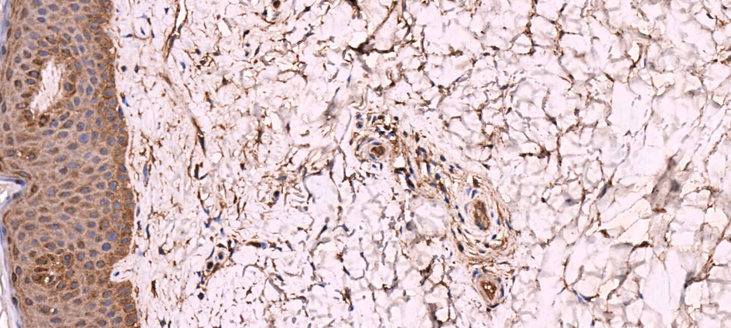
Application: Immunohistochemistry-ParaffinSample Tested: human skinSpecies: HumanVerified Customer | Posted 12/08/2016

There are no reviews that match your criteria.
Protocols
Find general support by application which include: protocols, troubleshooting, illustrated assays, videos and webinars.
- Antigen Retrieval Protocol (PIER)
- Antigen Retrieval for Frozen Sections Protocol
- Appropriate Fixation of IHC/ICC Samples
- Cellular Response to Hypoxia Protocols
- Chromogenic IHC Staining of Formalin-Fixed Paraffin-Embedded (FFPE) Tissue Protocol
- Chromogenic Immunohistochemistry Staining of Frozen Tissue
- ClariTSA™ Fluorophore Kits
- Detection & Visualization of Antibody Binding
- ELISA Sample Preparation & Collection Guide
- ELISA Troubleshooting Guide
- Fluorescent IHC Staining of Frozen Tissue Protocol
- Graphic Protocol for Heat-induced Epitope Retrieval
- Graphic Protocol for the Preparation and Fluorescent IHC Staining of Frozen Tissue Sections
- Graphic Protocol for the Preparation and Fluorescent IHC Staining of Paraffin-embedded Tissue Sections
- Graphic Protocol for the Preparation of Gelatin-coated Slides for Histological Tissue Sections
- How to Run an R&D Systems DuoSet ELISA
- How to Run an R&D Systems Quantikine ELISA
- How to Run an R&D Systems Quantikine™ QuicKit™ ELISA
- IHC Sample Preparation (Frozen sections vs Paraffin)
- Immunofluorescent IHC Staining of Formalin-Fixed Paraffin-Embedded (FFPE) Tissue Protocol
- Immunohistochemistry (IHC) and Immunocytochemistry (ICC) Protocols
- Immunohistochemistry Frozen Troubleshooting
- Immunohistochemistry Paraffin Troubleshooting
- Immunoprecipitation Protocol
- Preparing Samples for IHC/ICC Experiments
- Preventing Non-Specific Staining (Non-Specific Binding)
- Primary Antibody Selection & Optimization
- Protocol for Heat-Induced Epitope Retrieval (HIER)
- Protocol for Making a 4% Formaldehyde Solution in PBS
- Protocol for VisUCyte™ HRP Polymer Detection Reagent
- Protocol for the Preparation & Fixation of Cells on Coverslips
- Protocol for the Preparation and Chromogenic IHC Staining of Frozen Tissue Sections
- Protocol for the Preparation and Chromogenic IHC Staining of Frozen Tissue Sections - Graphic
- Protocol for the Preparation and Chromogenic IHC Staining of Paraffin-embedded Tissue Sections
- Protocol for the Preparation and Chromogenic IHC Staining of Paraffin-embedded Tissue Sections - Graphic
- Protocol for the Preparation and Fluorescent IHC Staining of Frozen Tissue Sections
- Protocol for the Preparation and Fluorescent IHC Staining of Paraffin-embedded Tissue Sections
- Protocol for the Preparation of Gelatin-coated Slides for Histological Tissue Sections
- Quantikine HS ELISA Kit Assay Principle, Alkaline Phosphatase
- Quantikine HS ELISA Kit Principle, Streptavidin-HRP Polymer
- R&D Systems Quality Control Western Blot Protocol
- Sandwich ELISA (Colorimetric) – Biotin/Streptavidin Detection Protocol
- Sandwich ELISA (Colorimetric) – Direct Detection Protocol
- TUNEL and Active Caspase-3 Detection by IHC/ICC Protocol
- The Importance of IHC/ICC Controls
- Troubleshooting Guide: ELISA
- Troubleshooting Guide: Immunohistochemistry
- Troubleshooting Guide: Western Blot Figures
- Western Blot Conditions
- Western Blot Protocol
- Western Blot Protocol for Cell Lysates
- Western Blot Troubleshooting
- Western Blot Troubleshooting Guide
- View all Protocols, Troubleshooting, Illustrated assays and Webinars
Loading...