beta Galactosidase Antibody - BSA Free
Novus Biologicals | Catalog # NB600-305
![Western Blot: beta Galactosidase Antibody [NB600-305]](https://resources.rndsystems.com/images/products/beta-Galactosidase-Antibody-Western-Blot-NB600-305-img0017.jpg "Western Blot: beta Galactosidase Antibody [NB600-305]")
Key Product Details
Species Reactivity
Validated:
Cited:
Applications
Validated:
Cited:
Label
Antibody Source
Format
Product Specifications
Immunogen
Reactivity Notes
Localization
Clonality
Host
Isotype
Description
Store vial at 4C prior to restoration. For extended storage aliquot contents and freeze at -20C or below. Avoid cycles of freezing and thawing. Centrifuge product if not completely clear after standing at room temperature. This product is stable for several weeks at 4C as an undiluted liquid. Dilute only prior to immediate use.
Scientific Data Images for beta Galactosidase Antibody - BSA Free
Western Blot: beta Galactosidase Antibody [NB600-305]
Western Blot: beta Galactosidase Antibody [NB600-305] - Analysis using FITC conjugate of NB600-305. 3) corresponding to 60 ng, 30 ng and 15 ng, respectively of b-Gal present in partially purified preparations (arrowhead). Lane 4 shows no cross reactivity with proteins present in a non-specific control E.coli lysate. Proteins were resolved on a 4-20% Tris-Glycine gel by SDS-PAGE and transferred to nitrocellulose and blocking using Blocking Buffer for Fluorescent Western Blotting. The membrane was probed with fluorescein conjugated anti-b-Galactosidase diluted to 1:10,000. Reaction occurred for 2 hours at room temperature.![Western Blot: beta Galactosidase Antibody [NB600-305]](https://resources.rndsystems.com/images/products/beta-Galactosidase-Antibody-Western-Blot-NB600-305-img0014.jpg "Western Blot: beta Galactosidase Antibody [NB600-305]")
Western Blot: beta Galactosidase Antibody [NB600-305]
Western Blot: beta Galactosidase Antibody [NB600-305] - Analysis of a band at 117 kDa (lane 1) corresponding to b-Gal present in a partially purified preparation (arrowhead). Approximately 1 ug of protein was resolved on a 4-20% Tris-Glycine gel by SDS-PAGE and transferred onto nitrocellulose. After blocking, the membrane was probed with the primary antibody diluted to 1:1,000. Reaction occurred overnight at 4 C followed by washes and reaction with a 1:10,000 dilution of IRDye 800 conjugated Gt-a-Rabbit IgG (H&L) MX10 for 45 min at room temperature (800 nm channel, green).![Western Blot: beta Galactosidase Antibody [NB600-305]](https://resources.rndsystems.com/images/products/beta-Galactosidase-Antibody-Western-Blot-NB600-305-img0015.jpg "Western Blot: beta Galactosidase Antibody [NB600-305]")
Western Blot: beta Galactosidase Antibody [NB600-305]
Western Blot: beta Galactosidase Antibody [NB600-305] - Lane 1 shows 80 ng and lane 2 shows 20 ng loaded onto gel. Results for non-reducing conditions of SDS-PAGE prior to transfer to nitrocellulose are shown on the left side of the figure; results obtainined under reducing conditions are shown on the right. Blots were blocked overnight at 4 C with Blocking Buffer for Fluorescent Western Blotting. The membrane was probed with anti-b-Galactosidase diluted to 1:10,000. Reaction occurred overnight at 4C. Dylight649 conjugated Gt-a-anti-Rabbit IgG was used for detection.![Western Blot: beta Galactosidase Antibody [NB600-305]](https://resources.rndsystems.com/images/products/beta-Galactosidase-Antibody-Western-Blot-NB600-305-img0016.jpg "Western Blot: beta Galactosidase Antibody [NB600-305]")
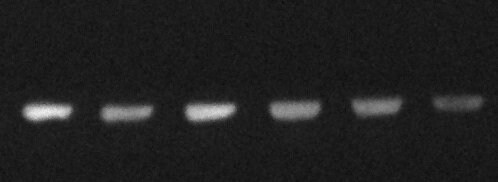
Western Blot: beta Galactosidase Antibody [NB600-305]
Western Blot: beta Galactosidase Antibody [NB600-305] - Shows a band at 117 kDa (lanes 1 - 3) corresponding to 60 ng, 30 ng and 15 ng, respectively of b-Gal present in partially purified preparations (arrowhead). Lane 4 shows no cross reactivity with proteins present in a non-specific control E.coli lysate. Proteins were resolved on a 4-20% Tris-Glycine gel by SDS-PAGE and transferred to nitrocellulose and blocking using Blocking Buffer for Fluorescent Western Blotting. The membrane was probed with fluorescein conjugated anti-b-Galactosidase diluted to 1:10,000. Reaction occurred for 2 hours at room temperature.
beta Galactosidase Antibody
Western Blot of Rabbit Anti-Beta-Galactosidase Antibody. Lane 1: Beta-Galactosidase Reduced [0.1ug]. Lane 2: Opal Prestained Molecular Weight Marker
beta Galactosidase Antibody
Western Blot of Rabbit Anti-Beta-Galactosidase Antibody. Lane 1: partially purified preparation b-Galactosidase [1.0ug]. Lane 2: Molecular Weight Marker. Primary Antibody: Anti-Beta-Galactosidase at 1:1000 overnight at 2-8C. Secondary Antibody: Goat Anti-Rabbit IgG IRDye(R)800Applications for beta Galactosidase Antibody - BSA Free
ELISA
Immunocytochemistry/ Immunofluorescence
Immunohistochemistry
Immunohistochemistry-Frozen
Immunohistochemistry-Paraffin
Western Blot
Use in Immunocytochemistry/immunofluorescence reported in scientific literature (PMID 23838935).
Use in Immunohistochemistry-Frozen reported in scientific literature (PMID 25799059)
Reviewed Applications
Read 1 review rated 5 using NB600-305 in the following applications:
Formulation, Preparation, and Storage
Purification
Reconstitution
Formulation
Format
Preservative
Concentration
Shipping
Stability & Storage
Calculators
Background: beta Galactosidase
Alternate Names
UniProt
Additional beta Galactosidase Products
Product Documents for beta Galactosidase Antibody - BSA Free
Certificate of Analysis
To download a Certificate of Analysis, please enter a lot or batch number in the search box below.
Product Specific Notices for beta Galactosidase Antibody - BSA Free
This product is for research use only and is not approved for use in humans or in clinical diagnosis. Primary Antibodies are guaranteed for 1 year from date of receipt.
Citations for beta Galactosidase Antibody - BSA Free
 Powered by Bioz
Customer Reviews for beta Galactosidase Antibody - BSA Free (1)
Have you used beta Galactosidase Antibody - BSA Free?
Submit a review and receive an Amazon gift card!
$25/€18/£15/$25CAN/¥2500 Yen for a review with an image
$10/€7/£6/$10CAN/¥1110 Yen for a review without an image
Submit a review
Customer Images
-
Application: Western BlotSample Tested: SH-SY5Y whole cell lysateSpecies: HumanVerified Customer | Posted 05/05/2019Samples from neuroblastoma cells (30 ug each) transfected with a plasmid containing the lacZ gene incubated with the bgal antibody NB600-305.Antibody concentration: 1:2,000 Incubation time/temp: overnight at 4C (shaking)

There are no reviews that match your criteria.
Protocols
Find general support by application which include: protocols, troubleshooting, illustrated assays, videos and webinars.
- Antigen Retrieval Protocol (PIER)
- Antigen Retrieval for Frozen Sections Protocol
- Appropriate Fixation of IHC/ICC Samples
- Cellular Response to Hypoxia Protocols
- Chromogenic IHC Staining of Formalin-Fixed Paraffin-Embedded (FFPE) Tissue Protocol
- Chromogenic Immunohistochemistry Staining of Frozen Tissue
- ClariTSA™ Fluorophore Kits
- Detection & Visualization of Antibody Binding
- ELISA Sample Preparation & Collection Guide
- ELISA Troubleshooting Guide
- Fluorescent IHC Staining of Frozen Tissue Protocol
- Graphic Protocol for Heat-induced Epitope Retrieval
- Graphic Protocol for the Preparation and Fluorescent IHC Staining of Frozen Tissue Sections
- Graphic Protocol for the Preparation and Fluorescent IHC Staining of Paraffin-embedded Tissue Sections
- Graphic Protocol for the Preparation of Gelatin-coated Slides for Histological Tissue Sections
- How to Run an R&D Systems DuoSet ELISA
- How to Run an R&D Systems Quantikine ELISA
- How to Run an R&D Systems Quantikine™ QuicKit™ ELISA
- ICC Cell Smear Protocol for Suspension Cells
- ICC Immunocytochemistry Protocol Videos
- ICC for Adherent Cells
- IHC Sample Preparation (Frozen sections vs Paraffin)
- Immunocytochemistry (ICC) Protocol
- Immunocytochemistry Troubleshooting
- Immunofluorescence of Organoids Embedded in Cultrex Basement Membrane Extract
- Immunofluorescent IHC Staining of Formalin-Fixed Paraffin-Embedded (FFPE) Tissue Protocol
- Immunohistochemistry (IHC) and Immunocytochemistry (ICC) Protocols
- Immunohistochemistry Frozen Troubleshooting
- Immunohistochemistry Paraffin Troubleshooting
- Immunoprecipitation Protocol
- Preparing Samples for IHC/ICC Experiments
- Preventing Non-Specific Staining (Non-Specific Binding)
- Primary Antibody Selection & Optimization
- Protocol for Heat-Induced Epitope Retrieval (HIER)
- Protocol for Making a 4% Formaldehyde Solution in PBS
- Protocol for VisUCyte™ HRP Polymer Detection Reagent
- Protocol for the Fluorescent ICC Staining of Cell Smears - Graphic
- Protocol for the Fluorescent ICC Staining of Cultured Cells on Coverslips - Graphic
- Protocol for the Preparation & Fixation of Cells on Coverslips
- Protocol for the Preparation and Chromogenic IHC Staining of Frozen Tissue Sections
- Protocol for the Preparation and Chromogenic IHC Staining of Frozen Tissue Sections - Graphic
- Protocol for the Preparation and Chromogenic IHC Staining of Paraffin-embedded Tissue Sections
- Protocol for the Preparation and Chromogenic IHC Staining of Paraffin-embedded Tissue Sections - Graphic
- Protocol for the Preparation and Fluorescent ICC Staining of Cells on Coverslips
- Protocol for the Preparation and Fluorescent ICC Staining of Non-adherent Cells
- Protocol for the Preparation and Fluorescent ICC Staining of Stem Cells on Coverslips
- Protocol for the Preparation and Fluorescent IHC Staining of Frozen Tissue Sections
- Protocol for the Preparation and Fluorescent IHC Staining of Paraffin-embedded Tissue Sections
- Protocol for the Preparation of Gelatin-coated Slides for Histological Tissue Sections
- Protocol for the Preparation of a Cell Smear for Non-adherent Cell ICC - Graphic
- Quantikine HS ELISA Kit Assay Principle, Alkaline Phosphatase
- Quantikine HS ELISA Kit Principle, Streptavidin-HRP Polymer
- R&D Systems Quality Control Western Blot Protocol
- Sandwich ELISA (Colorimetric) – Biotin/Streptavidin Detection Protocol
- Sandwich ELISA (Colorimetric) – Direct Detection Protocol
- TUNEL and Active Caspase-3 Detection by IHC/ICC Protocol
- The Importance of IHC/ICC Controls
- Troubleshooting Guide: ELISA
- Troubleshooting Guide: Immunohistochemistry
- Troubleshooting Guide: Western Blot Figures
- Western Blot Conditions
- Western Blot Protocol
- Western Blot Protocol for Cell Lysates
- Western Blot Troubleshooting
- Western Blot Troubleshooting Guide
- View all Protocols, Troubleshooting, Illustrated assays and Webinars
FAQs for beta Galactosidase Antibody - BSA Free
-
Q: I have a potential customer who is looking for an antibody to detect Beta Galactosidase in paraffin-embedded transgenic mouse samples. Would antibody NB600-305 work for their experiments? I looked at the reference http://www.pnas.org/content/101/14/4978.long but I could not find that they mentioned if the tissue samples used were frozen or fixed and embedded in paraffin. Can you help?
A: A couple of publications have mentioned about detail of IHC and you may use following links to have access to the same. For IHC-P, Li B, et al. VEGF and PlGF promote adult vasculogenesis by enhancing EPC recruitment and vessel formation at the site of tumor neovascularization.FASEB J. 2006 Jul;20(9):1495-7. Epub 2006 Jun 5. [PMID: 16754748]. For IHC-Fr, Li B, et al. TNFalpha accelerates monocyte to endothelial transdifferentiation in tumors by the induction of integrin alpha5 expression and adhesion to fibronectin. Mol Cancer Res. 2011 Jun;9(6):702-11. Epub 2011 May 2. [PMID:21536688].