beta Tubulin Antibody - BSA Free
Novus Biologicals | Catalog # NB600-936
![Immunocytochemistry/ Immunofluorescence: beta Tubulin Antibody - BSA Free [NB600-936]](https://resources.rndsystems.com/images/products/beta-Tubulin-Antibody-Immunocytochemistry-Immunofluorescence-NB600-936-img0021.jpg "Immunocytochemistry/ Immunofluorescence: beta Tubulin Antibody - BSA Free [NB600-936]")
Key Product Details
Validated by
Species Reactivity
Validated:
Cited:
Applications
Validated:
Cited:
Label
Antibody Source
Format
Product Specifications
Immunogen
Reactivity Notes
Localization
Marker
Clonality
Host
Isotype
Theoretical MW
Disclaimer note: The observed molecular weight of the protein may vary from the listed predicted molecular weight due to post translational modifications, post translation cleavages, relative charges, and other experimental factors.
Scientific Data Images for beta Tubulin Antibody - BSA Free
Immunocytochemistry/ Immunofluorescence: beta Tubulin Antibody - BSA Free [NB600-936]
Immunocytochemistry/Immunofluorescence: beta Tubulin Antibody [NB600-936] - MCF7 cells were fixed in 4% paraformaldehyde for 10 minutes and permeabilized in 0.05% Triton X-100 in PBS for 5 minutes. The cells were incubated with anti- (NB600-936) at 2 ug/ml overnight at 4C and detected with an anti-rabbit Dylight 488 (Green) at a 1:1000 dilution for 60 minutes. Nuclei were counterstained with DAPI (Blue). Cells were imaged using a 100X objective and digitally deconvolved.![Simple Western: beta Tubulin AntibodyBSA Free [NB600-936]](https://resources.rndsystems.com/images/products/beta-Tubulin-Antibody-Simple-Western-NB600-936-img0016.jpg "Simple Western: beta Tubulin AntibodyBSA Free [NB600-936]")
Simple Western: beta Tubulin AntibodyBSA Free [NB600-936]
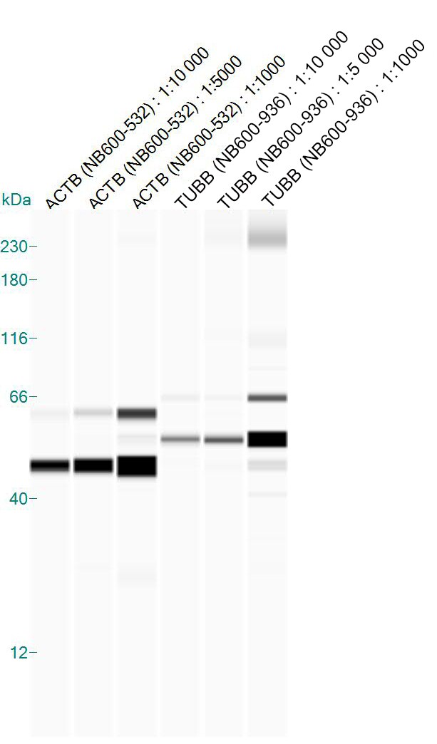
Simple Western: beta Tubulin Antibody [NB600-936] - Lane view shows a specific band for beta Tubulin in 0.05 mg/ml of MCF-7 lysate. This experiment was performed under reducing conditions using the 12-230 kDa separation system.![Western Blot: beta Tubulin AntibodyBSA Free [NB600-936]](https://resources.rndsystems.com/images/products/beta-Tubulin-Antibody-Western-Blot-NB600-936-img0006.jpg "Western Blot: beta Tubulin AntibodyBSA Free [NB600-936]")
Western Blot: beta Tubulin AntibodyBSA Free [NB600-936]
Western Blot: beta Tubulin Antibody [NB600-936] - Analysis of extracts from HeLa, A549 and Jurkat cells using NB600-936 Beta-tubulin antibody at 1:1000![Western Blot: beta Tubulin AntibodyBSA Free [NB600-936]](https://resources.rndsystems.com/images/products/beta-Tubulin-Antibody-Western-Blot-NB600-936-img0020.jpg "Western Blot: beta Tubulin AntibodyBSA Free [NB600-936]")
![Immunocytochemistry/ Immunofluorescence: beta Tubulin Antibody - BSA Free [NB600-936]](https://resources.rndsystems.com/images/products/beta-Tubulin-Antibody-Immunocytochemistry-Immunofluorescence-NB600-936-img0023.jpg "Immunocytochemistry/ Immunofluorescence: beta Tubulin Antibody - BSA Free [NB600-936]")
Immunocytochemistry/ Immunofluorescence: beta Tubulin Antibody - BSA Free [NB600-936]
Immunocytochemistry/Immunofluorescence: beta Tubulin Antibody [NB600-936] - PC12 cells were fixed in 4% paraformaldehyde for 10 minutes and permeabilized in 0.05% Triton X-100 in PBS for 5 minutes. The cells were incubated with anti-beta Tubulin Antibody (NB600-936) at 2 ug/ml overnight at 4C and detected with an anti-rabbit Dylight 488 (Green) at a 1:1000 dilution for 60 minutes. Nuclei were counterstained with DAPI (Blue). Cells were imaged using a 100X objective and digitally deconvolved.![Immunohistochemistry-Paraffin: beta Tubulin Antibody - BSA Free [NB600-936]](https://resources.rndsystems.com/images/products/beta-Tubulin-Antibody-Immunohistochemistry-Paraffin-NB600-936-img0011.jpg "Immunohistochemistry-Paraffin: beta Tubulin Antibody - BSA Free [NB600-936]")
Immunohistochemistry-Paraffin: beta Tubulin Antibody - BSA Free [NB600-936]
Immunohistochemistry-Paraffin: beta Tubulin Antibody [NB600-936] - Analysis of FFPE tissue section of normal human skin using 1:2000 dilution of beta Tubulin antibody. Intense cytoplasmic staining of beta Tubulin (TUBB) protein was observed in various cells of the epidermal as well as the dermal cells [10X Magnification].![Western Blot: beta Tubulin AntibodyBSA Free [NB600-936]](https://resources.rndsystems.com/images/products/beta-Tubulin-Antibody-Western-Blot-NB600-936-img0004.jpg "Western Blot: beta Tubulin AntibodyBSA Free [NB600-936]")
Western Blot: beta Tubulin AntibodyBSA Free [NB600-936]
Western Blot: beta Tubulin Antibody [NB600-936] - Analysis in HeLa whole cell lysate at a 1:1,000 dilution.![Western Blot: beta Tubulin AntibodyBSA Free [NB600-936]](https://resources.rndsystems.com/images/products/beta-Tubulin-Antibody-Western-Blot-NB600-936-img0024.jpg "Western Blot: beta Tubulin AntibodyBSA Free [NB600-936]")
Western Blot: beta Tubulin AntibodyBSA Free [NB600-936]
beta-Tubulin-Antibody-Western-Blot-NB600-936-img0024.jpg![Immunocytochemistry/ Immunofluorescence: beta Tubulin Antibody - BSA Free [NB600-936]](https://resources.rndsystems.com/images/products/beta-Tubulin-Antibody-Immunocytochemistry-Immunofluorescence-NB600-936-img0015.jpg "Immunocytochemistry/ Immunofluorescence: beta Tubulin Antibody - BSA Free [NB600-936]")
Immunocytochemistry/ Immunofluorescence: beta Tubulin Antibody - BSA Free [NB600-936]
Immunocytochemistry/Immunofluorescence: beta Tubulin Antibody [NB600-936] - Confocal immunofluorescent analysis of C2C12 cells using beta Tubulin antibody (NB600-936, 1:5). An Alexa Fluor 488-conjugated Goat to rabbit IgG was used as secondary antibody (green). Actin filaments were labeled with Alexa Fluor 568 phalloidin (red). DAPI was used to stain the cell nuclei (blue).![Immunocytochemistry/ Immunofluorescence: beta Tubulin Antibody - BSA Free [NB600-936]](https://resources.rndsystems.com/images/products/beta-Tubulin-Antibody-Immunocytochemistry-Immunofluorescence-NB600-936-img0001.jpg "Immunocytochemistry/ Immunofluorescence: beta Tubulin Antibody - BSA Free [NB600-936]")
Immunocytochemistry/ Immunofluorescence: beta Tubulin Antibody - BSA Free [NB600-936]
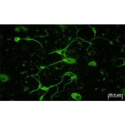
Immunocytochemistry/Immunofluorescence: beta Tubulin Antibody [NB600-936] - Analysis of beta Tubulin in mouse hippocampal primary culture. Image courtesy of product review by Lin Yi-Wen.![Immunocytochemistry/ Immunofluorescence: beta Tubulin Antibody - BSA Free [NB600-936]](https://resources.rndsystems.com/images/products/beta-Tubulin-Antibody-Immunocytochemistry-Immunofluorescence-NB600-936-img0022.jpg "Immunocytochemistry/ Immunofluorescence: beta Tubulin Antibody - BSA Free [NB600-936]")
Immunocytochemistry/ Immunofluorescence: beta Tubulin Antibody - BSA Free [NB600-936]
Immunocytochemistry/Immunofluorescence: beta Tubulin Antibody [NB600-936] - NIH3T3 cells were fixed in 4% paraformaldehyde for 10 minutes and permeabilized in 0.05% Triton X-100 in PBS for 5 minutes. The cells were incubated with anti-beta Tubulin Antibody (NB600-936) at 2 ug/ml overnight at 4C and detected with an anti-rabbit Dylight 488 (Green) at a 1:1000 dilution for 60 minutes. Nuclei were counterstained with DAPI (Blue). Cells were imaged using a 100X objective and digitally deconvolved.![Immunohistochemistry-Paraffin: beta Tubulin Antibody - BSA Free [NB600-936]](https://resources.rndsystems.com/images/products/beta-Tubulin-Antibody-Immunohistochemistry-Paraffin-NB600-936-img0014.jpg "Immunohistochemistry-Paraffin: beta Tubulin Antibody - BSA Free [NB600-936]")
Immunohistochemistry-Paraffin: beta Tubulin Antibody - BSA Free [NB600-936]
Immunohistochemistry-Paraffin: beta Tubulin Antibody [NB600-936] - Analysis of FFPE tissue section of human esophageal squamous cell carcinoma (SCC) using 1:2000 dilution of beta Tubulin antibody. Strong cytoplasmic immuno-positivity of beta Tubulin (TUBB) was observed in SCC cells as well as the associated tumor stromal cells [10X Magnification].![Immunohistochemistry-Paraffin: beta Tubulin Antibody - BSA Free [NB600-936]](https://resources.rndsystems.com/images/products/beta-Tubulin-Antibody-Immunohistochemistry-Paraffin-NB600-936-img0013.jpg "Immunohistochemistry-Paraffin: beta Tubulin Antibody - BSA Free [NB600-936]")
Immunohistochemistry-Paraffin: beta Tubulin Antibody - BSA Free [NB600-936]
Immunohistochemistry-Paraffin: beta Tubulin Antibody [NB600-936] - Analysis of FFPE tissue section of human esophageal squamous cell carcinoma (SCC) using 1:2000 dilution of beta Tubulin antibody. This representative image shows a cytoplasm specific staining of beta Tubulin (TUBB) in SCC cells [60X Magnification].![Immunohistochemistry-Paraffin: beta Tubulin Antibody - BSA Free [NB600-936]](https://resources.rndsystems.com/images/products/beta-Tubulin-Antibody-Immunohistochemistry-Paraffin-NB600-936-img0012.jpg "Immunohistochemistry-Paraffin: beta Tubulin Antibody - BSA Free [NB600-936]")
Immunohistochemistry-Paraffin: beta Tubulin Antibody - BSA Free [NB600-936]
Immunohistochemistry-Paraffin: beta Tubulin Antibody [NB600-936] - Analysis of FFPE tissue section of normal human brain using 1:2000 dilution of beta Tubulin antibody. The various brain cells depicted strong cytoplasmic immunoreactivity of beta Tubulin (TUBB) protein [10X Magnification].
Beta Tubulin in A431 Human Cell Line.
Beta Tubulin was detected in immersion fixed A431 human skin carcinoma cell line using Rabbit anti-beta Tubulin Affinity Purified Polyclonal Antibody conjugated to Alexa Fluor® 647 (Catalog # NB600-936AF647) (light blue) at 5 µg/mL overnight at 4C. Cells were counterstained with DAPI (blue). Cells were imaged using a 100X objective and digitally deconvolved.
Beta Tubulin in U-2 OS Human Cell Line.
Beta Tubulin was detected in immersion fixed U-2 OS human osteosarcoma cell line using Rabbit anti-beta Tubulin Affinity Purified Polyclonal Antibody conjugated to DyLight 488 (Catalog # NB600-936G) (green) at 5 µg/mL overnight at 4C. Cells were counterstained with DAPI (blue). Cells were imaged using a 100X objective and digitally deconvolved.
Western Blot: beta Tubulin Antibody - BSA Free [NB600-936] -
Western Blot: beta Tubulin Antibody - BSA Free [NB600-936] - Dual pathway inhibition regulates TK1 protein levels & results in greater p27 protein levels than single agents alone.Single agent PLX4032 resulted in activation of p-AKT Ser473 following 24 hours of exposure at two concentrations (100 nM, 1 µM). The addition of the dual PI3K/mTOR inhibitor BEZ235 blocks p-AKT Ser473 activation & resulted in a greater increase in p27 protein levels & diminished TK1 protein levels. Image collected & cropped by CiteAb from the following publication (https://pubmed.ncbi.nlm.nih.gov/25247710), licensed under a CC-BY license. Not internally tested by Novus Biologicals.
Western Blot: beta Tubulin Antibody - BSA Free [NB600-936] -
Western Blot: beta Tubulin Antibody - BSA Free [NB600-936] - Expression of pluripotency related markers & a sphere culture of BEAS-2B under normoxia.(a) HIF-2 alpha expression was detected in the nucleus of BEAS-2B under normoxia by western blotting. Beta tubulin & TATA binding protein (TBP) were used as protein markers for the cytosol & nucleus fraction, respectively. (b) Western blotting for Oct-4. (c) For knockdown of HIF-2 alpha, BEAS-2B cells were transfected with shRNA targeting HIF-2 alpha & sh-Luc was used as control. (d) Western blotting of Nanog & a culture of BEAS-2B containing floating spheres. Representative result from two or three experiments was presented. Image collected & cropped by CiteAb from the following publication (https://pubmed.ncbi.nlm.nih.gov/27373565), licensed under a CC-BY license. Not internally tested by Novus Biologicals.
Western Blot: beta Tubulin Antibody - BSA Free [NB600-936] -
Western Blot: beta Tubulin Antibody - BSA Free [NB600-936] - Expression of AAV transgene before & after experimental autoimmune uveoretinitis (EAU) induction. (A) B10.RIII mice were evaluated by funduscopy one month after an intravitreal injection of AAV delivering sGFP-TatM013v5 or secreted GFP (sGFP). As a control we evaluated mice that received an intravitreal injection of saline. Diffuse fluorescence indicates the secretion of sGFP & sGFP-TatM013v5 in the retina. (B) Western blot from retinas harvested 14 days after IRBP immunization. Membrane was probed with anti-GFP & anti-Tubulin antibodies. Image shows expression of sGFP on both sGFP & sGFP-TatM013v5 retina lysates. (n = 2–3 retina samples from different mice per group). Image collected & cropped by CiteAb from the following publication (https://pubmed.ncbi.nlm.nih.gov/31795515), licensed under a CC-BY license. Not internally tested by Novus Biologicals.
Western Blot: beta Tubulin Antibody - BSA Free [NB600-936] -
Western Blot: beta Tubulin Antibody - BSA Free [NB600-936] - Combined V600EBRAF & mTOR inhibition results in transcriptional control of TK1 protein levels in COLO 205 cells.(A) Western blot of COLO 205 cells treated with PP242 (250 nM) & increasing PLX4032. Similar to single agent PLX4032, p-MEK, but not p-ERK, was inhibited in a PLX4032-dependent manner. Consistent with mTORC1/mTORC2 inhibition, p-AKT Ser473, but not p-AKT Thr308, was inhibited. Unlike single agent PLX4032, which resulted in concentration-dependent activation of p-rpS6, combined treatment maintained p-rpS6 levels at essentially baseline levels except at the highest PLX4032 concentration. Similarly, DUSP6 levels were inversely related to p-ERK protein levels. With combined mTOR & V600EBRAF blockade, p27 & TK1 protein levels were inversely correlated & dramatically affected by PLX4032 exposure. (B) Similarly, TK1 mRNA was significantly reduced at PLX4032 concentrations as low as 10 nM. (C) Despite elevated p27 protein levels, p27 mRNA was unaffected by combined mTOR-V600EBRAF inhibition. Image collected & cropped by CiteAb from the following publication (https://pubmed.ncbi.nlm.nih.gov/25247710), licensed under a CC-BY license. Not internally tested by Novus Biologicals.
Western Blot: beta Tubulin Antibody - BSA Free [NB600-936] -
Western Blot: beta Tubulin Antibody - BSA Free [NB600-936] - [18F]-FLT PET reflects BEZ235-dependent inhibition of PI3K/mTOR activity in PLX4720 treated COLO 205 xenografts. Xenograft-bearing mice were imaged with [18F]-FLT PET on treatment day 4. (A) [18F]-FLT uptake was diminished in the combination treatment cohort relative to vehicle (p = 0.0087), but not single agent PLX4720- or BEZ235-treated cohorts. (B) Western blot of xenograft tissue harvested immediately following imaging illustrated elevated p-ERK & p-rpS6 levels in PLX4720-treated mice. Combining PLX4032 with BEZ235 resulted in reduced p-ERK & p-rpS6 protein levels. (C) TK1 levels, as measured by IHC, were reduced only in the combination treatment group in agreement with [18F]-FLT PET. (D) Consistent with in vitro studies, diminished TK1 levels, & consequently [18F]-FLT PET, correlated with elevated p27 that was elevated only in the combination treated group. Image collected & cropped by CiteAb from the following publication (https://pubmed.ncbi.nlm.nih.gov/25247710), licensed under a CC-BY license. Not internally tested by Novus Biologicals.
Western Blot: beta Tubulin Antibody - BSA Free [NB600-936] -
Western Blot: beta Tubulin Antibody - BSA Free [NB600-936] - PLX4720 exposure does not affect [18F]-FLT PET in COLO 205 xenografts, despite evidence of target inhibition & diminished [18F]-FDG uptake.(A) Representative transverse [18F]-FLT & [18F]-FDG PET images acquired after three daily treatments with vehicle or 60 mg/kg PLX4720 (tumor indicated by arrowhead). (B) Quantification of PET data illustrated similar [18F]-FLT uptake in vehicle-treated & PLX4720-treated tumors. Unlike [18F]-FLT PET, PLX4720 exposure elicited a significant reduction in [18F]-FDG uptake (p = 0.0006). (C) Western blot analysis of vehicle- & PLX4720-treated tumor tissue confirmed that PLX4720 had no effect on TK1 protein levels in agreement with [18F]-FLT PET. Target inhibition as measured by p-MEK levels was observed. However, similar to in vitro studies, PLX4720-treated COLO 205 xenografts exhibited elevated p-ERK & p-rpS6 protein levels relative to vehicle controls. Image collected & cropped by CiteAb from the following publication (https://pubmed.ncbi.nlm.nih.gov/25247710), licensed under a CC-BY license. Not internally tested by Novus Biologicals.
Western Blot: beta Tubulin Antibody - BSA Free [NB600-936] -
Western Blot: beta Tubulin Antibody - BSA Free [NB600-936] - Deletion of Phb1 affects lipid metabolism.Western blot (a) & quantification (b) of Acetyl-CoA carboxylase (ACC) & phosphorylated ACC (p-ACC) expression at P20. N = 6–7 animals per genotype. Unpaired two-tailed t-test [p-ACC (t = 0.4627, df = 11, p = 0.021), ACC (t = 1.355, df = 11, p = 0.26)]. Western blot (c) & quantification (d) of ACC & p-ACC expression at P40. N = 6–8 animals per genotype [p-ACC (t = 0.5447, df = 12, p = 0.42), ACC (t = 1.153, df = 12, p = 0.17)]. Unpaired two-tailed t-test. By RT-qPCR, we identified a significant downregulation of many enzymes involved with lipid biosynthesis at both P20 (e) & P40 (f): sterol regulatory element-binding protein 1 (Srebp1), 3-hydroxy-3-methylglutaryl-CoA reductase (Hmgcr), ATP citrate lyase (Acly), fatty acid synthase (FASN), acetyl-CoA carboxylase 2 (ACC2), N = 5 animals per genotype. Unpaired two-tailed t-test P20 [Srebp1 (t = 3.26, df = 8, p = 0.012), Hmgcr (t = 7.63, df = 8, p = 0.000061), Acly (t = 4.418, df = 8, 0.0022), FASN (t = 4.109, df = 8, p = 0.0034), ACC2 (t = 3.408, df = 8, p = 0.0092)]; P40 [Srebp1 (t = 7.551, df = 8, p = 0.000066), Hmgcr (t = 5.091, df = 8, p = 0.00094), Acly (t = 4.934, df = 8, p = 0.0011), FASN (t = 7.186, df = 8, p = 0.000094), ACC2 (t = 2.697, df = 8, p = 0.027)]. Data are presented as mean ± SEM. *p < 0.05; **p < 0.01; ***p < 0.001. n.s. non-significant. Image collected & cropped by CiteAb from the following publication (https://pubmed.ncbi.nlm.nih.gov/34078899), licensed under a CC-BY license. Not internally tested by Novus Biologicals.
Western Blot: beta Tubulin Antibody - BSA Free [NB600-936] -
Western Blot: beta Tubulin Antibody - BSA Free [NB600-936] - TK1 protein levels do not reflect p-ERK attenuation following inhibition of V600EBRAF inhibition in COLO 205 cells.COLO 205 cells were collected 48 hours of PLX 4032 exposure at 10 nM, 100 nM, 500 nM, 1 µM, or 5 µM. (A) Western blot analysis demonstrated target inhibition of p-MEK despite increased p-ERK levels. PI3K-mTOR signaling was elevated in a PLX 4032-dependent manner as exhibited by a steady rise in p-rpS6 levels. The ERK-phosphatase DUSP6 decreased in conjunction with mTOR signaling & was inversely proportional to p-ERK levels. A slight increase in p27 levels were observed concomitantly with only modest changes in TK1 levels, except at the highest dose of PLX4032. (B) Decreased TK1 mRNA levels were observed at all drug concentrations above 10 nM (p<0.05). Image collected & cropped by CiteAb from the following publication (https://pubmed.ncbi.nlm.nih.gov/25247710), licensed under a CC-BY license. Not internally tested by Novus Biologicals.
Beta Tubulin in NIH-3T3 Mouse Cell Line.
Beta Tubulin was detected in immersion fixed NIH-3T3 Mouse fibroblast cell line using Rabbit anti-beta Tubulin Affinity Purified Polyclonal Antibody conjugated to Alexa Fluor® 647 (Catalog # NB600-936AF647) (light blue) at 2 µg/mL overnight at 4C. Cells were counterstained with DAPI (blue). Cells were imaged using a 100X objective and digitally deconvolved.Applications for beta Tubulin Antibody - BSA Free
Immunocytochemistry/ Immunofluorescence
Immunohistochemistry
Immunohistochemistry-Paraffin
Simple Western
Western Blot
See Simple Western Antibody Database for Simple Western validation: separated by Size, antibody dilution of 1:500. Separated by Size-Wes, Sally Sue/Peggy Sue. SCW validated using murine pancreatic cancer cells.
Reviewed Applications
Read 7 reviews rated 4.4 using NB600-936 in the following applications:
Formulation, Preparation, and Storage
Purification
Formulation
Format
Preservative
Concentration
Shipping
Stability & Storage
Background: beta Tubulin
Long Name
Alternate Names
Gene Symbol
UniProt
Additional beta Tubulin Products
Product Documents for beta Tubulin Antibody - BSA Free
Certificate of Analysis
To download a Certificate of Analysis, please enter a lot or batch number in the search box below.
Product Specific Notices for beta Tubulin Antibody - BSA Free
This product is for research use only and is not approved for use in humans or in clinical diagnosis. Primary Antibodies are guaranteed for 1 year from date of receipt.
Citations for beta Tubulin Antibody - BSA Free
 Powered by Bioz
Customer Reviews for beta Tubulin Antibody - BSA Free (7)
Have you used beta Tubulin Antibody - BSA Free?
Submit a review and receive an Amazon gift card!
$25/€18/£15/$25CAN/¥2500 Yen for a review with an image
$10/€7/£6/$10CAN/¥1110 Yen for a review without an image
Submit a review
Customer Images
-(01-ml)_NB600-936_8571.jpg)
-(01-ml)_NB600-936_8221.bmp)
-(01-ml)_NB600-936_7861.jpg)
-
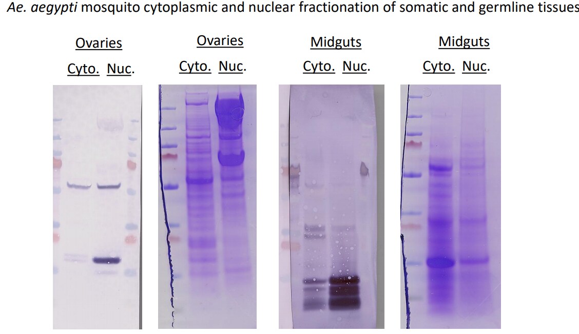
Application: Western BlotSample Tested: Ovary tissue, Midgut tissue and A549 Nuclear and cytoplasmic, Sample Amount: 30ugSpecies: Ae. aegyptiVerified Customer | Posted 05/22/2021Western and Commassie-stained SDS/PAGE gels of Ae. aegypti ovary or midgut tissues where cytoplasmic and nuclear fractions were obtainedTo test both anti-histone H3 and anti-beta tubulin as markers for nuclear or cytoplasmic fractions, respectively, in Ae. aegypti mosquitoes, a western blot was performed using midguts or ovaries dissected from mosquitoes 48h post-bloodmeal. The cytoplasmic or nuclear fractions from each tissue type were obtained and protein concentration was quantified by A260/280. Roughly 30ug of tissue was loaded onto SDS/PAGE gels that were either transferred to PVDF membranes or stained by Coomassie. Membranes were blocked in TBST 5% milk for 2h. Both anti-histone H3 and anti-beta tubulin were added 1:1000 in blocking buffer and incubated O/N shaking at 4C. Proteins were detected by Alkaline Phosphatase. In Ae. aegypti ovaries, anti-histone H3 bound to protein roughly 15kDa in the nuclear fraction of ovaries but not the cytoplasmic fraction, while anti-beta-tubulin bound to protein roughly 55kDa in both cytoplasmic and nuclear fractions. In Ae. aegypti midguts, anti-histone H3 bound to 3 proteins between 10-15kDa strongly in the nuclear fraction and faintly in the cytoplasmic fraction-perhaps due to nuclear contamination in the cytoplasmic fraction. anti-beta-tubulin did not react well in midgut fractions. The beta-tubulin antibody was not successful for differentiating cytoplasmic from nuclear fractions in Ae. aegypti tissues. For your reference, protein ladder = pageruler plus prestained ThermoFisher

-
Application: Simple WesternSample Tested: whole brain homogenates from miceSpecies: MouseVerified Customer | Posted 01/21/2016Simple Western: TUBB in mouse brain lysate
-
Application: Western BlotSample Tested:Species: MouseVerified Customer | Posted 07/01/2014A review for beta-tubulin antibody
-
Application: ImmunocytochemistrySample Tested:Species: MouseVerified Customer | Posted 06/12/2014Confocal immunofluorescent analysis of C2C12 cells using beta Tubulin antibody (NB600-936, 1:5).
-
Application: Western BlotSample Tested:Species: HumanVerified Customer | Posted 05/27/2014Western blot analysis of extracts from HeLa, A549 and Jurkat cells using beta Tubulin antibody (NB600-936, 1:1000).
-
Application: ImmunohistochemistrySample Tested: Hippocampal Primary CultureSpecies: MouseVerified Customer | Posted 01/13/2012
-
Application: Western BlotSample Tested: Chicken cell lineSpecies: OtherVerified Customer | Posted 11/09/2011
There are no reviews that match your criteria.
Protocols
View specific protocols for beta Tubulin Antibody - BSA Free (NB600-936):
Culture cells to appropriate density in 35 mm culture dishes or 6-well plates.
1. Remove culture medium and wash the cells briefly in PBS. Add 10% formalin to the dish and fix at room temperature for 10 minutes.
2. Remove the formalin and wash the cells in PBS.
3. Permeablize the cells with 0.1% Triton X100 or other suitable detergent for 10 min.
4. Remove the permeablization buffer and wash three times for 10 minutes each in PBS. Be sure to not let the specimen dry out.
5. To block nonspecific antibody binding, incubate in 10% normal goat serum from 1 hour to overnight at room temperature.
6. Add primary antibody at appropriate dilution and incubate overnight at 4C.
7. Remove primary antibody and replace with PBS. Wash three times for 10 minutes each.
8. Add secondary antibody at appropriate dilution. Incubate for 1 hour at room temperature.
9. Remove secondary antibody and replace with PBS. Wash three times for 10 minutes each.
10. Counter stain DNA with DAPi if required.
Antigen Unmasking:
Bring slides to a boil in 10 mM sodium citrate buffer (pH 6.0) then maintain at a sub-boiling temperature for 10 minutes. Cool slides on bench-top for 30 minutes (keep slides in the sodium citrate buffer all the time).
Staining:
1. Wash sections in deionized water three times for 5 minutes each.
2. Wash sections in PBS for 5 minutes.
3. Block each section with 100-400 ul blocking solution (1% BSA in PBS) for 1 hour at room temperature.
4. Remove blocking solution and add 100-400 ul diluted primary antibody. Incubate overnight at 4 C.
5. Remove antibody solution and wash sections in wash buffer three times for 5 minutes each.
6. Add 100-400 ul HRP polymer conjugated secondary antibody. Incubate 30 minutes at room temperature.
7. Wash sections three times in wash buffer for 5 minutes each.
8. Add 100-400 ul DAB substrate to each section and monitor staining closely.
9. As soon as the sections develop, immerse slides in deionized water.
10. Counterstain sections in hematoxylin.
11. Wash sections in deionized water two times for 5 minutes each.
12. Dehydrate sections.
13. Mount coverslips.
Western Blot Protocol
1. Perform SDS-PAGE on samples to be analyzed, loading 10-25 ug of total protein per lane.
2. Transfer proteins to PVDF membrane according to the instructions provided by the manufacturer of the membrane and transfer apparatus.
3. Stain the membrane with Ponceau S (or similar product) to assess transfer success, and mark molecular weight standards where appropriate.
4. Rinse the blot TBS -0.05% Tween 20 (TBST).
5. Block the membrane in 5% Non-fat milk in TBST (blocking buffer) for at least 1 hour.
6. Wash the membrane in TBST three times for 10 minutes each.
7. Dilute primary antibody in blocking buffer and incubate overnight at 4C with gentle rocking.
8. Wash the membrane in TBST three times for 10 minutes each.
9. Incubate the membrane in diluted HRP conjugated secondary antibody in blocking buffer (as per manufacturer's instructions) for 1 hour at room temperature.
10. Wash the blot in TBST three times for 10 minutes each (this step can be repeated as required to reduce background).
11. Apply the detection reagent of choice in accordance with the manufacturers instructions.
Find general support by application which include: protocols, troubleshooting, illustrated assays, videos and webinars.
- Antigen Retrieval Protocol (PIER)
- Antigen Retrieval for Frozen Sections Protocol
- Appropriate Fixation of IHC/ICC Samples
- Cellular Response to Hypoxia Protocols
- Chromogenic IHC Staining of Formalin-Fixed Paraffin-Embedded (FFPE) Tissue Protocol
- Chromogenic Immunohistochemistry Staining of Frozen Tissue
- ClariTSA™ Fluorophore Kits
- Detection & Visualization of Antibody Binding
- Fluorescent IHC Staining of Frozen Tissue Protocol
- Graphic Protocol for Heat-induced Epitope Retrieval
- Graphic Protocol for the Preparation and Fluorescent IHC Staining of Frozen Tissue Sections
- Graphic Protocol for the Preparation and Fluorescent IHC Staining of Paraffin-embedded Tissue Sections
- Graphic Protocol for the Preparation of Gelatin-coated Slides for Histological Tissue Sections
- ICC Cell Smear Protocol for Suspension Cells
- ICC Immunocytochemistry Protocol Videos
- ICC for Adherent Cells
- IHC Sample Preparation (Frozen sections vs Paraffin)
- Immunocytochemistry (ICC) Protocol
- Immunocytochemistry Troubleshooting
- Immunofluorescence of Organoids Embedded in Cultrex Basement Membrane Extract
- Immunofluorescent IHC Staining of Formalin-Fixed Paraffin-Embedded (FFPE) Tissue Protocol
- Immunohistochemistry (IHC) and Immunocytochemistry (ICC) Protocols
- Immunohistochemistry Frozen Troubleshooting
- Immunohistochemistry Paraffin Troubleshooting
- Preparing Samples for IHC/ICC Experiments
- Preventing Non-Specific Staining (Non-Specific Binding)
- Primary Antibody Selection & Optimization
- Protocol for Heat-Induced Epitope Retrieval (HIER)
- Protocol for Making a 4% Formaldehyde Solution in PBS
- Protocol for VisUCyte™ HRP Polymer Detection Reagent
- Protocol for the Fluorescent ICC Staining of Cell Smears - Graphic
- Protocol for the Fluorescent ICC Staining of Cultured Cells on Coverslips - Graphic
- Protocol for the Preparation & Fixation of Cells on Coverslips
- Protocol for the Preparation and Chromogenic IHC Staining of Frozen Tissue Sections
- Protocol for the Preparation and Chromogenic IHC Staining of Frozen Tissue Sections - Graphic
- Protocol for the Preparation and Chromogenic IHC Staining of Paraffin-embedded Tissue Sections
- Protocol for the Preparation and Chromogenic IHC Staining of Paraffin-embedded Tissue Sections - Graphic
- Protocol for the Preparation and Fluorescent ICC Staining of Cells on Coverslips
- Protocol for the Preparation and Fluorescent ICC Staining of Non-adherent Cells
- Protocol for the Preparation and Fluorescent ICC Staining of Stem Cells on Coverslips
- Protocol for the Preparation and Fluorescent IHC Staining of Frozen Tissue Sections
- Protocol for the Preparation and Fluorescent IHC Staining of Paraffin-embedded Tissue Sections
- Protocol for the Preparation of Gelatin-coated Slides for Histological Tissue Sections
- Protocol for the Preparation of a Cell Smear for Non-adherent Cell ICC - Graphic
- R&D Systems Quality Control Western Blot Protocol
- TUNEL and Active Caspase-3 Detection by IHC/ICC Protocol
- The Importance of IHC/ICC Controls
- Troubleshooting Guide: Immunohistochemistry
- Troubleshooting Guide: Western Blot Figures
- Western Blot Conditions
- Western Blot Protocol
- Western Blot Protocol for Cell Lysates
- Western Blot Troubleshooting
- Western Blot Troubleshooting Guide
- View all Protocols, Troubleshooting, Illustrated assays and Webinars
FAQs for beta Tubulin Antibody - BSA Free
-
Q: We are looking for an antibody which is a specific marker for Mouse Bronchial cilia in IHC(p) technique. Preferably not from a mouse host. Can you help?
A: According to the literature I have reviewed on bronchial cilia markers, the most commonly used marker is acetylated alpha tubulin (AcTub). Please see the relevant publications with images attached (Song et al. 2014, Schamberger et al. 2014). We have one antibody to AcTub that is validated for IHC-P and mouse (it is however also raised in mouse, hence mouse on mouse blocking step would be necessary): NB600-567. There are also some reports of using beta tubulin as cilial marker (Davis et al. 2015). For rabbit host antibodies, you may look at our best seller products like NB600-936 and NB100-56459