BST2 Antibody - BSA Free
Novus Biologicals | Catalog # NBP2-27154
![Western Blot: BST2 Antibody [NBP2-27154]](https://resources.rndsystems.com/images/products/BST2-Antibody-Western-Blot-NBP2-27154-img0002.jpg "Western Blot: BST2 Antibody [NBP2-27154]")
Key Product Details
Species Reactivity
Validated:
Human, Mouse, Rat, Primate
Cited:
Human, Mouse
Applications
Validated:
Immunohistochemistry, Immunohistochemistry-Paraffin, Immunohistochemistry-Frozen, Western Blot, Flow Cytometry, Flow (Cell Surface), Immunocytochemistry/ Immunofluorescence, Simple Western
Cited:
Immunohistochemistry-Paraffin, Western Blot, Flow Cytometry, Flow (Cell Surface), Immunocytochemistry/ Immunofluorescence
Label
Unconjugated
Antibody Source
Polyclonal Rabbit IgG
Format
BSA Free
Loading...
Product Specifications
Immunogen
A portion of amino acids 80-130 of human BST2 was used as the immunogen for this antibody.
Clonality
Polyclonal
Host
Rabbit
Isotype
IgG
Scientific Data Images for BST2 Antibody - BSA Free
Western Blot: BST2 Antibody [NBP2-27154]
Western Blot: BST2 Antibody [NBP2-27154] - Analysis using BST2 antibody. Human heart in the 1) absence and 2) presence of immunizing peptide, 3) human Jurkat, 4) mouse RAW lysate and 5) recombinant human BST2 protein probed with BST2 antibody at 4 ug/mL. Goat anti-rabbit IgG HRP secondary antibody and PicoTect ECL substrate solution were used for this test.![Immunohistochemistry-Frozen: BST2 Antibody [NBP2-27154]](https://resources.rndsystems.com/images/products/BST2-Antibody-Immunohistochemistry-Frozen-NBP2-27154-img0005.jpg "Immunohistochemistry-Frozen: BST2 Antibody [NBP2-27154]")
Immunohistochemistry-Frozen: BST2 Antibody [NBP2-27154]
Immunohistochemistry-Frozen: BST2 Antibody [NBP2-27154] - BST-2 (green) was detected in human skin (nevus) using Ang-2 antibody (1:50; 1 hour) and secondary anti-rabbit-FITC (1:500; 30 min) in PBS. Nuclei were stained with DAPI (blue). Tissue was fixed in acetone. Image from a verified customer review.![Immunohistochemistry-Paraffin: BST2 Antibody [NBP2-27154]](https://resources.rndsystems.com/images/products/BST2-Antibody-Immunohistochemistry-Paraffin-NBP2-27154-img0001.jpg "Immunohistochemistry-Paraffin: BST2 Antibody [NBP2-27154]")
Immunohistochemistry-Paraffin: BST2 Antibody [NBP2-27154]
Immunohistochemistry-Paraffin: BST2 Antibody [NBP2-27154] - Analysis of BST2 in human liver tissue using BST2 antibody at 5 ug/mL.![Simple Western: BST2 Antibody [NBP2-27154]](https://resources.rndsystems.com/images/products/BST2-Antibody-Simple-Western-NBP2-27154-img0003.jpg "Simple Western: BST2 Antibody [NBP2-27154]")
Simple Western: BST2 Antibody [NBP2-27154]
Simple Western: BST2 Antibody [NBP2-27154] - Simple Western lane view shows a specific band for BST2 in 0.5 mg/ml of Jurkat lysate. This experiment was performed under standard reducing conditions using the 12-230 kDa separation system.![Immunofluorescence: BST2 Antibody [NBP2-27154]](https://resources.rndsystems.com/images/products/BST2-Antibody-Immunofluorescence-NBP2-27154-img0004.jpg "Immunofluorescence: BST2 Antibody [NBP2-27154]")
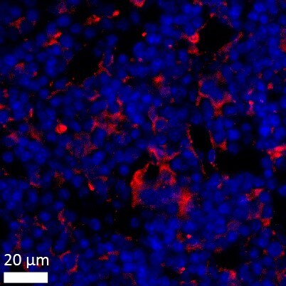
Immunofluorescence: BST2 Antibody [NBP2-27154]
Immunofluorescence: BST2 Antibody [NBP2-27154] - Mouse femur bone marrow section stained with BST2 antibody (red) and DAPI (blue). Image from a verified customer review.
Simple Western: BST2 Antibody [NBP2-27154] -
Simple Western: BST2 Antibody [NBP2-27154] - Lane view shows a specific band for BST2 using 0.5 mg/mL Jurkat cell lysate and antibody at 1:200. Electoropherogram image of corresponding Simple Western lane view. Image reported by internal validation.Applications for BST2 Antibody - BSA Free
Application
Recommended Usage
Flow (Cell Surface)
reported in scientific literature (PMID 21695198)
Immunohistochemistry-Frozen
reported by customer review
Immunohistochemistry-Paraffin
5 ug/mL
Simple Western
1:200
Western Blot
2 - 4 ug/mL
Application Notes
BST2 is a 180 amino acid protein with an observed molecular weight of approximately 30 kDa in Western blot (see Ishikawa et all, 1995 PMID 7607676).
In Simple Western only 10 - 15 uL of the recommended dilution is used per data point.
See Simple Western Antibody Database for Simple Western validation: Tested in Jurkat lysate 0.5 mg/mL, separated by Size, antibody dilution of 1:200, apparent MW was 21 kDa. Separated by Size-Wes, Sally Sue/Peggy Sue.
Reviewed Applications
Read 3 reviews rated 4 using NBP2-27154 in the following applications:
Flow Cytometry Panel Builder
Bio-Techne Knows Flow Cytometry
Save time and reduce costly mistakes by quickly finding compatible reagents using the Panel Builder Tool.
Advanced Features
- Spectra Viewer - Custom analysis of spectra from multiple fluorochromes
- Spillover Popups - Visualize the spectra of individual fluorochromes
- Antigen Density Selector - Match fluorochrome brightness with antigen density

Formulation, Preparation, and Storage
Purification
Protein A purified
Formulation
PBS
Format
BSA Free
Preservative
0.05% Sodium Azide
Concentration
1.0 mg/ml
Shipping
The product is shipped with polar packs. Upon receipt, store it immediately at the temperature recommended below.
Stability & Storage
Store at 4C short term. Aliquot and store at -20C long term. Avoid freeze-thaw cycles.
Background: BST-2/Tetherin
Long Name
Bone Marrow Stromal Cell Antigen 2
Alternate Names
BST2, CD317, HM1.24 Antigen, NPC-A-7, Tetherin
Gene Symbol
BST2
UniProt
Additional BST-2/Tetherin Products
Product Documents for BST2 Antibody - BSA Free
Certificate of Analysis
To download a Certificate of Analysis, please enter a lot or batch number in the search box below.
Product Specific Notices for BST2 Antibody - BSA Free
This product is for research use only and is not approved for use in humans or in clinical diagnosis. Primary Antibodies are guaranteed for 1 year from date of receipt.
Related Research Areas
Citations for BST2 Antibody - BSA Free
 Powered by Bioz
Customer Reviews for BST2 Antibody - BSA Free (3)
4 out of 5
3 Customer Ratings
Have you used BST2 Antibody - BSA Free?
Submit a review and receive an Amazon gift card!
$25/€18/£15/$25CAN/¥2500 Yen for a review with an image
$10/€7/£6/$10CAN/¥1110 Yen for a review without an image
Submit a review
Customer Images

Showing
1
-
3 of
3 reviews
Showing All
Filter By:
-
Application: Immunohistochemistry-FrozenSample Tested: Frozen human skin sectionsSpecies: HumanVerified Customer | Posted 07/18/2019BST-2 (green) was detected in human skin (nevus) using Ang-2 antibody (1:50; 1 hour) and secondary anti-rabbit-FITC (1:500; 30 min) in PBS. Nuclei were stained with Dapi (blue). Tissue was fixed in acetone.

-
Application: ImmunofluorescenceSample Tested:Species: HumanVerified Customer | Posted 01/04/2016
-
Application: ImmunofluorescenceSample Tested:Species: MouseVerified Customer | Posted 08/06/2015Immunohistochemistry of mouse femur bone marrow section stained with BST2 antibody (NBP2-27154) [red] and DAPI (blue)
There are no reviews that match your criteria.
Protocols
Find general support by application which include: protocols, troubleshooting, illustrated assays, videos and webinars.
- 7-Amino Actinomycin D (7-AAD) Cell Viability Flow Cytometry Protocol
- Antigen Retrieval Protocol (PIER)
- Antigen Retrieval for Frozen Sections Protocol
- Appropriate Fixation of IHC/ICC Samples
- Cellular Response to Hypoxia Protocols
- Chromogenic IHC Staining of Formalin-Fixed Paraffin-Embedded (FFPE) Tissue Protocol
- Chromogenic Immunohistochemistry Staining of Frozen Tissue
- ClariTSA™ Fluorophore Kits
- Detection & Visualization of Antibody Binding
- Extracellular Membrane Flow Cytometry Protocol
- Flow Cytometry Protocol for Cell Surface Markers
- Flow Cytometry Protocol for Staining Membrane Associated Proteins
- Flow Cytometry Staining Protocols
- Flow Cytometry Troubleshooting Guide
- Fluorescent IHC Staining of Frozen Tissue Protocol
- Graphic Protocol for Heat-induced Epitope Retrieval
- Graphic Protocol for the Preparation and Fluorescent IHC Staining of Frozen Tissue Sections
- Graphic Protocol for the Preparation and Fluorescent IHC Staining of Paraffin-embedded Tissue Sections
- Graphic Protocol for the Preparation of Gelatin-coated Slides for Histological Tissue Sections
- ICC Cell Smear Protocol for Suspension Cells
- ICC Immunocytochemistry Protocol Videos
- ICC for Adherent Cells
- IHC Sample Preparation (Frozen sections vs Paraffin)
- Immunocytochemistry (ICC) Protocol
- Immunocytochemistry Troubleshooting
- Immunofluorescence of Organoids Embedded in Cultrex Basement Membrane Extract
- Immunofluorescent IHC Staining of Formalin-Fixed Paraffin-Embedded (FFPE) Tissue Protocol
- Immunohistochemistry (IHC) and Immunocytochemistry (ICC) Protocols
- Immunohistochemistry Frozen Troubleshooting
- Immunohistochemistry Paraffin Troubleshooting
- Intracellular Flow Cytometry Protocol Using Alcohol (Methanol)
- Intracellular Flow Cytometry Protocol Using Detergents
- Intracellular Nuclear Staining Flow Cytometry Protocol Using Detergents
- Intracellular Staining Flow Cytometry Protocol Using Alcohol Permeabilization
- Intracellular Staining Flow Cytometry Protocol Using Detergents to Permeabilize Cells
- Preparing Samples for IHC/ICC Experiments
- Preventing Non-Specific Staining (Non-Specific Binding)
- Primary Antibody Selection & Optimization
- Propidium Iodide Cell Viability Flow Cytometry Protocol
- Protocol for Heat-Induced Epitope Retrieval (HIER)
- Protocol for Liperfluo
- Protocol for Making a 4% Formaldehyde Solution in PBS
- Protocol for VisUCyte™ HRP Polymer Detection Reagent
- Protocol for the Characterization of Human Th22 Cells
- Protocol for the Characterization of Human Th9 Cells
- Protocol for the Fluorescent ICC Staining of Cell Smears - Graphic
- Protocol for the Fluorescent ICC Staining of Cultured Cells on Coverslips - Graphic
- Protocol for the Preparation & Fixation of Cells on Coverslips
- Protocol for the Preparation and Chromogenic IHC Staining of Frozen Tissue Sections
- Protocol for the Preparation and Chromogenic IHC Staining of Frozen Tissue Sections - Graphic
- Protocol for the Preparation and Chromogenic IHC Staining of Paraffin-embedded Tissue Sections
- Protocol for the Preparation and Chromogenic IHC Staining of Paraffin-embedded Tissue Sections - Graphic
- Protocol for the Preparation and Fluorescent ICC Staining of Cells on Coverslips
- Protocol for the Preparation and Fluorescent ICC Staining of Non-adherent Cells
- Protocol for the Preparation and Fluorescent ICC Staining of Stem Cells on Coverslips
- Protocol for the Preparation and Fluorescent IHC Staining of Frozen Tissue Sections
- Protocol for the Preparation and Fluorescent IHC Staining of Paraffin-embedded Tissue Sections
- Protocol for the Preparation of Gelatin-coated Slides for Histological Tissue Sections
- Protocol for the Preparation of a Cell Smear for Non-adherent Cell ICC - Graphic
- Protocol: Annexin V and PI Staining by Flow Cytometry
- Protocol: Annexin V and PI Staining for Apoptosis by Flow Cytometry
- R&D Systems Quality Control Western Blot Protocol
- TUNEL and Active Caspase-3 Detection by IHC/ICC Protocol
- The Importance of IHC/ICC Controls
- Troubleshooting Guide: Fluorokine Flow Cytometry Kits
- Troubleshooting Guide: Immunohistochemistry
- Troubleshooting Guide: Western Blot Figures
- Western Blot Conditions
- Western Blot Protocol
- Western Blot Protocol for Cell Lysates
- Western Blot Troubleshooting
- Western Blot Troubleshooting Guide
- View all Protocols, Troubleshooting, Illustrated assays and Webinars
Loading...