![Western Blot: BTBD7 Antibody [NBP1-49652]](https://resources.rndsystems.com/images/products/BTBD7-Antibody-Western-Blot-NBP1-49652-img0004.jpg "Western Blot: BTBD7 Antibody [NBP1-49652]")
Key Product Details
Species Reactivity
Validated:
Human, Mouse
Cited:
Human, Mouse
Applications
Validated:
Immunohistochemistry, Immunohistochemistry-Paraffin, Western Blot, ELISA, Immunocytochemistry/ Immunofluorescence, Immunoprecipitation
Cited:
Immunohistochemistry-Paraffin, Western Blot
Label
Unconjugated
Antibody Source
Polyclonal Rabbit IgG
Loading...
Product Specifications
Immunogen
Synthetic peptide taken within amino acid region 1050-1150 on human BTBD7 protein.
Reactivity Notes
Mouse reactivity reported in scientific literature (PMID: 11231300)
Clonality
Polyclonal
Host
Rabbit
Isotype
IgG
Scientific Data Images for BTBD7 Antibody
Western Blot: BTBD7 Antibody [NBP1-49652]
Western Blot: BTBD7 Antibody [NBP1-49652] - Human 293T cells were infected by lentivirus overexpression of control or BTBD7 for 72h. Total cell lysates were subjected to Western blot. PVDF membrane was probed with BTBD7 Antibody. A specific band was detected for BTBD7 at approximately 138 kDa. Image from verified customer review.![Western Blot: BTBD7 Antibody [NBP1-49652]](https://resources.rndsystems.com/images/products/BTBD7-Antibody-Western-Blot-NBP1-49652-img0001.jpg "Western Blot: BTBD7 Antibody [NBP1-49652]")
Western Blot: BTBD7 Antibody [NBP1-49652]
Western Blot: BTBD7 Antibody [NBP1-49652] - Lane A: Immune Serum, Lane B: Preimmune. Apparent MW is 138 kDaApplications for BTBD7 Antibody
Application
Recommended Usage
ELISA
1:10000
Immunocytochemistry/ Immunofluorescence
1:200
Immunohistochemistry-Paraffin
1:200
Immunoprecipitation
1:200
Western Blot
1:5000
Application Notes
Use in IHC-Paraffin reported in scientific literature (PMID: 23325674).
Reviewed Applications
Read 1 review rated 4 using NBP1-49652 in the following applications:
Formulation, Preparation, and Storage
Purification
Immunogen affinity purified
Formulation
Tris/Glycine buffer, pH 7.4-7.8, HEPES,BSA 0.5%, glycerol 30%.
Preservative
0.02% Sodium Azide
Concentration
0.5 mg/ml
Shipping
The product is shipped with polar packs. Upon receipt, store it immediately at the temperature recommended below.
Stability & Storage
Store at -20C. Avoid freeze-thaw cycles.
Background: BTBD7
Alternate Names
BTB (POZ) domain containing 7, DKFZp686N0544, FLJ10648, FUP1BTB/POZ domain-containing protein 7, KIAA1525, MGC48310
Gene Symbol
BTBD7
UniProt
Additional BTBD7 Products
Product Documents for BTBD7 Antibody
Certificate of Analysis
To download a Certificate of Analysis, please enter a lot or batch number in the search box below.
Product Specific Notices for BTBD7 Antibody
This product is for research use only and is not approved for use in humans or in clinical diagnosis. Primary Antibodies are guaranteed for 1 year from date of receipt.
Citations for BTBD7 Antibody
 Powered by Bioz
Customer Reviews for BTBD7 Antibody (1)
4 out of 5
1 Customer Rating
Have you used BTBD7 Antibody?
Submit a review and receive an Amazon gift card!
$25/€18/£15/$25CAN/¥2500 Yen for a review with an image
$10/€7/£6/$10CAN/¥1110 Yen for a review without an image
Submit a review
Customer Images
Showing
1
-
1 of
1 review
Showing All
Filter By:
-
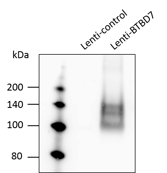
Application: Western BlotSample Tested: 293T cell lineSpecies: HumanVerified Customer | Posted 07/17/2018293T cells were infected by lentivirus overexpression of control or BTBD7 for 72h. Total cell lysates were subjected to western blot. PVDF membrane were probed with 1mm/ml Human BTBD7 Antibody. A specific band was detected for BTBD7 at approximately 138 kDa. This experiment was conducted under reducing conditions

There are no reviews that match your criteria.
Protocols
Find general support by application which include: protocols, troubleshooting, illustrated assays, videos and webinars.
- Antigen Retrieval Protocol (PIER)
- Antigen Retrieval for Frozen Sections Protocol
- Appropriate Fixation of IHC/ICC Samples
- Cellular Response to Hypoxia Protocols
- Chromogenic IHC Staining of Formalin-Fixed Paraffin-Embedded (FFPE) Tissue Protocol
- Chromogenic Immunohistochemistry Staining of Frozen Tissue
- ClariTSA™ Fluorophore Kits
- Detection & Visualization of Antibody Binding
- ELISA Sample Preparation & Collection Guide
- ELISA Troubleshooting Guide
- Fluorescent IHC Staining of Frozen Tissue Protocol
- Graphic Protocol for Heat-induced Epitope Retrieval
- Graphic Protocol for the Preparation and Fluorescent IHC Staining of Frozen Tissue Sections
- Graphic Protocol for the Preparation and Fluorescent IHC Staining of Paraffin-embedded Tissue Sections
- Graphic Protocol for the Preparation of Gelatin-coated Slides for Histological Tissue Sections
- How to Run an R&D Systems DuoSet ELISA
- How to Run an R&D Systems Quantikine ELISA
- How to Run an R&D Systems Quantikine™ QuicKit™ ELISA
- ICC Cell Smear Protocol for Suspension Cells
- ICC Immunocytochemistry Protocol Videos
- ICC for Adherent Cells
- IHC Sample Preparation (Frozen sections vs Paraffin)
- Immunocytochemistry (ICC) Protocol
- Immunocytochemistry Troubleshooting
- Immunofluorescence of Organoids Embedded in Cultrex Basement Membrane Extract
- Immunofluorescent IHC Staining of Formalin-Fixed Paraffin-Embedded (FFPE) Tissue Protocol
- Immunohistochemistry (IHC) and Immunocytochemistry (ICC) Protocols
- Immunohistochemistry Frozen Troubleshooting
- Immunohistochemistry Paraffin Troubleshooting
- Immunoprecipitation Protocol
- Preparing Samples for IHC/ICC Experiments
- Preventing Non-Specific Staining (Non-Specific Binding)
- Primary Antibody Selection & Optimization
- Protocol for Heat-Induced Epitope Retrieval (HIER)
- Protocol for Making a 4% Formaldehyde Solution in PBS
- Protocol for VisUCyte™ HRP Polymer Detection Reagent
- Protocol for the Fluorescent ICC Staining of Cell Smears - Graphic
- Protocol for the Fluorescent ICC Staining of Cultured Cells on Coverslips - Graphic
- Protocol for the Preparation & Fixation of Cells on Coverslips
- Protocol for the Preparation and Chromogenic IHC Staining of Frozen Tissue Sections
- Protocol for the Preparation and Chromogenic IHC Staining of Frozen Tissue Sections - Graphic
- Protocol for the Preparation and Chromogenic IHC Staining of Paraffin-embedded Tissue Sections
- Protocol for the Preparation and Chromogenic IHC Staining of Paraffin-embedded Tissue Sections - Graphic
- Protocol for the Preparation and Fluorescent ICC Staining of Cells on Coverslips
- Protocol for the Preparation and Fluorescent ICC Staining of Non-adherent Cells
- Protocol for the Preparation and Fluorescent ICC Staining of Stem Cells on Coverslips
- Protocol for the Preparation and Fluorescent IHC Staining of Frozen Tissue Sections
- Protocol for the Preparation and Fluorescent IHC Staining of Paraffin-embedded Tissue Sections
- Protocol for the Preparation of Gelatin-coated Slides for Histological Tissue Sections
- Protocol for the Preparation of a Cell Smear for Non-adherent Cell ICC - Graphic
- Quantikine HS ELISA Kit Assay Principle, Alkaline Phosphatase
- Quantikine HS ELISA Kit Principle, Streptavidin-HRP Polymer
- R&D Systems Quality Control Western Blot Protocol
- Sandwich ELISA (Colorimetric) – Biotin/Streptavidin Detection Protocol
- Sandwich ELISA (Colorimetric) – Direct Detection Protocol
- TUNEL and Active Caspase-3 Detection by IHC/ICC Protocol
- The Importance of IHC/ICC Controls
- Troubleshooting Guide: ELISA
- Troubleshooting Guide: Immunohistochemistry
- Troubleshooting Guide: Western Blot Figures
- Western Blot Conditions
- Western Blot Protocol
- Western Blot Protocol for Cell Lysates
- Western Blot Troubleshooting
- Western Blot Troubleshooting Guide
- View all Protocols, Troubleshooting, Illustrated assays and Webinars
Loading...