Calreticulin Antibody - BSA Free
Novus Biologicals | Catalog # NB600-103
![Knockout Validated: Calreticulin Antibody [NB600-103]](https://resources.rndsystems.com/images/products/Calreticulin-Antibody-Knockdown-Validated-NB600-103-img0008.jpg "Western Blot: Calreticulin Antibody [NB600-103]")
Loading...
Key Product Details
Validated by
Knockout/Knockdown
Species Reactivity
Validated:
Human, Mouse
Cited:
Human, Mouse
Applications
Validated:
Knockout Validated, Immunohistochemistry, Immunohistochemistry-Paraffin, Immunohistochemistry-Frozen, Western Blot, Immunocytochemistry/ Immunofluorescence, Simple Western, Knockdown Validated
Cited:
Immunohistochemistry-Paraffin, Immunohistochemistry-Frozen, Western Blot
Label
Unconjugated
Antibody Source
Polyclonal Rabbit IgG
Format
BSA Free
Loading...
Product Specifications
Immunogen
Calreticulin Antibody was developed against a full-length protein to mouse Calreticulin [UniProt# P14211]
Localization
Calreticulin staining is localized to the ER in most cell types and under most conditions.
Marker
Endoplasmic Reticulum Marker
Clonality
Polyclonal
Host
Rabbit
Isotype
IgG
Theoretical MW
48 kDa.
Disclaimer note: The observed molecular weight of the protein may vary from the listed predicted molecular weight due to post translational modifications, post translation cleavages, relative charges, and other experimental factors.
Disclaimer note: The observed molecular weight of the protein may vary from the listed predicted molecular weight due to post translational modifications, post translation cleavages, relative charges, and other experimental factors.
Scientific Data Images for Calreticulin Antibody - BSA Free
Western Blot: Calreticulin Antibody [NB600-103]
Western Blot: Calreticulin Antibody [NB600-103] - Western blot shows lysates of HeLa human cervical epithelial carcinoma parental cell line and Calreticulin knockout (KO) HeLa cell line. PVDF membrane was probed with 1:2500 of Rabbit Anti-Human Calreticulin Polyclonal Antibody (Catalog # NB600-103) followed by HRP-conjugated Anti-Rabbit IgG Secondary Antibody (Catalog #HAF008). Specific band was detected for Calreticulin at approximately 55 kDa (as indicated) in the parental HeLa cell line, but is not detectable in the knockout HeLa cell line. This experiment was conducted under reducing conditions.![Simple Western: Calreticulin Antibody [NB600-103]](https://resources.rndsystems.com/images/products/Calreticulin-Antibody-Simple-Western-NB600-103-img0006.jpg "Simple Western: Calreticulin Antibody [NB600-103]")
Simple Western: Calreticulin Antibody [NB600-103]
Simple Western: Calreticulin Antibody [NB600-103] - Lane view shows a specific band for Calreticulin in 0.2 mg/ml of HeLa lysate. This experiment was performed under reducing conditions using the 12-230 kDa separation system.![Immunocytochemistry/ Immunofluorescence: Calreticulin Antibody [NB600-103]](https://resources.rndsystems.com/images/products/Calreticulin-Antibody-Immunocytochemistry-Immunofluorescence-NB600-103-img0005.jpg "Immunocytochemistry/ Immunofluorescence: Calreticulin Antibody [NB600-103]")
Immunocytochemistry/ Immunofluorescence: Calreticulin Antibody [NB600-103]
Immunocytochemistry/Immunofluorescence: Calreticulin Antibody [NB600-103] - IF Confocal analysis of 3T3 cells using Calreticulin antibody (NB600-103, 1:20). An Alexa Fluor 488-conjugated Goat to rabbit IgG was used as secondary antibody (green). Actin filaments were labeled with Alexa Fluor 568 phalloidin (red). DAPI was used to stain the cell nuclei (blue).![Immunohistochemistry-Paraffin: Calreticulin Antibody [NB600-103]](https://resources.rndsystems.com/images/products/Calreticulin-Antibody-Immunohistochemistry-Paraffin-NB600-103-img0007.jpg "Immunohistochemistry-Paraffin: Calreticulin Antibody [NB600-103]")
Immunohistochemistry-Paraffin: Calreticulin Antibody [NB600-103]
Immunohistochemistry-Paraffin: Calreticulin Antibody [NB600-103] - Analysis of FFPE human prostate cancer using Calreticulin antibody at 1:20 on a Bond Rx autostainer (Leica Biosystems). The assay involved 20 minutes of heat induced antigen retrieval (HIER) using 10mM sodium citrate buffer (pH 6.0) and endogenous peroxidase quenching with peroxide block. The sections were incubated with primary antibody for 30 minutes and Bond Polymer Refine Detection (Leica Biosystems) with DAB was used for signal development followed by counterstaining with hematoxylin. Whole slide scanning and capturing of representative images was performed using Aperio AT2 (Leica Biosystems). Cytoplasmic staining in glands was observed. Staining was performed by Histowiz.![Western Blot: Calreticulin Antibody [NB600-103]](https://resources.rndsystems.com/images/products/Calreticulin-Antibody-Western-Blot-NB600-103-img0003.jpg "Western Blot: Calreticulin Antibody [NB600-103]")
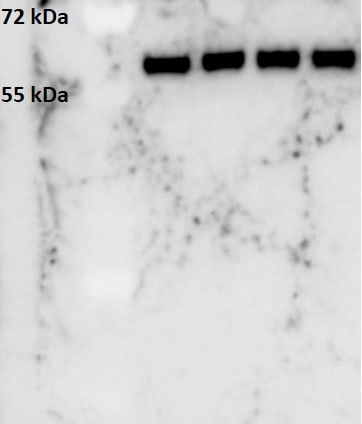
Western Blot: Calreticulin Antibody [NB600-103]
Western Blot: Calreticulin Antibody [NB600-103] - Detection of calreticulin in human kidney lysate using NB600-103 at 1:1,000. ECL exposure, 10 seconds.![Western Blot: Calreticulin Antibody [NB600-103]](https://resources.rndsystems.com/images/products/Calreticulin-Antibody-Western-Blot-NB600-103-img0004.jpg "Western Blot: Calreticulin Antibody [NB600-103]")
Western Blot: Calreticulin Antibody [NB600-103]
Western Blot: Calreticulin Antibody [NB600-103] - Analysis of HeLa and C6 cell lysate using calreticulin antibody at 1:1000.Applications for Calreticulin Antibody - BSA Free
Application
Recommended Usage
Immunocytochemistry/ Immunofluorescence
1:20-1:100. Use reported by customer review
Immunohistochemistry
1:20
Immunohistochemistry-Frozen
reported in scientific literature (PMID 26643212)
Immunohistochemistry-Paraffin
1:20
Knockdown Validated
1:2500
Simple Western
1:500
Western Blot
1:1000-1:10000
Application Notes
A band at approx. 55 kDa is seen in Western Blot. Note that Calreticulin has several reported isoforms that range from 48-62 kDa.
In Simple Western only 10 - 15 uL of the recommended dilution is used per data point.
See Simple Western Antibody Database for Simple Western validation: Tested in HeLa lysate 0.2 mg/mL, separated by Size, antibody dilution of 1:500, apparent MW was 45 kDa. Separated by Size-Wes, Sally Sue/Peggy Sue.
Reviewed Applications
Read 3 reviews rated 4.7 using NB600-103 in the following applications:
Formulation, Preparation, and Storage
Purification
Unpurified
Formulation
Whole antisera
Format
BSA Free
Preservative
0.1% Sodium Azide
Concentration
This product is unpurified. The exact concentration of antibody is not quantifiable.
Shipping
The product is shipped with polar packs. Upon receipt, store it immediately at the temperature recommended below.
Stability & Storage
Store at 4C short term. Aliquot and store at -20C long term. Avoid freeze-thaw cycles.
Background: Calreticulin
Given its role in multiple biological processes, it makes sense that calreticulin is implicated in both healthy and disease states. Studies have found that calreticulin mutations were present in patients with myeloproliferative neoplasms (MFN) and essential thrombocythaemia (ET) (4). Calreticulin expression is typically upregulated in most cancer lines, however it is downregulated in some tissues including cervical carcinomas, prostate cancer, and human colon adenocarcinoma (3). High expression of calreticulin on cancer cells is related to tumor cell phagocytosis and is often correlated with, and counteracted by, elevated CD47 expression to prevent cancer cell phagocytosis (3). Calreticulin mutations can serve as a major diagnostic biomarker for MFN and ET and additionally the calreticulin gene may be a potential target for cancer therapeutics (3,4).
References
1. Michalak, M., Groenendyk, J., Szabo, E., Gold, L. I., & Opas, M. (2009). Calreticulin, a multi-process calcium-buffering chaperone of the endoplasmic reticulum. The Biochemical Journal. https://doi.org/10.1042/BJ20081847
2. Fucikova, J., Spisek, R., Kroemer, G., & Galluzzi, L. (2021). Calreticulin and cancer. Cell Research. https://doi.org/10.1038/s41422-020-0383-9
3. Sun, J., Mu, H., Dai, K., & Yi, L. (2017). Calreticulin: a potential anti-cancer therapeutic target. Die Pharmazie. https://doi.org/10.1691/ph.2017.7031
4. Prins, D., Gonzalez Arias, C., Klampfl, T., Grinfeld, J., & Green, A. R. (2020). Mutant Calreticulin in the Myeloproliferative Neoplasms. HemaSphere. https://doi.org/10.1097/HS9.0000000000000333
Alternate Names
Autoantigen Ro, CALR, Calregulin, cC1qR, CRT, SSA, Vasostatin
Entrez Gene IDs
811 (Human)
Gene Symbol
CALR
UniProt
Additional Calreticulin Products
Product Documents for Calreticulin Antibody - BSA Free
Certificate of Analysis
To download a Certificate of Analysis, please enter a lot or batch number in the search box below.
Product Specific Notices for Calreticulin Antibody - BSA Free
This product is for research use only and is not approved for use in humans or in clinical diagnosis. Primary Antibodies are guaranteed for 1 year from date of receipt.
Related Research Areas
Citations for Calreticulin Antibody - BSA Free
 Powered by Bioz
Customer Reviews for Calreticulin Antibody - BSA Free (3)
4.7 out of 5
3 Customer Ratings
Have you used Calreticulin Antibody - BSA Free?
Submit a review and receive an Amazon gift card!
$25/€18/£15/$25CAN/¥2500 Yen for a review with an image
$10/€7/£6/$10CAN/¥1110 Yen for a review without an image
Submit a review
Customer Images
-(01-ml)_NB600-103_8191.bmp)
-(01-ml)_NB600-103_8186.jpg)
Showing
1
-
3 of
3 reviews
Showing All
Filter By:
-
Application: Western BlotSample Tested: Spinal cordSpecies: MouseVerified Customer | Posted 01/07/2019Careticulin band can be observed at about 60 kDa.Whole spinal cord extracts, 30 ug of protein loaded. Primary antibody: 1/500, one hour secondary antibody: 1/1000, one hour

-
Application: ImmunocytochemistrySample Tested:Species: MouseVerified Customer | Posted 06/11/2014IF Confocal analysis of 3T3 cells using Calreticulin antibody (NB600-103, 1:20).
-
Application: Western BlotSample Tested:Species: HumanVerified Customer | Posted 06/11/2014Western blot analysis of extracts from HeLa and C6 cells using Calreticulin antibody (NB600-103, 1:1000).
There are no reviews that match your criteria.
Protocols
View specific protocols for Calreticulin Antibody - BSA Free (NB600-103):
Western Blot Protocol
1. Perform SDS-PAGE (4-12%) on samples to be analyzed, loading 40 ug of total protein per lane.
2. Transfer proteins to Nitrocellulose according to the instructions provided by the manufacturer of the transfer apparatus.
3. Rinse membrane with dH2O and then stain the blot using ponceau S for 1-2 minutes to access the transfer of proteins onto the nitrocellulose membrane. Rinse the blot in water to remove excess stain and mark the lane locations and locations of molecular weight markers using a pencil.
4. Rinse the blot in TBS for approximately 5 minutes.
5. Block the membrane using 5% non-fat dry milk + 1% BSA in TBS for 1 hour at room temperature.
6. Rinse the membrane in dH2O and then wash the membrane in wash buffer [TBS + 0.1% Tween] 3 times for 10 minutes each.
7. Dilute the rabbit anti-calreticulin primary antibody (NB 600-103) in blocking buffer and incubate 1.5 hours at room temperature.
8. Rinse the membrane in dH2O and then wash the membrane in wash buffer [TBS + 0.1% Tween] 3 times for 10 minutes each.
9. Apply the diluted rabbit-IgG HRP-conjugated secondary antibody in blocking buffer (as per manufacturer's instructions) and incubate 1 hour at room temperature.
10. Wash the blot in wash buffer [TBS + 0.1% Tween] 3 times for 10 minutes each (this step can be repeated as required to reduce background).
11. Apply the detection reagent of choice in accordance with the manufacturer's instructions (Pierce's ECL).
Note: Tween-20 can be added to the blocking or antibody dilution buffer at a final concentration of 0.05-0.2%, provided it does not interfere with antibody-antigen binding.
Find general support by application which include: protocols, troubleshooting, illustrated assays, videos and webinars.
- Antigen Retrieval Protocol (PIER)
- Antigen Retrieval for Frozen Sections Protocol
- Appropriate Fixation of IHC/ICC Samples
- Cellular Response to Hypoxia Protocols
- Chromogenic IHC Staining of Formalin-Fixed Paraffin-Embedded (FFPE) Tissue Protocol
- Chromogenic Immunohistochemistry Staining of Frozen Tissue
- ClariTSA™ Fluorophore Kits
- Detection & Visualization of Antibody Binding
- Fluorescent IHC Staining of Frozen Tissue Protocol
- Graphic Protocol for Heat-induced Epitope Retrieval
- Graphic Protocol for the Preparation and Fluorescent IHC Staining of Frozen Tissue Sections
- Graphic Protocol for the Preparation and Fluorescent IHC Staining of Paraffin-embedded Tissue Sections
- Graphic Protocol for the Preparation of Gelatin-coated Slides for Histological Tissue Sections
- ICC Cell Smear Protocol for Suspension Cells
- ICC Immunocytochemistry Protocol Videos
- ICC for Adherent Cells
- IHC Sample Preparation (Frozen sections vs Paraffin)
- Immunocytochemistry (ICC) Protocol
- Immunocytochemistry Troubleshooting
- Immunofluorescence of Organoids Embedded in Cultrex Basement Membrane Extract
- Immunofluorescent IHC Staining of Formalin-Fixed Paraffin-Embedded (FFPE) Tissue Protocol
- Immunohistochemistry (IHC) and Immunocytochemistry (ICC) Protocols
- Immunohistochemistry Frozen Troubleshooting
- Immunohistochemistry Paraffin Troubleshooting
- Preparing Samples for IHC/ICC Experiments
- Preventing Non-Specific Staining (Non-Specific Binding)
- Primary Antibody Selection & Optimization
- Protocol for Heat-Induced Epitope Retrieval (HIER)
- Protocol for Making a 4% Formaldehyde Solution in PBS
- Protocol for VisUCyte™ HRP Polymer Detection Reagent
- Protocol for the Fluorescent ICC Staining of Cell Smears - Graphic
- Protocol for the Fluorescent ICC Staining of Cultured Cells on Coverslips - Graphic
- Protocol for the Preparation & Fixation of Cells on Coverslips
- Protocol for the Preparation and Chromogenic IHC Staining of Frozen Tissue Sections
- Protocol for the Preparation and Chromogenic IHC Staining of Frozen Tissue Sections - Graphic
- Protocol for the Preparation and Chromogenic IHC Staining of Paraffin-embedded Tissue Sections
- Protocol for the Preparation and Chromogenic IHC Staining of Paraffin-embedded Tissue Sections - Graphic
- Protocol for the Preparation and Fluorescent ICC Staining of Cells on Coverslips
- Protocol for the Preparation and Fluorescent ICC Staining of Non-adherent Cells
- Protocol for the Preparation and Fluorescent ICC Staining of Stem Cells on Coverslips
- Protocol for the Preparation and Fluorescent IHC Staining of Frozen Tissue Sections
- Protocol for the Preparation and Fluorescent IHC Staining of Paraffin-embedded Tissue Sections
- Protocol for the Preparation of Gelatin-coated Slides for Histological Tissue Sections
- Protocol for the Preparation of a Cell Smear for Non-adherent Cell ICC - Graphic
- R&D Systems Quality Control Western Blot Protocol
- TUNEL and Active Caspase-3 Detection by IHC/ICC Protocol
- The Importance of IHC/ICC Controls
- Troubleshooting Guide: Immunohistochemistry
- Troubleshooting Guide: Western Blot Figures
- Western Blot Conditions
- Western Blot Protocol
- Western Blot Protocol for Cell Lysates
- Western Blot Troubleshooting
- Western Blot Troubleshooting Guide
- View all Protocols, Troubleshooting, Illustrated assays and Webinars
Loading...