Caspase-3 Antibody (31A1067) - (Pro and Active) - BSA Free
Novus Biologicals | Catalog # NB100-56708
Key Product Details
Validated by
Biological Validation
Species Reactivity
Validated:
Human, Mouse, Rat, Porcine, Chicken, Chinese Hamster, Mammal
Cited:
Human, Mouse, Rat, Avian - Chicken, Hamster - Cricetulus (Chinese Hamster), Mammal, Rabbit
Applications
Validated:
Knockout Validated, Immunohistochemistry, Immunohistochemistry-Paraffin, Immunohistochemistry-Frozen, Immunohistochemistry Free-Floating, Western Blot, Immunoblotting, Flow Cytometry, Immunocytochemistry/ Immunofluorescence, Simple Western, Hematoxylin and Eosin Stain, Electron Microscopy, Knockdown Validated, CyTOF-ready
Cited:
Immunohistochemistry, Immunohistochemistry-Paraffin, Immunohistochemistry-Frozen, Immunohistochemistry Free-Floating, Western Blot, Immunoblotting, Flow Cytometry, Immunocytochemistry/ Immunofluorescence, Simple Western, Hematoxylin and Eosin Stain, IF/IHC, Electron Microscopy, Knockdown Validated
Label
Unconjugated
Antibody Source
Monoclonal Mouse IgG1 kappa Clone # 31A1067
Format
BSA Free
Loading...
Product Specifications
Immunogen
This Caspase-3 Antibody (31A1067) - (Pro and Active) was developed against full-length recombinant human caspase-3 protein.
Reactivity Notes
Chicken reactivity reported in scientific literature (PMID: 30298003).
Specificity
The antibody detects both pro Caspase-3 (~32 kDa) and the large subunit of the active/cleaved form (~14-21 kDa) of Caspase-3.
Clonality
Monoclonal
Host
Mouse
Isotype
IgG1 kappa
Theoretical MW
31.7 kDa.
Disclaimer note: The observed molecular weight of the protein may vary from the listed predicted molecular weight due to post translational modifications, post translation cleavages, relative charges, and other experimental factors.
Disclaimer note: The observed molecular weight of the protein may vary from the listed predicted molecular weight due to post translational modifications, post translation cleavages, relative charges, and other experimental factors.
Scientific Data Images for Caspase-3 Antibody (31A1067) - (Pro and Active) - BSA Free

Western Blot Detection of Pro and Active Caspase-3 in Whole Cell Protein From Treated Jurkat Cells
Image of Caspase-3 Antibody (31A1067) - (Pro and Active). Whole cell protein from Jurkat cells treated with and without 2 uM staurosporine as indicated was separated on a 4-15% gel by SDS-PAGE, transferred to 0.2 um PVDF membrane and blocked in 5% non-fat milk in TBST. The membrane was probed with 5 ug/ml anti-Caspase 3 in 1% milk, and detected with an anti-mouse HRP secondary antibody using a Femto sensitivity chemiluminescence reagent. Note the detection of both pro-caspase 3 at 35 kDa and the cleaved active caspase 3 at 15-17 kDa.
Immunohistochemical Staining of Caspase-3 in Paraffin Embedded Human Spleen
Tissue section of human spleen using 1:200 dilution of Caspase-3 antibody (clone 31A1067). The staining was developed with HRP labeled anti-mouse IgG secondary antibody and DAB reagent, and nuclei of cells were counter-stained with hematoxylin. This Caspase 3 antibody generated primarily a specific cytoplasmic staining in a subset of spleenocytes with some nuclear signal in a few cells.
Detection of Caspase-3 in HeLa Cell Lysate by Simple Western
Simple Western lane view shows a specific band for Caspase-3 Antibody (31A1067) - (Pro and Active) in 0.1 mg/ml of HeLa lysate. This experiment was performed under reducing conditions using the 12-230kDa separation system.
Immunohistochemical Analysis of Caspase-3 in Paraffin Embedded Human Bladder
Caspase-3 was detected in immersion fixed paraffin-embedded sections of human bladder tissue using 1:50 dilution of mouse aCaspase-3 Antibody (31A1067) - (Pro and Active) (NB100-56708), for 1 hour at room temperature followed by anti-mouse IgG VisUCyte HRP polymer(VC001). Tissue was stained using DAB (brown) and counterstained with hematoxylin (blue).
Western Blot Analysis of Caspase-3 in TSGH 8301 Cells After Multiple Treatment Types
Caspase-3-Antibody-31A1067-Pro-and-Active-Western-Blot-NB100-56708-img0044.jpg
Flow Cytometry of NIH3T3 Cells Stained with Alexa Fluor 700 Conjugated Caspase-3 Antibody
An intracellular stain was performed on NIH3T3 cells with Caspase-3 Antibody (31A1067) - (Pro and Active) Antibody NB100-56708AF700 (blue) and a matched isotype control (orange). Cells were fixed with 4% PFA and then permeabilized with 0.1% saponin. Cells were incubated in an antibody dilution of 10 ug/mL for 30 minutes at room temperature. Both antibodies were conjugated to Alexa Fluor 700.
Western Blotting of Caspase-3 in VP-16 Treated and Untreated Jurkat Cells
Lysates of Jurkat human acute T cell leukemia cell line untreated (-) or treated (+) with VP-16. PVDF membrane was probed with 0.1 ug/mL of mouse monoclonal Caspase-3 Antibody (31A1067) - (Pro and Active) (NB100-56708, Novus Biologicals) followed by 1:2000 dilution donkey anti-mouse IgG.
Flow Cytometry of HeLa Cells Stained with Caspase-3 Antibody
An intracellular stain was performed on HeLa cells with Caspase-3 Antibody (31A1067) - (Pro and Active) NB100-56708 (blue) and a matched isotype control (orange). Cells were fixed with 4% PFA and then permeablized with 0.1% saponin. Cells were incubated in an antibody dilution of 5 ug/mL for 30 minutes at room temperature, followed by mouse F(ab)2 IgG (H+L) APC-conjugated secondary antibody (F0101B, R&D Systems). - (Pro and Active) [IMGENEX: IMG-144A] [NB100-56708] -")
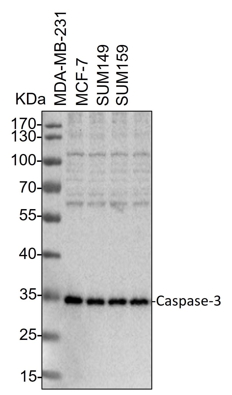
Western Blot: Mouse Monoclonal Caspase-3 Antibody (31A1067) - (Pro and Active) [IMGENEX: IMG-144A] [NB100-56708] -
Western Blot: Mouse Monoclonal Caspase-3 Antibody (31A1067) - (Pro and Active) [IMGENEX: IMG-144A] [NB100-56708] - Whole cell lysates from MDA-MB-231, MCF-7, SUM149 and SUM159 cells were loaded with 50 ug/lane. 10% SDS-PAGE. Caspase-3 Antibody (NB100-56708) was used for primary antibody: 1:2000, 4℃, overnight. Image from a verified customer review. - (Pro and Active) - BSA Free [NB100-56708] -")
Immunocytochemistry/ Immunofluorescence: Caspase-3 Antibody (31A1067) - (Pro and Active) - BSA Free [NB100-56708] -
Immunocytochemistry/ Immunofluorescence: Caspase-3 Antibody (31A1067) - (Pro and Active) - BSA Free [NB100-56708] - rOPN administration influenced the interaction & balance between Beclin 1 & Caspase‐3 at 24 h after SAH. Double immunofluorescence staining of Caspase‐3 & Beclin 1 in Sham group, SAH + Vehicle group & SAH + rOPN group at 24 h after SAH induction. Sample size is 9, n = 3 per group. Localization of Caspase‐3 can be cytoplasmic & nuclear. Staining in the nucleus is considered to be an indication of active Caspase‐3. The dashed lines & the red box on brain slice images indicate the locations observed. Vehicle, phosphate‐buffered saline; Scale bar = 50 μm Image collected & cropped by CiteAb from the following publication (https://pubmed.ncbi.nlm.nih.gov/31436915), licensed under a CC-BY license. Not internally tested by Novus Biologicals. - (Pro and Active) - BSA Free [NB100-56708] -")
Western Blot: Caspase-3 Antibody (31A1067) - (Pro and Active) - BSA Free [NB100-56708] -
Western Blot: Caspase-3 Antibody (31A1067) - (Pro and Active) - BSA Free [NB100-56708] - PEM induces YIPF2 upregulation & apoptosis in NSCLC cells.a, b H1792 (a) & H1299 (b) NSCLC cells were treated with PEM at various concentrations (0–10.0 μM) for 36 h. Cell lysates were analyzed by Western blotting with antibodies against YIPF2 & ACTB. c Overexpression of YIPF2 in H1299 cells in the presence or absence of PEM at 5.0 μM for 36 h. Cell lysates were analyzed by Western blotting with antibodies against YIPF2, CASP8, CASP3, PARP1 & ACTB. d Knockdown of YIPF2 expression by YIPF2–1 siRNA in A549 NSCLC cells in the presence or absence of PEM at 5.0 μM for 36 h. Cell lysates were analyzed by Western blotting with antibodies against YIPF2, CASP8, CASP3, PARP1 & ACTB. Image collected & cropped by CiteAb from the following publication (https://pubmed.ncbi.nlm.nih.gov/32303681), licensed under a CC-BY license. Not internally tested by Novus Biologicals. - (Pro and Active) - BSA Free [NB100-56708] -")
Immunocytochemistry/ Immunofluorescence: Caspase-3 Antibody (31A1067) - (Pro and Active) - BSA Free [NB100-56708] -
Immunocytochemistry/ Immunofluorescence: Caspase-3 Antibody (31A1067) - (Pro and Active) - BSA Free [NB100-56708] - Lipofection of top broccoletti-miR candidates does not influence basal & induced apoptosis.(A) BxPc-3 & Bx-Gem cells were transfected as described in Figure 3. Seventy-two hours later, the cells were stained with Annexin V-PE & 7-AAD, followed by FACS analysis. The percentage of Annexin V- & 7-AAD-positive cells is shown. (B) Lipofected BxPc-3 & Bx-Gem cells were stained with an antibody specific for the proliferation marker Ki-67 (green) or the apoptosis marker cleaved fragment of caspase-3 (red), which indicates apoptosis. Representative images at ×100 magnification are shown. The percentage of Ki-67- or caspase-3-positive cells was counted in 18 visual fields, & the means ± SD are shown in the diagram below. (C) BxPc-3 & Bx-Gem were lipofected as described above, & at 24 h later, the cells were treated with gemcitabine (10 nM) or were left untreated. Ninety-six hours after gemcitabine treatment, viability was determined by MTT assay. The data are presented as the means ± SD (**P < 0.01). Image collected & cropped by CiteAb from the following publication (https://pubmed.ncbi.nlm.nih.gov/32292571), licensed under a CC-BY license. Not internally tested by Novus Biologicals. - (Pro and Active) - BSA Free [NB100-56708] -")
Western Blot: Caspase-3 Antibody (31A1067) - (Pro and Active) - BSA Free [NB100-56708] -
Western Blot: Caspase-3 Antibody (31A1067) - (Pro and Active) - BSA Free [NB100-56708] - PEM induces apoptosis of NSCLC cells via YIPF2-TNFRSF10B axis.a, b A549 (a) & H1792 (b) cells were treated with PEM at 5.0 μM for the indicated times (0, 6, 12, 24, 36 & 48 h). Cell lysates were analyzed by Western blotting with antibodies against YIPF2, TNFRSF10B & ACTB. c A549 cells were treated with doxorubicin (DOX) at various concentrations (0–2.0 μM) for 18 h. Cell lysates were analyzed by Western blotting with antibodies against YIPF2, TNFRSF10B & ACTB. d Overexpression of YIPF2 in H1792 & H1299 cells in the presence or absence of PEM at 5.0 μM for 36 h. Cell lysates were analyzed by Western blotting with antibodies against YIPF2, TNFRSF10B & ACTB. e Knockdown of YIPF2 expression by YIPF2–1 siRNA in A549 & H1792 cells in the presence or absence of PEM at 5.0 μM for 36 h. Cell lysates were analyzed by Western blotting with antibodies against YIPF2, TNFRSF10B & ACTB. f Overexpression of YIPF2 in H1299 cells (left) or Knockdown of YIPF2 expression by YIPF2–1 siRNA in H1792 cells (right) in the presence or absence of PEM at 5.0 μM for 36 h. Cell lysates were analyzed by Western blotting with antibodies against YIPF2, TNFRSF10A & ACTB. g Three A549 cell lines (Ctrl, YIPF2, YIPF2 + siTNFRSF10B) in the presence or absence of PEM at 5.0 μM for 36 h. Cell lysates were analyzed by Western blotting with antibodies against YIPF2, TNFRSF10B, CASP8, CASP3 & ACTB. h Three H1299 cell lines (Ctrl, siYIPF2–1, siYIPF2–1 + TNFRSF10B (short isoform)) in the presence or absence of PEM at 5.0 μM for 36 h. Cell lysates were analyzed by Western blotting with antibodies against YIPF2, TNFRSF10B, CASP8, CASP3 & ACTB. Image collected & cropped by CiteAb from the following publication (https://pubmed.ncbi.nlm.nih.gov/32303681), licensed under a CC-BY license. Not internally tested by Novus Biologicals. - (Pro and Active) - BSA Free [NB100-56708] -")
Immunocytochemistry/ Immunofluorescence: Caspase-3 Antibody (31A1067) - (Pro and Active) - BSA Free [NB100-56708] -
Immunocytochemistry/ Immunofluorescence: Caspase-3 Antibody (31A1067) - (Pro and Active) - BSA Free [NB100-56708] - Cell numbers positively stained for NeuN, DCX, PCNA & caspase-3 in the subgranular region of the dentate gyrus in WT & RanBP9−/− (KO) mice.(A), DAPI-stained brain sections to show the highlighted subgranular zone within the dentate gyrus region of the hippocampus used for cell counts shown in B. (B), Representative brain sections stained with anti-NeuN, anti-DCX, anti-PCNA, anti-capsase-3 & counter stained with DAPI. Cell counting revealed significantly decreased numbers of NeuN positive cells in RanBP9−/− (KO) brains (22%) compared to WT controls. However DCX, PCNA & caspase positive cell numbers were not significantly altered. In each group, n = 3, data presented as mean± SEM. **, p<0.01 by Student’s t-test. Image collected & cropped by CiteAb from the following publication (https://pubmed.ncbi.nlm.nih.gov/23840553), licensed under a CC-BY license. Not internally tested by Novus Biologicals. - (Pro and Active) - BSA Free [NB100-56708] -")
Western Blot: Caspase-3 Antibody (31A1067) - (Pro and Active) - BSA Free [NB100-56708] -
Western Blot: Caspase-3 Antibody (31A1067) - (Pro and Active) - BSA Free [NB100-56708] - NFATc1 regulates DcR3 expression at a transcriptional level. (A,B) Immunoblot analysis of whole-cell lysates & quantitative real-time-PCR assaying relative DcR3 mRNA expression of ACHN & 769-P cells 24 h after treatment with cyclosporin A (CsA, 25 μM) or Tacrolimus (FK-506, 50 μM) (A); 48 h post transfection with NFATc1 or an empty vector control (neo) (B). Expression data were normalized to internal 18S rRNA expression (mean ± SEM; n=3; *p<0.05, **p<0.01, ***p<0.001; T-test). (C) Immunoblot analysis of cytoplasmic & nuclear fractions of ACHN & 769-P cells after treatment with LY294002 (50 μM), Everolimus (1 μM), or Cyclosporine A (25 μM). (D) Relative NFATc1–luciferase reporter activity of ACHN & 769-P cells 24 h post transfection with myrAkt or an empty vector control (neo) (mean ± SEM; n=3; *p<0.05, **p<0.01; T-test). Image collected & cropped by CiteAb from the following publication (https://pubmed.ncbi.nlm.nih.gov/24107265), licensed under a CC-BY license. Not internally tested by Novus Biologicals. - (Pro and Active) - BSA Free [NB100-56708] -")
Western Blot: Caspase-3 Antibody (31A1067) - (Pro and Active) - BSA Free [NB100-56708] -
Western Blot: Caspase-3 Antibody (31A1067) - (Pro and Active) - BSA Free [NB100-56708] - rOPN administration elevated the expression of autophagy‐related proteins while suppressing apoptosis in rat brain at 24 h after SAH. The effects of rOPN on expression levels of (A) Beclin 1, mean ± SD is 1.016 ± 0.2262 in Sham group, 2.874 ± 1.147 in SAH + Vehicle group, 4.963 ± 2.05 in SAH + rOPN group, F = 15.52, (B) ATG5, mean ± SD is 0.8908 ± 0.2545 in Sham group, 2.332 ± 0.6431 in SAH + Vehicle group, 4.364 ± 1.309 in SAH + rOPN group, F = 25.02, (C) LC3, mean ± SD is 1 ± 0.1845 in Sham group, 1.755 ± 0.3017 in SAH + Vehicle group, 2.7 ± 0.7957 in SAH + rOPN group, F = 17.23, (D) Cleaved Caspase‐3, mean ± SD is 1.008 ± 0.186 in Sham group, 4.112 ± 1.528 in SAH + Vehicle group, 2.291 ± 0.6268 in SAH + rOPN group, F = 15.86, (E) Bax, mean ± SD is 1.006 ± 0.321 in Sham group, 37.47 ± 10.86 in SAH + Vehicle group, 23.83 ± 8.143 in SAH + rOPN group, F = 33.13, (F) Bcl‐2, mean ± SD is 1.005 ± 0.2736 in Sham group, 0.2309 ± 0.1257 in SAH + Vehicle group, 0.7843 ± 0.2278 in SAH + rOPN group, F = 20.1, in the left hemisphere of rat brain at 24 h after SAH. Sample size is 18, n = 6 per group. Data were presented as mean ± SD. *P < .05, ***P < .001 vs Sham group; #P < .05, ##P <.01 vs SAH + Vehicle group Image collected & cropped by CiteAb from the following publication (https://pubmed.ncbi.nlm.nih.gov/31436915), licensed under a CC-BY license. Not internally tested by Novus Biologicals. - (Pro and Active) - BSA Free [NB100-56708] -")
Western Blot: Caspase-3 Antibody (31A1067) - (Pro and Active) - BSA Free [NB100-56708] -
Western Blot: Caspase-3 Antibody (31A1067) - (Pro and Active) - BSA Free [NB100-56708] - RAB8 suppresses PEM-induced apoptosis of NSCLC cells by promoting the removing of TNFRSF10B from plasma membrane to cytoplasm.a, b Knockdown of RAB8 expression by RAB8 siRNA in H1792 (a) & A549 (b) cells in the presence or absence of PEM at 5.0 μM for 36 h. The surface expression of TNFRSF10B was confirmed by flow cytometry analyses. c Overexpression of RAB8 in H1299 cells in the presence or absence of PEM at 5.0 μM for 36 h. Cell lysates were analyzed by Western blotting with antibodies against RAB8, TNFRSF10B, CASP8, CASP3, PARP1 & ACTB. d Knockdown of RAB8 expression by RAB8 siRNA in H1792 cells in the presence or absence of PEM at 5.0 μM for 36 h. Cell lysates were analyzed by Western blotting with antibodies against RAB8, TNFRSF10B, CASP8, CASP3, PARP1 & ACTB. Image collected & cropped by CiteAb from the following publication (https://pubmed.ncbi.nlm.nih.gov/32303681), licensed under a CC-BY license. Not internally tested by Novus Biologicals. - (Pro and Active) - BSA Free [NB100-56708] -")
Western Blot: Caspase-3 Antibody (31A1067) - (Pro and Active) - BSA Free [NB100-56708] -
Western Blot: Caspase-3 Antibody (31A1067) - (Pro and Active) - BSA Free [NB100-56708] - RAB8 suppresses PEM-induced apoptosis of NSCLC cells by promoting the removing of TNFRSF10B from plasma membrane to cytoplasm.a, b Knockdown of RAB8 expression by RAB8 siRNA in H1792 (a) & A549 (b) cells in the presence or absence of PEM at 5.0 μM for 36 h. The surface expression of TNFRSF10B was confirmed by flow cytometry analyses. c Overexpression of RAB8 in H1299 cells in the presence or absence of PEM at 5.0 μM for 36 h. Cell lysates were analyzed by Western blotting with antibodies against RAB8, TNFRSF10B, CASP8, CASP3, PARP1 & ACTB. d Knockdown of RAB8 expression by RAB8 siRNA in H1792 cells in the presence or absence of PEM at 5.0 μM for 36 h. Cell lysates were analyzed by Western blotting with antibodies against RAB8, TNFRSF10B, CASP8, CASP3, PARP1 & ACTB. Image collected & cropped by CiteAb from the following publication (https://pubmed.ncbi.nlm.nih.gov/32303681), licensed under a CC-BY license. Not internally tested by Novus Biologicals.Applications for Caspase-3 Antibody (31A1067) - (Pro and Active) - BSA Free
Application
Recommended Usage
Electron Microscopy
reported in scientific literature (PMID 27450722)
Flow Cytometry
reported in scientific literature (PMID 27429862)
Hematoxylin and Eosin Stain
reported in scientific literature (PMID 28186963)
Immunoblotting
reported in scientific literature (PMID 28500555)
Immunocytochemistry/ Immunofluorescence
reported in scientific literature (PMID 23840553)
Immunohistochemistry
1:10 - 1:500
Immunohistochemistry Free-Floating
reported in scientific literature (PMID 31771656)
Immunohistochemistry-Frozen
1:10 - 1:500. Use reported by customer review
Immunohistochemistry-Paraffin
1:10 - 1:500. Use reported in scientific literature (PMID 28500555)
Knockdown Validated
reported in scientific literature (PMID 32867814)
Simple Western
1:50
Western Blot
1 - 5 ug/ml
Application Notes
The large subunit of the cleaved form may appear as one or two or even as a stack of bands depending on the presence or absence of the Caspase-3 pro-domain. It is highly recommended that a maximum sensitivity ECL substrate (Femto sensitive) be used for efficient detection of this antibody in Western blot applications. In Simple Western only 10 - 15 ul of the recommended dilution is used per data point.
See Simple Western Antibody Database for Simple Western validation: Tested in Ovaries, separated by Size, antibody dilution of 1:200, apparent MW was 38,32,23 kDa. Separated by Size-Wes, Sally Sue/Peggy Sue. This antibody is CyTOF ready.
See Simple Western Antibody Database for Simple Western validation: Tested in Ovaries, separated by Size, antibody dilution of 1:200, apparent MW was 38,32,23 kDa. Separated by Size-Wes, Sally Sue/Peggy Sue. This antibody is CyTOF ready.
Reviewed Applications
Read 3 reviews rated 4.7 using NB100-56708 in the following applications:
Flow Cytometry Panel Builder
Bio-Techne Knows Flow Cytometry
Save time and reduce costly mistakes by quickly finding compatible reagents using the Panel Builder Tool.
Advanced Features
- Spectra Viewer - Custom analysis of spectra from multiple fluorochromes
- Spillover Popups - Visualize the spectra of individual fluorochromes
- Antigen Density Selector - Match fluorochrome brightness with antigen density

Formulation, Preparation, and Storage
Purification
Protein G purified
Formulation
PBS
Format
BSA Free
Preservative
0.05% Sodium Azide
Concentration
1 mg/ml
Shipping
The product is shipped with polar packs. Upon receipt, store it immediately at the temperature recommended below.
Stability & Storage
Store at 4C short term. Aliquot and store at -20C long term. Avoid freeze-thaw cycles.
Background: Caspase-3
References
1.Mu, N., Lei, Y., Wang, Y., Wang, Y., Duan, Q., Ma, G.,... Su, L. (2019). Inhibition of SIRT1/2 upregulates HSPA5 acetylation and induces pro-survival autophagy via ATF4-DDIT4-mTORC1 axis in human lung cancer cells. Apoptosis, 24(9-10), 798-811. doi:10.1007/s10495-019-01559-3
2.Sun, C. M., Enkhjargal, B., Reis, C., Zhou, K. R., Xie, Z. Y., Wu, L. Y.,... Zhang, J. H. (2019). Osteopontin attenuates early brain injury through regulating autophagy-apoptosis interaction after subarachnoid hemorrhage in rats. CNS Neurosci Ther, 25(10), 1162-1172. doi:10.1111/cns.13199
3.Louneva, N., Cohen, J. W., Han, L. Y., Talbot, K., Wilson, R. S., Bennett, D. A.,... Arnold, S. E. (2008). Caspase-3 is enriched in postsynaptic densities and increased in Alzheimer's disease. Am J Pathol, 173(5), 1488-1495. doi:10.2353/ajpath.2008.080434
Alternate Names
Apopain, CASP3, Caspase3, CPP32, LICE-1, YAMA
Gene Symbol
CASP3
Additional Caspase-3 Products
Product Documents for Caspase-3 Antibody (31A1067) - (Pro and Active) - BSA Free
Certificate of Analysis
To download a Certificate of Analysis, please enter a lot or batch number in the search box below.
Product Specific Notices for Caspase-3 Antibody (31A1067) - (Pro and Active) - BSA Free
This product is for research use only and is not approved for use in humans or in clinical diagnosis. Primary Antibodies are guaranteed for 1 year from date of receipt.
Related Research Areas
Citations for Caspase-3 Antibody (31A1067) - (Pro and Active) - BSA Free
 Powered by Bioz
Customer Reviews for Caspase-3 Antibody (31A1067) - (Pro and Active) - BSA Free (3)
4.7 out of 5
3 Customer Ratings
Have you used Caspase-3 Antibody (31A1067) - (Pro and Active) - BSA Free?
Submit a review and receive an Amazon gift card!
$25/€18/£15/$25CAN/¥2500 Yen for a review with an image
$10/€7/£6/$10CAN/¥1110 Yen for a review without an image
Submit a review
Customer Images
Showing
1
-
3 of
3 reviews
Showing All
Filter By:
-
Application: Western BlotVerified Customer | Posted 12/20/2023Western Blot: whole cell lysates from MDA-MB-231, MCF-7, SUM149 and SUM159 cells were loaded with 50 ug/lane. 10% SDS-PAGE. Caspase-3 Antibody (NB100-56708) was used for primary antibody: 1:2000, 4℃, overnight.

-
Application: Western BlotSample Tested: Jurkat human acute T cell leukemia cell lineSpecies: HumanVerified Customer | Posted 01/24/2022Jurkat human acute T cell leukemia cell linePrimary antibody dilution 1:1000
-
Application: Immunohistochemistry-FrozenSample Tested:Species: MouseVerified Customer | Posted 04/23/2014
There are no reviews that match your criteria.
Protocols
Find general support by application which include: protocols, troubleshooting, illustrated assays, videos and webinars.
- 7-Amino Actinomycin D (7-AAD) Cell Viability Flow Cytometry Protocol
- Antigen Retrieval Protocol (PIER)
- Antigen Retrieval for Frozen Sections Protocol
- Appropriate Fixation of IHC/ICC Samples
- Cellular Response to Hypoxia Protocols
- Chromogenic IHC Staining of Formalin-Fixed Paraffin-Embedded (FFPE) Tissue Protocol
- Chromogenic Immunohistochemistry Staining of Frozen Tissue
- ClariTSA™ Fluorophore Kits
- Detection & Visualization of Antibody Binding
- Extracellular Membrane Flow Cytometry Protocol
- Flow Cytometry Protocol for Cell Surface Markers
- Flow Cytometry Protocol for Staining Membrane Associated Proteins
- Flow Cytometry Staining Protocols
- Flow Cytometry Troubleshooting Guide
- Fluorescent IHC Staining of Frozen Tissue Protocol
- Graphic Protocol for Heat-induced Epitope Retrieval
- Graphic Protocol for the Preparation and Fluorescent IHC Staining of Frozen Tissue Sections
- Graphic Protocol for the Preparation and Fluorescent IHC Staining of Paraffin-embedded Tissue Sections
- Graphic Protocol for the Preparation of Gelatin-coated Slides for Histological Tissue Sections
- ICC Cell Smear Protocol for Suspension Cells
- ICC Immunocytochemistry Protocol Videos
- ICC for Adherent Cells
- IHC Sample Preparation (Frozen sections vs Paraffin)
- Immunocytochemistry (ICC) Protocol
- Immunocytochemistry Troubleshooting
- Immunofluorescence of Organoids Embedded in Cultrex Basement Membrane Extract
- Immunofluorescent IHC Staining of Formalin-Fixed Paraffin-Embedded (FFPE) Tissue Protocol
- Immunohistochemistry (IHC) and Immunocytochemistry (ICC) Protocols
- Immunohistochemistry Frozen Troubleshooting
- Immunohistochemistry Paraffin Troubleshooting
- Intracellular Flow Cytometry Protocol Using Alcohol (Methanol)
- Intracellular Flow Cytometry Protocol Using Detergents
- Intracellular Nuclear Staining Flow Cytometry Protocol Using Detergents
- Intracellular Staining Flow Cytometry Protocol Using Alcohol Permeabilization
- Intracellular Staining Flow Cytometry Protocol Using Detergents to Permeabilize Cells
- Preparing Samples for IHC/ICC Experiments
- Preventing Non-Specific Staining (Non-Specific Binding)
- Primary Antibody Selection & Optimization
- Propidium Iodide Cell Viability Flow Cytometry Protocol
- Protocol for Heat-Induced Epitope Retrieval (HIER)
- Protocol for Liperfluo
- Protocol for Making a 4% Formaldehyde Solution in PBS
- Protocol for VisUCyte™ HRP Polymer Detection Reagent
- Protocol for the Characterization of Human Th22 Cells
- Protocol for the Characterization of Human Th9 Cells
- Protocol for the Fluorescent ICC Staining of Cell Smears - Graphic
- Protocol for the Fluorescent ICC Staining of Cultured Cells on Coverslips - Graphic
- Protocol for the Preparation & Fixation of Cells on Coverslips
- Protocol for the Preparation and Chromogenic IHC Staining of Frozen Tissue Sections
- Protocol for the Preparation and Chromogenic IHC Staining of Frozen Tissue Sections - Graphic
- Protocol for the Preparation and Chromogenic IHC Staining of Paraffin-embedded Tissue Sections
- Protocol for the Preparation and Chromogenic IHC Staining of Paraffin-embedded Tissue Sections - Graphic
- Protocol for the Preparation and Fluorescent ICC Staining of Cells on Coverslips
- Protocol for the Preparation and Fluorescent ICC Staining of Non-adherent Cells
- Protocol for the Preparation and Fluorescent ICC Staining of Stem Cells on Coverslips
- Protocol for the Preparation and Fluorescent IHC Staining of Frozen Tissue Sections
- Protocol for the Preparation and Fluorescent IHC Staining of Paraffin-embedded Tissue Sections
- Protocol for the Preparation of Gelatin-coated Slides for Histological Tissue Sections
- Protocol for the Preparation of a Cell Smear for Non-adherent Cell ICC - Graphic
- Protocol: Annexin V and PI Staining by Flow Cytometry
- Protocol: Annexin V and PI Staining for Apoptosis by Flow Cytometry
- R&D Systems Quality Control Western Blot Protocol
- TUNEL and Active Caspase-3 Detection by IHC/ICC Protocol
- The Importance of IHC/ICC Controls
- Troubleshooting Guide: Fluorokine Flow Cytometry Kits
- Troubleshooting Guide: Immunohistochemistry
- Troubleshooting Guide: Western Blot Figures
- Western Blot Conditions
- Western Blot Protocol
- Western Blot Protocol for Cell Lysates
- Western Blot Troubleshooting
- Western Blot Troubleshooting Guide
- View all Protocols, Troubleshooting, Illustrated assays and Webinars
Loading...
Associated Pathways

