Caspase-3 Antibody (CPP32 4-1-18) - BSA Free
Novus Biologicals | Catalog # NB500-210
Key Product Details
Validated by
Biological Validation
Species Reactivity
Validated:
Human, Mouse, Rat
Cited:
Human, Mouse
Applications
Validated:
Immunohistochemistry, Immunohistochemistry-Paraffin, Immunohistochemistry-Frozen, Western Blot, Immunocytochemistry/ Immunofluorescence, Simple Western, Immunoprecipitation
Cited:
Immunohistochemistry-Paraffin, Western Blot, IF/IHC, IHC-F
Label
Unconjugated
Antibody Source
Monoclonal Mouse IgG2a Kappa Clone # CPP32 4-1-18
Format
BSA Free
Loading...
Product Specifications
Immunogen
This Caspase-3 Antibody (CPP32 4-1-18) was developed against full-length recombinant human Caspase 3 [UniProt# P42574].
Clonality
Monoclonal
Host
Mouse
Isotype
IgG2a Kappa
Theoretical MW
31.7 kDa.
Disclaimer note: The observed molecular weight of the protein may vary from the listed predicted molecular weight due to post translational modifications, post translation cleavages, relative charges, and other experimental factors.
Disclaimer note: The observed molecular weight of the protein may vary from the listed predicted molecular weight due to post translational modifications, post translation cleavages, relative charges, and other experimental factors.
Scientific Data Images for Caspase-3 Antibody (CPP32 4-1-18) - BSA Free
![Western Blot: Caspase-3 Antibody (CPP32 4-1-18)BSA Free [NB500-210]](https://resources.rndsystems.com/images/products/Caspase-3-Antibody-CPP32-4-1-18-Western-Blot-NB500-210-img0008.jpg "Western Blot: Caspase-3 Antibody (CPP32 4-1-18)BSA Free [NB500-210]")
![Simple Western: Caspase-3 Antibody (CPP32 4-1-18)BSA Free [NB500-210]](https://resources.rndsystems.com/images/products/Caspase-3-Antibody-CPP32-4-1-18-Simple-Western-NB500-210-img0005.jpg "Simple Western: Caspase-3 Antibody (CPP32 4-1-18)BSA Free [NB500-210]")
Simple Western: Caspase-3 Antibody (CPP32 4-1-18)BSA Free [NB500-210]
Simple Western: Caspase-3 Antibody (CPP32 4-1-18) [NB500-210] - Lane view shows a specific band for Caspase 3 in 0.5 mg/ml of Hek293 lysate. This experiment was performed under reducing conditions using the 12-230 kDa separation system.![Immunohistochemistry-Paraffin: Caspase-3 Antibody (CPP32 4-1-18) - BSA Free [NB500-210]](https://resources.rndsystems.com/images/products/Caspase-3-Antibody-CPP32-4-1-18-Immunohistochemistry-Paraffin-NB500-210-img0007.jpg "Immunohistochemistry-Paraffin: Caspase-3 Antibody (CPP32 4-1-18) - BSA Free [NB500-210]")
Immunohistochemistry-Paraffin: Caspase-3 Antibody (CPP32 4-1-18) - BSA Free [NB500-210]
Immunohistochemistry-Paraffin: Caspase-3 Antibody (CPP32 4-1-18) [NB500-210] - Analysis of a FFPE human spleen section using 1:200 dilution of. The staining was developed using HRP conjugated anti-mouse secondary antibody and DAB reagent. This Caspase 3 antibody generated a specific staining in the cytoplasm of various spleenocytes.![Immunohistochemistry-Paraffin: Caspase-3 Antibody (CPP32 4-1-18) - BSA Free [NB500-210]](https://resources.rndsystems.com/images/products/Caspase-3-Antibody-CPP32-4-1-18-Immunohistochemistry-Paraffin-NB500-210-img0006.jpg "Immunohistochemistry-Paraffin: Caspase-3 Antibody (CPP32 4-1-18) - BSA Free [NB500-210]")
Immunohistochemistry-Paraffin: Caspase-3 Antibody (CPP32 4-1-18) - BSA Free [NB500-210]
Immunohistochemistry-Paraffin: Caspase-3 Antibody (CPP32 4-1-18) [NB500-210] - Caspase-3 was detected in immersion fixed paraffin-embedded sections of human bladder 1:300 dilution of mouse monoclonal Caspase-3 Antibody (CPP32 4-1-18) (NB500-210, Novus Biologicals), for 1 hour at room temperature followed by anti-mouse IgG VisUCyte HRP polymer(VC001). Tissue was stained using DAB (brown) and counterstained with hematoxylin (blue).![Immunohistochemistry-Frozen: Caspase-3 Antibody (CPP32 4-1-18) - BSA Free [NB500-210]](https://resources.rndsystems.com/images/products/Caspase-3-Antibody-CPP32-4-1-18-BSA-Free-Immunohistochemistry-Frozen-NB500-210-img0009.jpg "Immunohistochemistry-Frozen: Caspase-3 Antibody (CPP32 4-1-18) - BSA Free [NB500-210]")
Immunohistochemistry-Frozen: Caspase-3 Antibody (CPP32 4-1-18) - BSA Free [NB500-210]
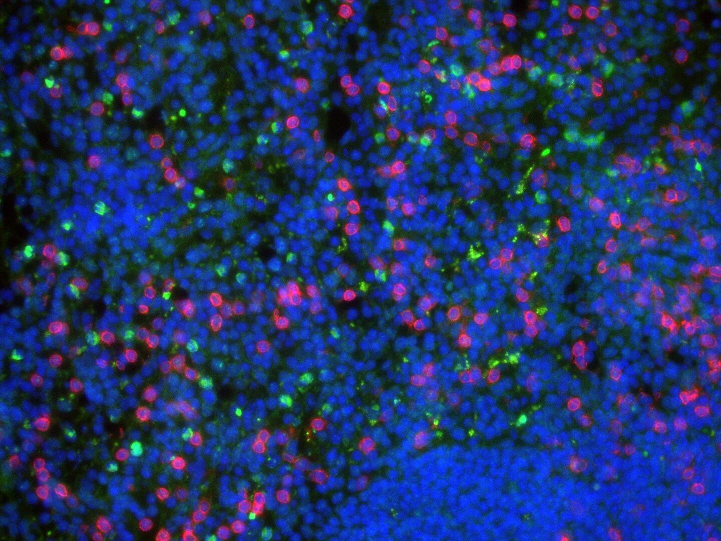
Immunohistochemistry-Frozen: Caspase-3 Antibody (CPP32 4-1-18) - BSA Free [NB500-210] - Analysis of murine spleen with Caspase-3 (green), CD3 (red) and DAPI (blue). Image from verified customer review.![Western Blot: Caspase-3 Antibody (CPP32 4-1-18)BSA Free [NB500-210]](https://resources.rndsystems.com/images/products/Caspase-3-Antibody-CPP32-4-1-18-Western-Blot-NB500-210-img0004.jpg "Western Blot: Caspase-3 Antibody (CPP32 4-1-18)BSA Free [NB500-210]")
Western Blot: Caspase-3 Antibody (CPP32 4-1-18)BSA Free [NB500-210]
Western Blot: Caspase-3 Antibody (CPP32 4-1-18) [NB500-210] - Detection of Caspase (19 and 35 kDa) from HEK293 cell extract using (NB500-210). Lanes 1 and 2 contain inactive and active Caspase, respectively.![Immunohistochemistry-Paraffin: Caspase-3 Antibody (CPP32 4-1-18) - BSA Free [NB500-210]](https://resources.rndsystems.com/images/products/Caspase-3-Antibody-CPP32-4-1-18-Immunohistochemistry-Paraffin-NB500-210-img0001.jpg "Immunohistochemistry-Paraffin: Caspase-3 Antibody (CPP32 4-1-18) - BSA Free [NB500-210]")
Immunohistochemistry-Paraffin: Caspase-3 Antibody (CPP32 4-1-18) - BSA Free [NB500-210]
Immunohistochemistry-Paraffin: Caspase-3 Antibody (CPP32 4-1-18) [NB500-210] - Rat epithelial cells of the tongue base. Antigen retrieval method: Citrate buffer.Applications for Caspase-3 Antibody (CPP32 4-1-18) - BSA Free
Application
Recommended Usage
Immunocytochemistry/ Immunofluorescence
2 ug/ml
Immunohistochemistry
1:200-1:500
Immunohistochemistry-Frozen
reported in scientific literature (PMID 31242448)
Immunohistochemistry-Paraffin
1:10-1:500
Immunoprecipitation
2 ug / mg lysate
Simple Western
1:2000
Western Blot
1:500-1:1000
Application Notes
A band is seen at ~32 kDa for the inactive form of Caspase 3 and ~17-22 kDa for the active form of Caspase 3 in Western Blot. (See protocol for additional information).
In Simple Western only 10 - 15 uL of the recommended dilution is used per data point.
See Simple Western Antibody Database for Simple Western validation: Tested in Hek293 lysate 0.5 mg/mL, separated by Size, antibody dilution of 1:2000, apparent MW was 40 kDa. Separated by Size-Wes, Sally Sue/Peggy Sue.
Reviewed Applications
Read 4 reviews rated 4 using NB500-210 in the following applications:
Formulation, Preparation, and Storage
Purification
Protein G purified
Formulation
PBS
Format
BSA Free
Preservative
0.02% Sodium Azide
Concentration
1.0 mg/ml
Shipping
The product is shipped with polar packs. Upon receipt, store it immediately at the temperature recommended below.
Stability & Storage
Store at 4C short term. Aliquot and store at -20C long term. Avoid freeze-thaw cycles.
Background: Caspase-3
References
1.Mu, N., Lei, Y., Wang, Y., Wang, Y., Duan, Q., Ma, G.,... Su, L. (2019). Inhibition of SIRT1/2 upregulates HSPA5 acetylation and induces pro-survival autophagy via ATF4-DDIT4-mTORC1 axis in human lung cancer cells. Apoptosis, 24(9-10), 798-811. doi:10.1007/s10495-019-01559-3
2.Sun, C. M., Enkhjargal, B., Reis, C., Zhou, K. R., Xie, Z. Y., Wu, L. Y.,... Zhang, J. H. (2019). Osteopontin attenuates early brain injury through regulating autophagy-apoptosis interaction after subarachnoid hemorrhage in rats. CNS Neurosci Ther, 25(10), 1162-1172. doi:10.1111/cns.13199
3.Louneva, N., Cohen, J. W., Han, L. Y., Talbot, K., Wilson, R. S., Bennett, D. A.,... Arnold, S. E. (2008). Caspase-3 is enriched in postsynaptic densities and increased in Alzheimer's disease. Am J Pathol, 173(5), 1488-1495. doi:10.2353/ajpath.2008.080434
Alternate Names
Apopain, CASP3, Caspase3, CPP32, LICE-1, YAMA
Gene Symbol
CASP3
UniProt
Additional Caspase-3 Products
Product Documents for Caspase-3 Antibody (CPP32 4-1-18) - BSA Free
Certificate of Analysis
To download a Certificate of Analysis, please enter a lot or batch number in the search box below.
Product Specific Notices for Caspase-3 Antibody (CPP32 4-1-18) - BSA Free
This product is for research use only and is not approved for use in humans or in clinical diagnosis. Primary Antibodies are guaranteed for 1 year from date of receipt.
Related Research Areas
Citations for Caspase-3 Antibody (CPP32 4-1-18) - BSA Free
 Powered by Bioz
Customer Reviews for Caspase-3 Antibody (CPP32 4-1-18) - BSA Free (4)
4 out of 5
4 Customer Ratings
Have you used Caspase-3 Antibody (CPP32 4-1-18) - BSA Free?
Submit a review and receive an Amazon gift card!
$25/€18/£15/$25CAN/¥2500 Yen for a review with an image
$10/€7/£6/$10CAN/¥1110 Yen for a review without an image
Submit a review
Customer Images
Showing
1
-
4 of
4 reviews
Showing All
Filter By:
-
Application: Immunohistochemistry-FrozenSample Tested: Mouse SpleenSpecies: MouseVerified Customer | Posted 12/05/2022Murine spleen with Caspase 3 (green), CD3 (red) and DAPI (blue)

-
Application: Western BlotSample Tested: Human cancer cell whole cell lysateSpecies: HumanVerified Customer | Posted 08/23/2015
-
Application: Western BlotSample Tested: Human HepatoblastomaSpecies: HumanVerified Customer | Posted 12/21/2011
-
Application: Western BlotSample Tested: HumanSpecies: HumanVerified Customer | Posted 11/21/2011
There are no reviews that match your criteria.
Protocols
View specific protocols for Caspase-3 Antibody (CPP32 4-1-18) - BSA Free (NB500-210):
Procedure Guide for NB 500-210 Monoclonal anti-Caspase 3
Western Protocol
1) Run a 10-*15% SDS polyacrylamide gel, loading ~20 ug of cell extract per lane.
2) Transfer the proteins to a membrane.
3) Block the membrane in PT-T20 [20 mM Tris-HCl, pH 7.4 / 150 mM NaCl/0.5% Tween 20] + 5% NFDM [non-fat dry milk], for 3 hour at room temperature (RT), shaking gently.
4) Rinse the membrane twice with PT-T20.
5) Incubate the membrane in anti-Caspase 3 [NB 500-210], diluted 1:500-1:1,000 in PT-T20 + 5% NFDM, for 60 minutes at RT.
6) Wash the membrane for 15 minutes, 3 times, in PT-T20 at RT.
7) Incubate the membrane in anti-mouse conjugated to HRP (secondary antibody), diluted in PT-T20 + 5% NFDM for 60 minutes at RT.
8) Wash the membrane for 15 minutes, 3 times, in PT-T20 at RT.
9) Develop membrane in chemiluminescent reagents, as instructed by kit-vendor.
*Positive control(s): Human kidney 293 cells
To Activate the Cell Extracts:
Bring the extract to a final concentration of 5 mM dATP.
Incubate the extracts at 37C for 15-30 minutes.
Find general support by application which include: protocols, troubleshooting, illustrated assays, videos and webinars.
- Antigen Retrieval Protocol (PIER)
- Antigen Retrieval for Frozen Sections Protocol
- Appropriate Fixation of IHC/ICC Samples
- Cellular Response to Hypoxia Protocols
- Chromogenic IHC Staining of Formalin-Fixed Paraffin-Embedded (FFPE) Tissue Protocol
- Chromogenic Immunohistochemistry Staining of Frozen Tissue
- ClariTSA™ Fluorophore Kits
- Detection & Visualization of Antibody Binding
- Fluorescent IHC Staining of Frozen Tissue Protocol
- Graphic Protocol for Heat-induced Epitope Retrieval
- Graphic Protocol for the Preparation and Fluorescent IHC Staining of Frozen Tissue Sections
- Graphic Protocol for the Preparation and Fluorescent IHC Staining of Paraffin-embedded Tissue Sections
- Graphic Protocol for the Preparation of Gelatin-coated Slides for Histological Tissue Sections
- ICC Cell Smear Protocol for Suspension Cells
- ICC Immunocytochemistry Protocol Videos
- ICC for Adherent Cells
- IHC Sample Preparation (Frozen sections vs Paraffin)
- Immunocytochemistry (ICC) Protocol
- Immunocytochemistry Troubleshooting
- Immunofluorescence of Organoids Embedded in Cultrex Basement Membrane Extract
- Immunofluorescent IHC Staining of Formalin-Fixed Paraffin-Embedded (FFPE) Tissue Protocol
- Immunohistochemistry (IHC) and Immunocytochemistry (ICC) Protocols
- Immunohistochemistry Frozen Troubleshooting
- Immunohistochemistry Paraffin Troubleshooting
- Immunoprecipitation Protocol
- Preparing Samples for IHC/ICC Experiments
- Preventing Non-Specific Staining (Non-Specific Binding)
- Primary Antibody Selection & Optimization
- Protocol for Heat-Induced Epitope Retrieval (HIER)
- Protocol for Making a 4% Formaldehyde Solution in PBS
- Protocol for VisUCyte™ HRP Polymer Detection Reagent
- Protocol for the Fluorescent ICC Staining of Cell Smears - Graphic
- Protocol for the Fluorescent ICC Staining of Cultured Cells on Coverslips - Graphic
- Protocol for the Preparation & Fixation of Cells on Coverslips
- Protocol for the Preparation and Chromogenic IHC Staining of Frozen Tissue Sections
- Protocol for the Preparation and Chromogenic IHC Staining of Frozen Tissue Sections - Graphic
- Protocol for the Preparation and Chromogenic IHC Staining of Paraffin-embedded Tissue Sections
- Protocol for the Preparation and Chromogenic IHC Staining of Paraffin-embedded Tissue Sections - Graphic
- Protocol for the Preparation and Fluorescent ICC Staining of Cells on Coverslips
- Protocol for the Preparation and Fluorescent ICC Staining of Non-adherent Cells
- Protocol for the Preparation and Fluorescent ICC Staining of Stem Cells on Coverslips
- Protocol for the Preparation and Fluorescent IHC Staining of Frozen Tissue Sections
- Protocol for the Preparation and Fluorescent IHC Staining of Paraffin-embedded Tissue Sections
- Protocol for the Preparation of Gelatin-coated Slides for Histological Tissue Sections
- Protocol for the Preparation of a Cell Smear for Non-adherent Cell ICC - Graphic
- R&D Systems Quality Control Western Blot Protocol
- TUNEL and Active Caspase-3 Detection by IHC/ICC Protocol
- The Importance of IHC/ICC Controls
- Troubleshooting Guide: Immunohistochemistry
- Troubleshooting Guide: Western Blot Figures
- Western Blot Conditions
- Western Blot Protocol
- Western Blot Protocol for Cell Lysates
- Western Blot Troubleshooting
- Western Blot Troubleshooting Guide
- View all Protocols, Troubleshooting, Illustrated assays and Webinars
FAQs for Caspase-3 Antibody (CPP32 4-1-18) - BSA Free
Showing
1
-
2 of
2 FAQs
Showing All
-
Q: I am looking for a mouse antibody against active caspase 3. This antibody you have is one of the few mouse anti-caspase 3 antibodies that I can find, but the information about it does not specify whether it is against the active form of the protein. I must have an antibody against active caspase 3, can you tell me whether this is the case for your antibody (NB500-210).
A: I have pulled up information about this antibody and it appears that it will detect both the active and non-active forms of the protein. I don't know if you are looking for one that is only specific to the active and will not detect the inactive, or if as long as it detects the active it will work.
-
Q: Which is the best retrieval buffer for this antibody to IHC? In addition, which is the dilution for it?
A:
Antigen retrieval should be performed using citrate buffer at a pH of 6.0. Our protocol for this can be found here: Antigen Retrieval Protocol.
-
Q: I am looking for a mouse antibody against active caspase 3. This antibody you have is one of the few mouse anti-caspase 3 antibodies that I can find, but the information about it does not specify whether it is against the active form of the protein. I must have an antibody against active caspase 3, can you tell me whether this is the case for your antibody (NB500-210).
A: I have pulled up information about this antibody and it appears that it will detect both the active and non-active forms of the protein. I don't know if you are looking for one that is only specific to the active and will not detect the inactive, or if as long as it detects the active it will work.
-
Q: Which is the best retrieval buffer for this antibody to IHC? In addition, which is the dilution for it?
A:
Antigen retrieval should be performed using citrate buffer at a pH of 6.0. Our protocol for this can be found here: Antigen Retrieval Protocol.
Loading...
Associated Pathways

