Caspase-9 Antibody (LAP6 96-2-22) - BSA Free
Novus Biologicals | Catalog # NB500-209
![Western Blot: Caspase-9 Antibody (LAP6 96-2-22)BSA Free [NB500-209]](https://resources.rndsystems.com/images/products/Caspase-9-Antibody-LAP6-96-2-22-Western-Blot-NB500-209-img0006.jpg "Western Blot: Caspase-9 Antibody (LAP6 96-2-22)BSA Free [NB500-209]")
Key Product Details
Validated by
Species Reactivity
Validated:
Cited:
Applications
Validated:
Cited:
Label
Antibody Source
Format
Product Specifications
Immunogen
Localization
Clonality
Host
Isotype
Scientific Data Images for Caspase-9 Antibody (LAP6 96-2-22) - BSA Free
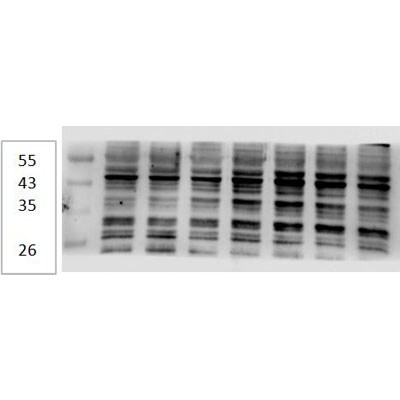
Western Blot: Caspase-9 Antibody (LAP6 96-2-22)BSA Free [NB500-209]
Western Blot: Caspase 9 Antibody (LAP6 96-2-22) [NB500-209] - Western blot analysis of Jurkat (A) and HeLa (B) cell lysate using Caspase 9 (NB500-209) antibody at 1:500.![Immunocytochemistry/ Immunofluorescence: Caspase-9 Antibody (LAP6 96-2-22) - BSA Free [NB500-209]](https://resources.rndsystems.com/images/products/Caspase-9-Antibody-LAP6-96-2-22-Immunocytochemistry-Immunofluorescence-NB500-209-img0003.jpg "Immunocytochemistry/ Immunofluorescence: Caspase-9 Antibody (LAP6 96-2-22) - BSA Free [NB500-209]")
Immunocytochemistry/ Immunofluorescence: Caspase-9 Antibody (LAP6 96-2-22) - BSA Free [NB500-209]
Immunocytochemistry/Immunofluorescence: Caspase 9 Antibody (LAP6 96-2-22) [NB500-209] - The Caspase 9 antibody was tested in HeLa cells against Dylight 488 (Green). Alpha-tubulin and nuclei were counterstained against Dylight 550 (Red) and DAPI (Blue).![Immunohistochemistry-Paraffin: Caspase-9 Antibody (LAP6 96-2-22) - BSA Free [NB500-209]](https://resources.rndsystems.com/images/products/Caspase-9-Antibody-LAP6-96-2-22-Immunohistochemistry-Paraffin-NB500-209-img0008.jpg "Immunohistochemistry-Paraffin: Caspase-9 Antibody (LAP6 96-2-22) - BSA Free [NB500-209]")
Immunohistochemistry-Paraffin: Caspase-9 Antibody (LAP6 96-2-22) - BSA Free [NB500-209]
Immunohistochemistry-Paraffin: Caspase 9 Antibody (LAP6 96-2-22) [NB500-209] - IHC analysis of paraffin-embedded human breast carcinomausing Caspase 9 antibody at 1:25. Image from verified customer review.![Western Blot: Caspase-9 Antibody (LAP6 96-2-22)BSA Free [NB500-209]](https://resources.rndsystems.com/images/products/Caspase-9-Antibody-LAP6-96-2-22-Western-Blot-NB500-209-img0002.jpg "Western Blot: Caspase-9 Antibody (LAP6 96-2-22)BSA Free [NB500-209]")
Western Blot: Caspase-9 Antibody (LAP6 96-2-22)BSA Free [NB500-209]
Western Blot: Caspase 9 Antibody (LAP6 96-2-22) [NB500-209] - The pro and active forms of Caspase 9 detected in active 293 cell lysate (lane 2). Lane 1: inactive 293 cell lysate.![Simple Western: Caspase-9 Antibody (LAP6 96-2-22)BSA Free [NB500-209]](https://resources.rndsystems.com/images/products/Caspase-9-Antibody-LAP6-96-2-22-Simple-Western-NB500-209-img0010.jpg "Simple Western: Caspase-9 Antibody (LAP6 96-2-22)BSA Free [NB500-209]")
Simple Western: Caspase-9 Antibody (LAP6 96-2-22)BSA Free [NB500-209]
Simple Western: Caspase-9 Antibody (LAP6 96-2-22) [NB500-209] - Western lane view shows lysates of Jurkat human acute T cell leukemia cell line untreated (-) or treated (+) with 1 mM Staurosporine (STS) for 3 hours, loaded at 0.2 mg/mL. A specific band was detected for Caspase 9 at approximately 53 kDa (as indicated) using 20 ug/mL of Mouse Anti-Caspase 9 Monoclonal Antibody (Catalog # NB500-209). This experiment was conducted under reducing conditions and using the 12-230 kDa separation system.Applications for Caspase-9 Antibody (LAP6 96-2-22) - BSA Free
Immunocytochemistry/ Immunofluorescence
Immunohistochemistry
Immunohistochemistry-Paraffin
Simple Western
Western Blot
Reviewed Applications
Read 4 reviews rated 4.8 using NB500-209 in the following applications:
Formulation, Preparation, and Storage
Purification
Formulation
Format
Preservative
Concentration
Shipping
Stability & Storage
Background: Caspase-9
Additional Caspase-9 Products
Product Documents for Caspase-9 Antibody (LAP6 96-2-22) - BSA Free
Certificate of Analysis
To download a Certificate of Analysis, please enter a lot or batch number in the search box below.
Product Specific Notices for Caspase-9 Antibody (LAP6 96-2-22) - BSA Free
This product is for research use only and is not approved for use in humans or in clinical diagnosis. Primary Antibodies are guaranteed for 1 year from date of receipt.
Related Research Areas
Citations for Caspase-9 Antibody (LAP6 96-2-22) - BSA Free
 Powered by Bioz
Customer Reviews for Caspase-9 Antibody (LAP6 96-2-22) - BSA Free (4)
Have you used Caspase-9 Antibody (LAP6 96-2-22) - BSA Free?
Submit a review and receive an Amazon gift card!
$25/€18/£15/$25CAN/¥2500 Yen for a review with an image
$10/€7/£6/$10CAN/¥1110 Yen for a review without an image
Submit a review
Customer Images
-(02-ml)_NB500-209_9681.jpg)
-(02-ml)_NB500-209_9676.jpg)
-
Application: Immunohistochemistry-ParaffinSample Tested:Species: HumanVerified Customer | Posted 08/29/2014IHC analysis of paraffin-embedded human breast carcinoma

-
Application: Western BlotSample Tested:Species: HumanVerified Customer | Posted 08/29/2014Western blot analysis of extracts from C6 cells at 1:1000
-
Application: Western BlotSample Tested: COLO-205 Cell LineSpecies: HumanVerified Customer | Posted 01/13/2012
-
Application: Western BlotSample Tested: Human cellSpecies: HumanVerified Customer | Posted 12/23/2011
There are no reviews that match your criteria.
Protocols
View specific protocols for Caspase-9 Antibody (LAP6 96-2-22) - BSA Free (NB500-209):
Immunocytochemistry Protocol
Culture cells to appropriate density in 35 mm culture dishes or 6-well plates.
1. Remove culture medium and add 10% formalin to the dish. Fix at room temperature for 30 minutes.
2. Remove the formalin and add ice cold methanol. Incubate for 5-10 minutes.
3. Remove methanol and add washing solution (i.e. PBS). Be sure to not let the specimen dry out. Wash three times for 10 minutes.
4. To block nonspecific antibody binding incubate in 10% normal goat serum from 1 hour to overnight at room temperature.
5. Add primary antibody at appropriate dilution and incubate at room temperature from 2 hours to overnight at room temperature.
6. Remove primary antibody and replace with washing solution. Wash three times for 10 minutes.
7. Add secondary antibody at appropriate dilution. Incubate for 1 hour at room temperature.
8. Remove antibody and replace with wash solution, then wash for 10 minutes. Add Hoechst 33258 to wash solution at 1:25,0000 and incubate for 10 minutes. Wash a third time for 10 minutes.
9. Cells can be viewed directly after washing. The plates can also be stored in PBS containing Azide covered in Parafilm (TM). Cells can also be cover-slipped using Fluoromount, with appropriate sealing.
*The above information is only intended as a guide. The researcher should determine what protocol best meets their needs. Please follow safe laboratory procedures.
Western Blot Protocol
1. Perform SDS-PAGE on samples to be analyzed, loading 10-25 ug of total protein per lane.
2. Transfer proteins to PVDF membrane according to the instructions provided by the manufacturer of the membrane and transfer apparatus.
3. Stain the membrane with Ponceau S (or similar product) to assess transfer success, and mark molecular weight standards where appropriate.
4. Rinse the blot TBS -0.05% Tween 20 (TBST).
5. Block the membrane in 5% Non-fat milk in TBST (blocking buffer) for at least 1 hour.
6. Wash the membrane in TBST three times for 10 minutes each.
7. Dilute primary antibody in blocking buffer and incubate overnight at 4C with gentle rocking.
8. Wash the membrane in TBST three times for 10 minutes each.
9. Incubate the membrane in diluted HRP conjugated secondary antibody in blocking buffer (as per manufacturer's instructions) for 1 hour at room temperature.
10. Wash the blot in TBST three times for 10 minutes each (this step can be repeated as required to reduce background).
11. Apply the detection reagent of choice in accordance with the manufacturers instructions.
Find general support by application which include: protocols, troubleshooting, illustrated assays, videos and webinars.
- Antigen Retrieval Protocol (PIER)
- Antigen Retrieval for Frozen Sections Protocol
- Appropriate Fixation of IHC/ICC Samples
- Cellular Response to Hypoxia Protocols
- Chromogenic IHC Staining of Formalin-Fixed Paraffin-Embedded (FFPE) Tissue Protocol
- Chromogenic Immunohistochemistry Staining of Frozen Tissue
- ClariTSA™ Fluorophore Kits
- Detection & Visualization of Antibody Binding
- Fluorescent IHC Staining of Frozen Tissue Protocol
- Graphic Protocol for Heat-induced Epitope Retrieval
- Graphic Protocol for the Preparation and Fluorescent IHC Staining of Frozen Tissue Sections
- Graphic Protocol for the Preparation and Fluorescent IHC Staining of Paraffin-embedded Tissue Sections
- Graphic Protocol for the Preparation of Gelatin-coated Slides for Histological Tissue Sections
- ICC Cell Smear Protocol for Suspension Cells
- ICC Immunocytochemistry Protocol Videos
- ICC for Adherent Cells
- IHC Sample Preparation (Frozen sections vs Paraffin)
- Immunocytochemistry (ICC) Protocol
- Immunocytochemistry Troubleshooting
- Immunofluorescence of Organoids Embedded in Cultrex Basement Membrane Extract
- Immunofluorescent IHC Staining of Formalin-Fixed Paraffin-Embedded (FFPE) Tissue Protocol
- Immunohistochemistry (IHC) and Immunocytochemistry (ICC) Protocols
- Immunohistochemistry Frozen Troubleshooting
- Immunohistochemistry Paraffin Troubleshooting
- Preparing Samples for IHC/ICC Experiments
- Preventing Non-Specific Staining (Non-Specific Binding)
- Primary Antibody Selection & Optimization
- Protocol for Heat-Induced Epitope Retrieval (HIER)
- Protocol for Making a 4% Formaldehyde Solution in PBS
- Protocol for VisUCyte™ HRP Polymer Detection Reagent
- Protocol for the Fluorescent ICC Staining of Cell Smears - Graphic
- Protocol for the Fluorescent ICC Staining of Cultured Cells on Coverslips - Graphic
- Protocol for the Preparation & Fixation of Cells on Coverslips
- Protocol for the Preparation and Chromogenic IHC Staining of Frozen Tissue Sections
- Protocol for the Preparation and Chromogenic IHC Staining of Frozen Tissue Sections - Graphic
- Protocol for the Preparation and Chromogenic IHC Staining of Paraffin-embedded Tissue Sections
- Protocol for the Preparation and Chromogenic IHC Staining of Paraffin-embedded Tissue Sections - Graphic
- Protocol for the Preparation and Fluorescent ICC Staining of Cells on Coverslips
- Protocol for the Preparation and Fluorescent ICC Staining of Non-adherent Cells
- Protocol for the Preparation and Fluorescent ICC Staining of Stem Cells on Coverslips
- Protocol for the Preparation and Fluorescent IHC Staining of Frozen Tissue Sections
- Protocol for the Preparation and Fluorescent IHC Staining of Paraffin-embedded Tissue Sections
- Protocol for the Preparation of Gelatin-coated Slides for Histological Tissue Sections
- Protocol for the Preparation of a Cell Smear for Non-adherent Cell ICC - Graphic
- R&D Systems Quality Control Western Blot Protocol
- TUNEL and Active Caspase-3 Detection by IHC/ICC Protocol
- The Importance of IHC/ICC Controls
- Troubleshooting Guide: Immunohistochemistry
- Troubleshooting Guide: Western Blot Figures
- Western Blot Conditions
- Western Blot Protocol
- Western Blot Protocol for Cell Lysates
- Western Blot Troubleshooting
- Western Blot Troubleshooting Guide
- View all Protocols, Troubleshooting, Illustrated assays and Webinars
FAQs for Caspase-9 Antibody (LAP6 96-2-22) - BSA Free
-
Q: Could I use HeLa cell lysate as a positive control for Caspase 9 primary antibodies in WB?
A: After looking through our testing it does appear you should be fine using HeLa as a positive control cell lysate for Caspase 9 in Western Blot, as we have used this sample successfully in our QC tests and shown expression of Caspase 9.
-
Q: Does NB500-209 both recognize human and rat active form of caspase 9 (subunit p35 or subunit p10)? Besides that please let me know if NB500-209 also recognize pro-form caspase 9?
A: Caspase 9 Antibody (LAP6 96-2-22) # NB500-209 is human specific and do not recognize rat samples. It is expected to detect pro as well as activated form of caspase 9. In our QC data using 293 cell lysates, we observed active subunit p35 only not subunit p10. Both of these antibodies were tested on lysates of 293 cells of human origin (also called Human Embryonic Kidney 293 cells). We did not validate them on rat samples as the antibodies are Human specific.
-
Q: Could I use HeLa cell lysate as a positive control for Caspase 9 primary antibodies in WB?
A: After looking through our testing it does appear you should be fine using HeLa as a positive control cell lysate for Caspase 9 in Western Blot, as we have used this sample successfully in our QC tests and shown expression of Caspase 9.
-
Q: Does NB500-209 both recognize human and rat active form of caspase 9 (subunit p35 or subunit p10)? Besides that please let me know if NB500-209 also recognize pro-form caspase 9?
A: Caspase 9 Antibody (LAP6 96-2-22) # NB500-209 is human specific and do not recognize rat samples. It is expected to detect pro as well as activated form of caspase 9. In our QC data using 293 cell lysates, we observed active subunit p35 only not subunit p10. Both of these antibodies were tested on lysates of 293 cells of human origin (also called Human Embryonic Kidney 293 cells). We did not validate them on rat samples as the antibodies are Human specific.
Associated Pathways

