Caveolin-1 Antibody (7C8) - BSA Free
Novus Biologicals | Catalog # NB100-615
Key Product Details
Species Reactivity
Validated:
Cited:
Applications
Validated:
Cited:
Label
Antibody Source
Format
Product Specifications
Immunogen
Localization
Marker
Specificity
Clonality
Host
Isotype
Theoretical MW
Disclaimer note: The observed molecular weight of the protein may vary from the listed predicted molecular weight due to post translational modifications, post translation cleavages, relative charges, and other experimental factors.
Scientific Data Images for Caveolin-1 Antibody (7C8) - BSA Free
![Western Blot: Caveolin-1 Antibody (7C8) [NB100-615]](https://resources.rndsystems.com/images/products/Caveolin-1-Antibody-7C8-Western-Blot-NB100-615-img0010.jpg "Western Blot: Caveolin-1 Antibody (7C8) [NB100-615]")
Western Blot: Caveolin-1 Antibody (7C8) [NB100-615]
Western Blot: Caveolin-1 Antibody (7C8) [NB100-615] - Detection of caveolin in 3T3 cell lysates (50 ug). Lanes 1 and 2: 1:4000. Lanes 3 and 4: 1:1000. Detection by ECL: 5 minute exposure.![Immunocytochemistry/ Immunofluorescence: Caveolin-1 Antibody (7C8) [NB100-615]](https://resources.rndsystems.com/images/products/Caveolin-1-Antibody-7C8-Immunocytochemistry-Immunofluorescence-NB100-615-img0014.jpg "Immunocytochemistry/ Immunofluorescence: Caveolin-1 Antibody (7C8) [NB100-615]")
Immunocytochemistry/ Immunofluorescence: Caveolin-1 Antibody (7C8) [NB100-615]
Caveolin-1-Antibody-7C8-Immunocytochemistry-Immunofluorescence-NB100-615-img0014.jpg![Immunohistochemistry-Frozen: Caveolin-1 Antibody (7C8) [NB100-615]](https://resources.rndsystems.com/images/products/Caveolin-1-Antibody-7C8-Immunohistochemistry-Frozen-NB100-615-img0015.jpg "Immunohistochemistry-Frozen: Caveolin-1 Antibody (7C8) [NB100-615]")
Immunohistochemistry-Frozen: Caveolin-1 Antibody (7C8) [NB100-615]
Immunohistochemistry-Frozen: Caveolin-1 Antibody (7C8) [NB100-615] - Immunofluorescence of human adipose tissue. Primary antibody at 1:250. IHC-Fr image submitted by a verified customer review.![Flow Cytometry: Caveolin-1 Antibody (7C8) [NB100-615]](https://resources.rndsystems.com/images/products/Caveolin-1-Antibody-7C8-Flow-Cytometry-NB100-615-img0012.jpg "Flow Cytometry: Caveolin-1 Antibody (7C8) [NB100-615]")
Flow Cytometry: Caveolin-1 Antibody (7C8) [NB100-615]
Flow Cytometry: Caveolin-1 Antibody (7C8) [NB100-615] - An intracellular stain was performed on HeLa cells with Caveolin-1 Antibody (7C8) NB100-615APC (blue) and a matched isotype control (orange). Cells were fixed with 4% PFA and then permeabilized with 0.1% saponin. Cells were incubated in an antibody dilution of 1 ug/mL for 30 minutes at room temperature. Both antibodies were conjugated to allophycocyanin.![Immunocytochemistry/ Immunofluorescence: Caveolin-1 Antibody (7C8) [NB100-615]](https://resources.rndsystems.com/images/products/Caveolin-1-Antibody-7C8-Immunocytochemistry-Immunofluorescence-NB100-615-img0009.jpg "Immunocytochemistry/ Immunofluorescence: Caveolin-1 Antibody (7C8) [NB100-615]")
Immunocytochemistry/ Immunofluorescence: Caveolin-1 Antibody (7C8) [NB100-615]
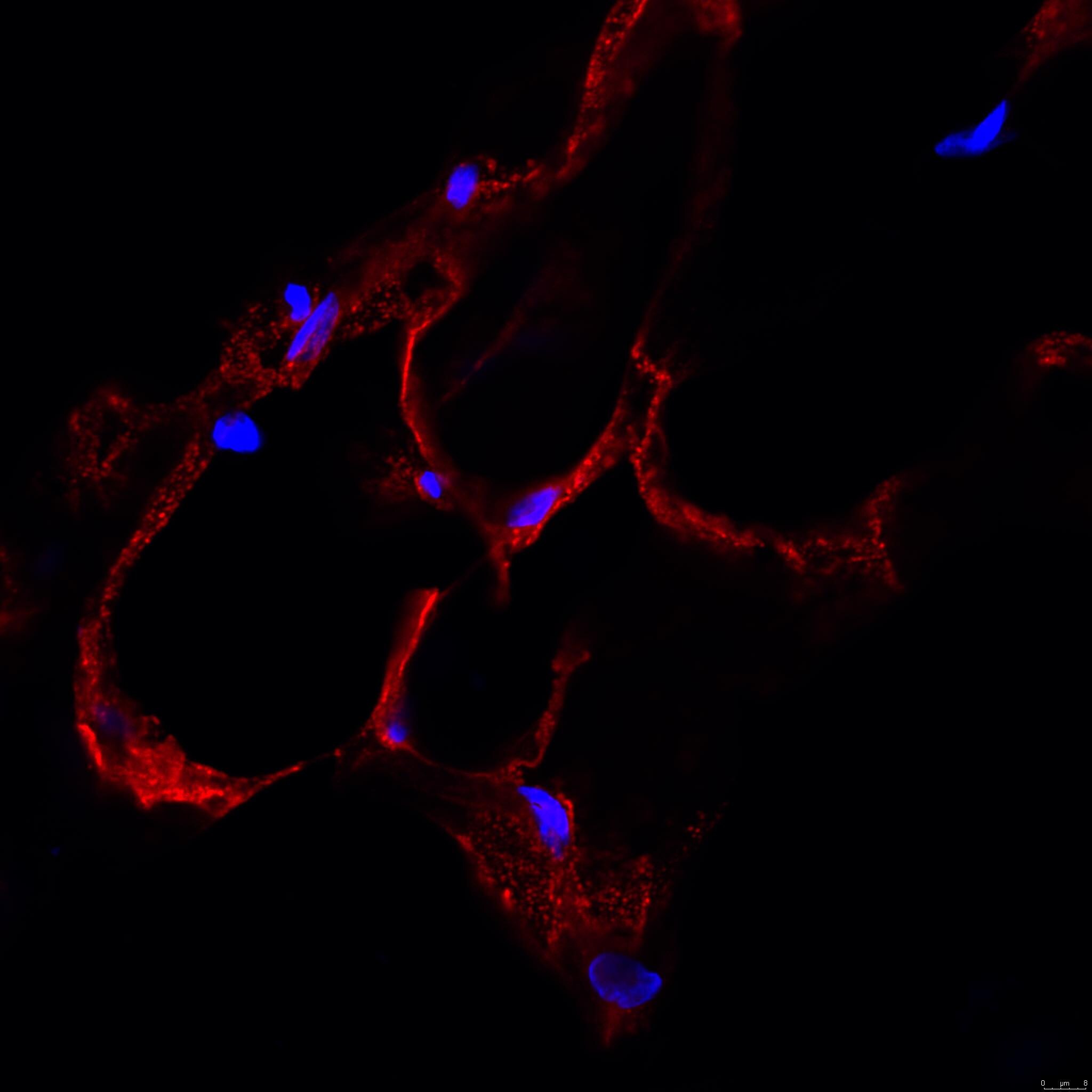
Immunocytochemistry/Immunofluorescence: Caveolin-1 Antibody (7C8) [NB100-615] - Antibody at 1:200 dilution (ON incubation) on EaHy926 endothelial cell line showing a clear localization in lipid raft/membrane. Photo courtesy of Alberto Davalos, Yale School of Medicine.![Immunocytochemistry/ Immunofluorescence: Caveolin-1 Antibody (7C8) [NB100-615]](https://resources.rndsystems.com/images/products/Caveolin-1-Antibody-7C8-Immunocytochemistry-Immunofluorescence-NB100-615-img0013.jpg "Immunocytochemistry/ Immunofluorescence: Caveolin-1 Antibody (7C8) [NB100-615]")
Immunocytochemistry/ Immunofluorescence: Caveolin-1 Antibody (7C8) [NB100-615]
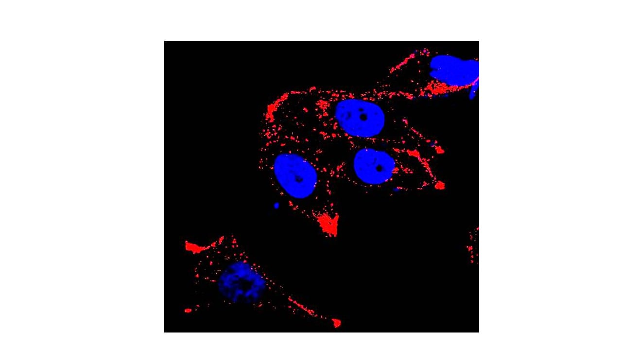
Immunocytochemistry/Immunofluorescence: Caveolin-1 Antibody (7C8) [NB100-615] - HeLa cells were fixed for 10 minutes using 10% formalin and then permeabilized for 5 minutes using 1X PBS + 0.05% Triton X-100. The cells were incubated with anti-Caveolin-1 [7C8] conjugated to Alexa Fluor 488 (NB100-615AF488) at 20 ug/mL for 1 hour at room temperature. Nuclei were counterstained with DAPI (Blue). Cells were imaged using a 40X objective.![Flow Cytometry: Caveolin-1 Antibody (7C8) [NB100-615]](https://resources.rndsystems.com/images/products/Caveolin-1-Antibody-7C8-Flow-Cytometry-NB100-615-img0008.jpg "Flow Cytometry: Caveolin-1 Antibody (7C8) [NB100-615]")
Flow Cytometry: Caveolin-1 Antibody (7C8) [NB100-615]
Flow Cytometry: Caveolin-1 Antibody (7C8) [NB100-615] - Intracellular flow cytometric staining of 1 x 10^6 CHO (A) and HEK-293 (B) cells using Caveolin 1 antibody (dark blue). Isotype control shown in orange. An antibody concentration of 1 ug/1x10^6 cells was used.![Flow Cytometry: Caveolin-1 Antibody (7C8) [NB100-615]](https://resources.rndsystems.com/images/products/Caveolin-1-Antibody-7C8-Flow-Cytometry-NB100-615-img0011.jpg "Flow Cytometry: Caveolin-1 Antibody (7C8) [NB100-615]")
Flow Cytometry: Caveolin-1 Antibody (7C8) [NB100-615]
Flow Cytometry: Caveolin-1 Antibody (7C8) [NB100-615] - An intracellular stain was performed on HeLa cells with Caveolin-1 Antibody (7C8) NB100-615 (blue) and a matched isotype control (orange). Cells were fixed with 4% PFA and then permeablized with 0.1% saponin. Cells were incubated in an antibody dilution of 5 ug/mL for 30 minutes at room temperature, followed by mouse F(ab)2 IgG (H+L) APC-conjugated secondary antibody (F0101B, R&D Systems).Applications for Caveolin-1 Antibody (7C8) - BSA Free
Flow Cytometry
Immunoblotting
Immunocytochemistry/ Immunofluorescence
Immunohistochemistry
Immunohistochemistry-Frozen
Immunohistochemistry-Paraffin
Immunoprecipitation
Western Blot
Reviewed Applications
Read 3 reviews rated 5 using NB100-615 in the following applications:
Flow Cytometry Panel Builder
Bio-Techne Knows Flow Cytometry
Save time and reduce costly mistakes by quickly finding compatible reagents using the Panel Builder Tool.
Advanced Features
- Spectra Viewer - Custom analysis of spectra from multiple fluorochromes
- Spillover Popups - Visualize the spectra of individual fluorochromes
- Antigen Density Selector - Match fluorochrome brightness with antigen density

Formulation, Preparation, and Storage
Purification
Formulation
Format
Preservative
Concentration
Shipping
Stability & Storage
Background: Caveolin-1
Alternate Names
Gene Symbol
Additional Caveolin-1 Products
Product Documents for Caveolin-1 Antibody (7C8) - BSA Free
Certificate of Analysis
To download a Certificate of Analysis, please enter a lot or batch number in the search box below.
Product Specific Notices for Caveolin-1 Antibody (7C8) - BSA Free
This product is for research use only and is not approved for use in humans or in clinical diagnosis. Primary Antibodies are guaranteed for 1 year from date of receipt.
Related Research Areas
Citations for Caveolin-1 Antibody (7C8) - BSA Free
 Powered by Bioz
Customer Reviews for Caveolin-1 Antibody (7C8) - BSA Free (3)
Have you used Caveolin-1 Antibody (7C8) - BSA Free?
Submit a review and receive an Amazon gift card!
$25/€18/£15/$25CAN/¥2500 Yen for a review with an image
$10/€7/£6/$10CAN/¥1110 Yen for a review without an image
Submit a review
Customer Images
-
Application: Immunohistochemistry-FrozenSample Tested: Adipose tissueSpecies: HumanVerified Customer | Posted 03/05/2021Immunofluorescence of human adipose tissueAntibody dilution 1:250

-
Application: ImmunocytochemistrySample Tested: Human prostate cancer cell lineSpecies: HumanVerified Customer | Posted 08/12/2015Immunocytochemistry of caveolin 1
-
Application: ImmunofluorescenceSample Tested: See PMID: 24348251Species: HumanVerified Customer | Posted 12/19/2014
There are no reviews that match your criteria.
Protocols
View specific protocols for Caveolin-1 Antibody (7C8) - BSA Free (NB100-615):
Western Blot Protocol
1. Perform SDS-PAGE (4-12%) on samples to be analyzed, loading 50ug of total protein per lane.
2. Transfer proteins to Nitrocellulose according to the instructions provided by the manufacturer of the transfer apparatus.
3. Stain the blot using ponceau S for 1-2 minutes to access the transfer of proteins onto the nitrocellulose membrane. Rinse the blot in water to remove excess stain and mark the lane locations and locations of molecular weight markers using a pencil.
4. Rinse the blot in TBS for approximately 5 minutes.
5. Block the membrane using 5% non-fat dry milk in TBS for 1 hour.
6. Dilute the mouse anti-Caveolin primary antibody (NB 100-615) in blocking buffer and incubate 3 hours at room temperature.
7. Wash the membrane in water for 5 minutes and apply the diluted mouse-IgG HRP-conjugated secondary antibody in blocking buffer (as per manufacturer's instructions) and incubate 1 hour at room temperature.
8. Wash the blot in TBS containing 0.05-0.1% Tween-20 for 10-20 minutes.
9. Wash the blot in type I water for an additional 10-20 minutes (this step can be repeated as required to reduce background).
10. Apply the detection reagent of choice in accordance with the manufacturer's instructions (Amersham's ECL is the standard reagent used at Novus Biologicals).
Note: Tween-20 can be added to the blocking buffer at a final concentration of 0.05-0.2%, provided it does not interfere with antibody-antigen binding.
Find general support by application which include: protocols, troubleshooting, illustrated assays, videos and webinars.
- 7-Amino Actinomycin D (7-AAD) Cell Viability Flow Cytometry Protocol
- Antigen Retrieval Protocol (PIER)
- Antigen Retrieval for Frozen Sections Protocol
- Appropriate Fixation of IHC/ICC Samples
- Cellular Response to Hypoxia Protocols
- Chromogenic IHC Staining of Formalin-Fixed Paraffin-Embedded (FFPE) Tissue Protocol
- Chromogenic Immunohistochemistry Staining of Frozen Tissue
- ClariTSA™ Fluorophore Kits
- Detection & Visualization of Antibody Binding
- Extracellular Membrane Flow Cytometry Protocol
- Flow Cytometry Protocol for Cell Surface Markers
- Flow Cytometry Protocol for Staining Membrane Associated Proteins
- Flow Cytometry Staining Protocols
- Flow Cytometry Troubleshooting Guide
- Fluorescent IHC Staining of Frozen Tissue Protocol
- Graphic Protocol for Heat-induced Epitope Retrieval
- Graphic Protocol for the Preparation and Fluorescent IHC Staining of Frozen Tissue Sections
- Graphic Protocol for the Preparation and Fluorescent IHC Staining of Paraffin-embedded Tissue Sections
- Graphic Protocol for the Preparation of Gelatin-coated Slides for Histological Tissue Sections
- ICC Cell Smear Protocol for Suspension Cells
- ICC Immunocytochemistry Protocol Videos
- ICC for Adherent Cells
- IHC Sample Preparation (Frozen sections vs Paraffin)
- Immunocytochemistry (ICC) Protocol
- Immunocytochemistry Troubleshooting
- Immunofluorescence of Organoids Embedded in Cultrex Basement Membrane Extract
- Immunofluorescent IHC Staining of Formalin-Fixed Paraffin-Embedded (FFPE) Tissue Protocol
- Immunohistochemistry (IHC) and Immunocytochemistry (ICC) Protocols
- Immunohistochemistry Frozen Troubleshooting
- Immunohistochemistry Paraffin Troubleshooting
- Immunoprecipitation Protocol
- Intracellular Flow Cytometry Protocol Using Alcohol (Methanol)
- Intracellular Flow Cytometry Protocol Using Detergents
- Intracellular Nuclear Staining Flow Cytometry Protocol Using Detergents
- Intracellular Staining Flow Cytometry Protocol Using Alcohol Permeabilization
- Intracellular Staining Flow Cytometry Protocol Using Detergents to Permeabilize Cells
- Preparing Samples for IHC/ICC Experiments
- Preventing Non-Specific Staining (Non-Specific Binding)
- Primary Antibody Selection & Optimization
- Propidium Iodide Cell Viability Flow Cytometry Protocol
- Protocol for Heat-Induced Epitope Retrieval (HIER)
- Protocol for Liperfluo
- Protocol for Making a 4% Formaldehyde Solution in PBS
- Protocol for VisUCyte™ HRP Polymer Detection Reagent
- Protocol for the Characterization of Human Th22 Cells
- Protocol for the Characterization of Human Th9 Cells
- Protocol for the Fluorescent ICC Staining of Cell Smears - Graphic
- Protocol for the Fluorescent ICC Staining of Cultured Cells on Coverslips - Graphic
- Protocol for the Preparation & Fixation of Cells on Coverslips
- Protocol for the Preparation and Chromogenic IHC Staining of Frozen Tissue Sections
- Protocol for the Preparation and Chromogenic IHC Staining of Frozen Tissue Sections - Graphic
- Protocol for the Preparation and Chromogenic IHC Staining of Paraffin-embedded Tissue Sections
- Protocol for the Preparation and Chromogenic IHC Staining of Paraffin-embedded Tissue Sections - Graphic
- Protocol for the Preparation and Fluorescent ICC Staining of Cells on Coverslips
- Protocol for the Preparation and Fluorescent ICC Staining of Non-adherent Cells
- Protocol for the Preparation and Fluorescent ICC Staining of Stem Cells on Coverslips
- Protocol for the Preparation and Fluorescent IHC Staining of Frozen Tissue Sections
- Protocol for the Preparation and Fluorescent IHC Staining of Paraffin-embedded Tissue Sections
- Protocol for the Preparation of Gelatin-coated Slides for Histological Tissue Sections
- Protocol for the Preparation of a Cell Smear for Non-adherent Cell ICC - Graphic
- Protocol: Annexin V and PI Staining by Flow Cytometry
- Protocol: Annexin V and PI Staining for Apoptosis by Flow Cytometry
- R&D Systems Quality Control Western Blot Protocol
- TUNEL and Active Caspase-3 Detection by IHC/ICC Protocol
- The Importance of IHC/ICC Controls
- Troubleshooting Guide: Fluorokine Flow Cytometry Kits
- Troubleshooting Guide: Immunohistochemistry
- Troubleshooting Guide: Western Blot Figures
- Western Blot Conditions
- Western Blot Protocol
- Western Blot Protocol for Cell Lysates
- Western Blot Troubleshooting
- Western Blot Troubleshooting Guide
- View all Protocols, Troubleshooting, Illustrated assays and Webinars
FAQs for Caveolin-1 Antibody (7C8) - BSA Free
-
Q: What is the sodium azide percentage for this product?
A: 0.05% sodium azide