CCL2/MCP1 Antibody - BSA Free
Novus Biologicals | Catalog # NBP1-07035
![Western Blot: CCL2/MCP1 AntibodyBSA Free [NBP1-07035]](https://resources.rndsystems.com/images/products/CCL2-MCP1-Antibody-Western-Blot-NBP1-07035-img0014.jpg "Western Blot: CCL2/MCP1 AntibodyBSA Free [NBP1-07035]")
Key Product Details
Validated by
Biological Validation
Species Reactivity
Validated:
Human, Mouse, Rat
Cited:
Human, Mouse, Rat
Applications
Validated:
Immunohistochemistry, Immunohistochemistry-Paraffin, Western Blot, Block/Neutralize, Immunocytochemistry/ Immunofluorescence
Cited:
Immunohistochemistry, Immunohistochemistry-Paraffin, Immunohistochemistry-Frozen, Western Blot, Block/Neutralize, Immunocytochemistry/ Immunofluorescence, IF/IHC
Label
Unconjugated
Antibody Source
Polyclonal Rabbit IgG
Format
BSA Free
Loading...
Product Specifications
Immunogen
Synthetic peptide made to an internal portion of rat MCP1 (within residues 15-40). [Swiss-Prot# P14844]
Localization
Secreted, secretory vesicles
Clonality
Polyclonal
Host
Rabbit
Isotype
IgG
Theoretical MW
16 kDa.
Disclaimer note: The observed molecular weight of the protein may vary from the listed predicted molecular weight due to post translational modifications, post translation cleavages, relative charges, and other experimental factors.
Disclaimer note: The observed molecular weight of the protein may vary from the listed predicted molecular weight due to post translational modifications, post translation cleavages, relative charges, and other experimental factors.
Scientific Data Images for CCL2/MCP1 Antibody - BSA Free
![Immunohistochemistry-Paraffin: CCL2/MCP1 Antibody - BSA Free [NBP1-07035]](https://resources.rndsystems.com/images/products/CCL2-MCP1-Antibody-Immunohistochemistry-Paraffin-NBP1-07035-img0007.jpg "Immunohistochemistry-Paraffin: CCL2/MCP1 Antibody - BSA Free [NBP1-07035]")
Immunohistochemistry-Paraffin: CCL2/MCP1 Antibody - BSA Free [NBP1-07035]
Immunohistochemistry-Paraffin: CCL2/MCP1 Antibody [NBP1-07035] - IHC analysis of a formalin-fixed paraffin-embedded (FFPE) human breast carcinoma tissue section using 1:1000 dilution of CCL2/MCP1 antibody (NBP1-07035) on a Bond Rx autostainer (Leica Biosystems). The assay involved 20 minutes of heat induced antigen retrieval (HIER) with 10mM sodium citrate buffer (pH 6.0) and endogenous peroxidase quenching using peroxide block. The sections were incubated with primary antibody for 30 minutes. Bond Polymer Refine Detection (Leica Biosystems) and DAB were used for signal detection which followed counterstaining with hematoxylin. Whole slide scanning and capturing of representative images (20X) were performed using Aperio AT2 (Leica Biosystems). This antibody generated a diffused cytoplasmic staining of CCL2 antigen in the cancer cells, stromal cells as well as the endothelial cells. The stroma itself showed a weak immunopositivity for CCL2. Staining was performed by Histowiz.![Western Blot: CCL2/MCP1 AntibodyBSA Free [NBP1-07035]](https://resources.rndsystems.com/images/products/CCL2-MCP1-Antibody-Western-Blot-NBP1-07035-img0013.jpg "Western Blot: CCL2/MCP1 AntibodyBSA Free [NBP1-07035]")
![Immunocytochemistry/ Immunofluorescence: CCL2/MCP1 Antibody - BSA Free [NBP1-07035]](https://resources.rndsystems.com/images/products/CCL2-MCP1-Antibody-Immunocytochemistry-Immunofluorescence-NBP1-07035-img0006.jpg "Immunocytochemistry/ Immunofluorescence: CCL2/MCP1 Antibody - BSA Free [NBP1-07035]")
Immunocytochemistry/ Immunofluorescence: CCL2/MCP1 Antibody - BSA Free [NBP1-07035]
Immunocytochemistry/Immunofluorescence: CCL2/MCP1 Antibody [NBP1-07035] - MCP1 antibody was tested in HeLa cells with Dylight 488 (green). Nuclei and alpha-tubulin were counterstained with DAPI (blue) and Dylight 550 (red).![Immunohistochemistry-Paraffin: CCL2/MCP1 Antibody - BSA Free [NBP1-07035]](https://resources.rndsystems.com/images/products/CCL2-MCP1-Antibody-Immunohistochemistry-Paraffin-NBP1-07035-img0008.jpg "Immunohistochemistry-Paraffin: CCL2/MCP1 Antibody - BSA Free [NBP1-07035]")
Immunohistochemistry-Paraffin: CCL2/MCP1 Antibody - BSA Free [NBP1-07035]
Immunohistochemistry-Paraffin: CCL2/MCP1 Antibody [NBP1-07035] - IHC analysis of a formalin-fixed paraffin-embedded (FFPE) human breast carcinoma tissue section using 1:1000 dilution of CCL2/MCP1 antibody (NBP1-07035) on a Bond Rx autostainer (Leica Biosystems). The assay involved 20 minutes of heat induced antigen retrieval (HIER) with 10mM sodium citrate buffer (pH 6.0) and endogenous peroxidase quenching using peroxide block. The sections were incubated with primary antibody for 30 minutes. Bond Polymer Refine Detection (Leica Biosystems) and DAB were used for signal detection which followed counterstaining with hematoxylin. Whole slide scanning and capturing of representative images (20X) were performed using Aperio AT2 (Leica Biosystems). This antibody generated a diffused cytoplasmic staining of CCL2 antigen in the cancer cells, stromal cells as well as the endothelial cells. The stroma itself showed a weak immunopositivity for CCL2. Staining was performed by Histowiz.![Western Blot: CCL2/MCP1 AntibodyBSA Free [NBP1-07035]](https://resources.rndsystems.com/images/products/CCL2-MCP1-Antibody-Western-Blot-NBP1-07035-img0009.jpg "Western Blot: CCL2/MCP1 AntibodyBSA Free [NBP1-07035]")
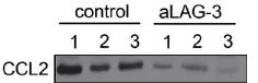
Western Blot: CCL2/MCP1 AntibodyBSA Free [NBP1-07035]
Western Blot: CCL2/MCP1 Antibody [NBP1-07035] - CCL2 expression level was evaluated in control group and aLAG-3 group.
Western Blot: CCL2/MCP1 Antibody - BSA Free [NBP1-07035] -
Western Blot: CCL2/MCP1 Antibody - BSA Free [NBP1-07035] - Ox-LDL induced macrophage foam cell formation, SIRT1 inhibition, autophagy impairment, & MCP-1 production in THP-1 cellsHuman THP-1 macrophages were exposed to 0, 20, 40, 60, & 80 μg/mL of ox-LDL for 24 hrs. Treated cells were photographed using light microscopy (A). The THP-1 macrophage-derived foam cell formation was determined using ORO staining method (B) & (C). Western blot for SIRT1, LC3, Beclin1, p62, & MCP-1 proteins were analyzed from the ox-LDL-stimulated THP-1 cells. beta -actin was used as loading control (D-I). Scale bar: 20 μm. Bar graph indicates the mean ± SD (n = 3). *P < 0.05 & **P < 0.01 vs. Cont group (0 μg/mL of ox-LDL). Image collected & cropped by CiteAb from the following publication (https://www.oncotarget.com/lookup/doi/10.18632/oncotarget.17691), licensed under a CC-BY license. Not internally tested by Novus Biologicals.
Western Blot: CCL2/MCP1 Antibody - BSA Free [NBP1-07035] -
Western Blot: CCL2/MCP1 Antibody - BSA Free [NBP1-07035] - Inhibition of autophagy with 3-MA led to increased foam cell formation & MCP-1 productionHuman THP-1 macrophages were pretreated with 3-MA (5 μM) for 2 hrs, & then exposed to 80 μg/mL of ox-LDL for an additional 24 hrs. Western blot for LC3, Beclin1, p62, & MCP-1 proteins were analyzed from the ox-LDL-stimulated THP-1 cells. beta -actin was used as loading control (A-E). THP-1 macrophage-derived foam cell formation was determined using ORO staining method (F-G). Scale bar: 40 μm. Bar graph indicates the mean ± SD (n = 3). *P < 0.05 & **P < 0.01 vs. Cont group; #P < 0.05 & ##P < 0.01 vs. 3-MA group; &P < 0.05 & &&P < 0.01 represent significant differences between ox-LDL group & 3-MA+ox-LDL group. Image collected & cropped by CiteAb from the following publication (https://www.oncotarget.com/lookup/doi/10.18632/oncotarget.17691), licensed under a CC-BY license. Not internally tested by Novus Biologicals.
Immunocytochemistry/ Immunofluorescence: CCL2/MCP1 Antibody - BSA Free [NBP1-07035] -
Immunocytochemistry/ Immunofluorescence: CCL2/MCP1 Antibody - BSA Free [NBP1-07035] - CCL2-ir cellular distribution in ependymal layers attached to the CC. A, B CCL2-ir (CCL2 panels, arrows) & GFAP (GFAP panels, arrowheads) double immunostained structures (Merged panels, arrow-arrowheads) are seen inlaid in the ependymal layers attached to the CC in both saline & LPS injected mice. C A CCL2-ir & Iba-1 double labeled soma (Merged, arrow-arrowhead) is seen at a corner of lateral ventricle, which seems be just next to an invaginated choroid plexus between the CC & basal ganglion (framed areas & insets). D, E Hyper-ramified & amoeba like Iba-1 labeled cells (Iba-1 panel, opened arrowheads) are observed in the CC of LPS injected mice, indicating their activated states. The CCL2-ir & Iba-1 double labeled structures are also regarded in the ependymal layers (Merged, arrow-arrowheads). Scare bar = 100 µm in all A– E Image collected & cropped by CiteAb from the following publication (https://pubmed.ncbi.nlm.nih.gov/35354428), licensed under a CC-BY license. Not internally tested by Novus Biologicals.
Western Blot: CCL2/MCP1 Antibody - BSA Free [NBP1-07035] -
Western Blot: CCL2/MCP1 Antibody - BSA Free [NBP1-07035] - Inhibition of autophagy using Atg5 siRNA aggravated foam cell formation & MCP-1 expressionHuman THP-1 macrophages were pretreated with Atg5 siRNA (20 μM) for 24 hrs, & then exposed to 80 μg/mL of ox-LDL for an additional 24 hrs. Western blot forAtg5, LC3, Beclin1, p62, & MCP-1 proteins were analyzed from the ox-LDL-stimulated THP-1 cells. beta -actin was used as loading control (A-F) & (I). THP-1 macrophage-derived foam cell formation was determined using ORO staining method (G) & (H). Scale bar: 40 μm. Bar graph indicates the mean ± SD (n = 3). *P < 0.05 & **P < 0.01 vs. NC siRNA group; #P < 0.05 & ##P < 0.01 vs. Atg5 siRNA group; &P < 0.05 & &&P < 0.01 represent significant differences between NC siRNA+ox-LDL group & Atg5 siRNA+ox-LDL group. Image collected & cropped by CiteAb from the following publication (https://www.oncotarget.com/lookup/doi/10.18632/oncotarget.17691), licensed under a CC-BY license. Not internally tested by Novus Biologicals.
Western Blot: CCL2/MCP1 Antibody - BSA Free [NBP1-07035] -
Western Blot: CCL2/MCP1 Antibody - BSA Free [NBP1-07035] - Inhibition of SIRT1 using EX-527 or SIRT1 siRNA transfection enhanced MCP-1 expression & foam cell formationHuman THP-1 macrophages were pretreated with EX-527 (2 μM, for 2 hrs) or SIRT1 siRNA (20 μM, for 24 hrs), & then exposed to 80 μg/mL of ox-LDL for an additional 24 hrs. Western blot for SIRT1 & MCP-1 proteins were analyzed from the ox-LDL-stimulated THP-1 cells. beta -actin was used as loading control (A-C) & (F-H). THP-1 macrophage-derived foam cell formation was determined using ORO staining method (D-E) & (I-J). Scale bar: 40 μm. Bar graph indicates the mean ± SD (n = 3). *P < 0.05 & **P < 0.01 vs. Cont group (NC siRNA group); #P < 0.05 & ##P < 0.01 vs. EX-527 group (SIRT1 siRNA group); &P < 0.05 & &&P < 0.01 represent significant differences between ox-LDL group (NC siRNA+ox-LDL group) & EX-527+ox-LDL group (SIRT1 siRNA+ox-LDL group). Image collected & cropped by CiteAb from the following publication (https://www.oncotarget.com/lookup/doi/10.18632/oncotarget.17691), licensed under a CC-BY license. Not internally tested by Novus Biologicals.
Western Blot: CCL2/MCP1 Antibody - BSA Free [NBP1-07035] -
Western Blot: CCL2/MCP1 Antibody - BSA Free [NBP1-07035] - Overexpression of SIRT1 using adenoviral transfection reversed ox-LDL-induced macrophage foam cell formation & autophagy impairment in THP-1 cellsHuman THP-1 macrophages were transfected by SIRT1 over-expressing adenovirus (HBAD-SIRT1) or NC adenovirus (HBAD-GFP) for 24 hrs & then exposed to 80 μg/mL of ox-LDL for an additional 24 hrs. The transfected THP-1 cells were observed using an inverted fluorescence microscope (A) & then were harvested for transfection efficiency analysis by Western blot method (B). THP-1 macrophage-derived foam cell formation was determined using ORO staining method (C) & (D). Western blot for LC3, Beclin1, p62, & Atg5 proteins & immunoprecipitation for acetyl-Lys Atg5 were analyzed from the ox-LDL-stimulated THP-1 cells. beta -actin was used as loading control (E-L). Scale bar: 40 μm. Bar graph indicates the mean ± SD (n = 3). *P < 0.05 & **P < 0.01 vs. HBAD-GFP group; #P < 0.05 & ##P < 0.01 vs. HBAD-SIRT1 group; &P < 0.05 & &&P < 0.01 represent significant differences between HBAD-GFP+ox-LDL group & HBAD-SIRT1+ox-LDL group. Image collected & cropped by CiteAb from the following publication (https://www.oncotarget.com/lookup/doi/10.18632/oncotarget.17691), licensed under a CC-BY license. Not internally tested by Novus Biologicals.Applications for CCL2/MCP1 Antibody - BSA Free
Application
Recommended Usage
Block/Neutralize
reported in scientific literature (PMID 22778093)
Immunocytochemistry/ Immunofluorescence
1:2000
Immunohistochemistry
1:500-1:1000
Immunohistochemistry-Paraffin
1:500-1:1000
Western Blot
1:1000. Use reported in scientific literature (PMID 22402584)
Application Notes
In Western blot, a band is seen at ~16 kDa. In ICC/IF secretory vesicles staining was observed in HeLa cells.
Reviewed Applications
Read 1 review rated 5 using NBP1-07035 in the following applications:
Formulation, Preparation, and Storage
Purification
Immunogen affinity purified
Formulation
PBS
Format
BSA Free
Preservative
0.02% Sodium Azide
Concentration
1 mg/ml
Shipping
The product is shipped with polar packs. Upon receipt, store it immediately at the temperature recommended below.
Stability & Storage
Store at 4C short term. Aliquot and store at -20C long term. Avoid freeze-thaw cycles.
Background: CCL2/MCP1
Alternate Names
C-C motif chemokine 2, chemokine (C-C motif) ligand 2, GDCF-2, HC11Monocyte secretory protein JE, MCAFMonocyte chemotactic and activating factor, MCP1 Monocyte chemotactic protein 1, MCP-1 Small-inducible cytokine A2, MGC9434, Monocyte chemoattractant protein 1, monocyte chemoattractant protein-1, SCYA2HSMCR30, small inducible cytokine A2 (monocyte chemotactic protein 1, homologous tomouse Sig-je), small inducible cytokine subfamily A (Cys-Cys), member 2, SMC-CF
Gene Symbol
CCL2
Additional CCL2/MCP1 Products
Product Documents for CCL2/MCP1 Antibody - BSA Free
Certificate of Analysis
To download a Certificate of Analysis, please enter a lot or batch number in the search box below.
Product Specific Notices for CCL2/MCP1 Antibody - BSA Free
This product is for research use only and is not approved for use in humans or in clinical diagnosis. Primary Antibodies are guaranteed for 1 year from date of receipt.
Citations for CCL2/MCP1 Antibody - BSA Free
 Powered by Bioz
Customer Reviews for CCL2/MCP1 Antibody - BSA Free (1)
5 out of 5
1 Customer Rating
Have you used CCL2/MCP1 Antibody - BSA Free?
Submit a review and receive an Amazon gift card!
$25/€18/£15/$25CAN/¥2500 Yen for a review with an image
$10/€7/£6/$10CAN/¥1110 Yen for a review without an image
Submit a review
Customer Images
Showing
1
-
1 of
1 review
Showing All
Filter By:
-
Application: Western BlotSample Tested: Mouse Head and neck squamous cell carcinoma (HNSCC)Species: MouseVerified Customer | Posted 10/23/2017CCL2 expression level was evaluated in control group and aLAG-3 group.

There are no reviews that match your criteria.
Protocols
View specific protocols for CCL2/MCP1 Antibody - BSA Free (NBP1-07035):
Immunocytochemistry Protocol
Culture cells to appropriate density in 35 mm culture dishes or 6-well plates.
1. Remove culture medium and add 10% formalin to the dish. Fix at room temperature for 30 minutes.
2. Remove the formalin and add ice cold methanol. Incubate for 5-10 minutes.
3. Remove methanol and add washing solution (i.e. PBS). Be sure to not let the specimen dry out. Wash three times for 10 minutes.
4. To block nonspecific antibody binding incubate in 10% normal goat serum from 1 hour to overnight at room temperature.
5. Add primary antibody at appropriate dilution and incubate at room temperature from 2 hours to overnight at room temperature.
6. Remove primary antibody and replace with washing solution. Wash three times for 10 minutes.
7. Add secondary antibody at appropriate dilution. Incubate for 1 hour at room temperature.
8. Remove antibody and replace with wash solution, then wash for 10 minutes. Add Hoechst 33258 to wash solution at 1:25,0000 and incubate for 10 minutes. Wash a third time for 10 minutes.
9. Cells can be viewed directly after washing. The plates can also be stored in PBS containing Azide covered in Parafilm (TM). Cells can also be cover-slipped using Fluoromount, with appropriate sealing.
*The above information is only intended as a guide. The researcher should determine what protocol best meets their needs. Please follow safe laboratory procedures.
Procedure Guide for NBP1-07035 - MCP-1 Antibody
Western Blot Protocol
1. Perform SDS-PAGE (4-12% MOPS) on samples to be analyzed, loading 40 ug of total protein per lane.
2. Transfer proteins to Nitrocellulose according to the instructions provided by the manufacturer of the transfer
apparatus.
3. Rinse membrane with dH2O and then stain the blot using Ponceau S for 1-2 minutes to access the transfer of
proteins onto the nitrocellulose membrane. Rinse the blot in water to remove excess stain and mark the lane locations
and locations of molecular weight markers using a pencil.
4. Rinse the blot in TBS for approximately 5 minutes.
5. Block the membrane using 5% NFDM + 1% BSA in TBS + Tween, 1 hour at RT.
6. Rinse the membrane in dH2O and then wash the membrane in wash buffer [TBS + 0.1% Tween] 3 times for 10
minutes each.
7. Dilute the rabbit anti-MCP-1 primary antibody (NBP1-07035) in blocking buffer and incubate 1 hour at room
temperature.
8. Rinse the membrane in dH2O and then wash the membrane in wash buffer [TBS + 0.1% Tween] 3 times for 10
minutes each.
9. Apply the diluted rabbit-IgG HRP-conjugated secondary antibody in blocking buffer (as per manufacturers
instructions) and incubate 1 hour at room temperature.
10. Wash the blot in wash buffer [TBS + 0.1% Tween] 3 times for 10 minutes each (this step can be repeated as
required to reduce background).
11. Apply the detection reagent of choice in accordance with the manufacturers instructions (Pierce ECL).
Note: Tween-20 can be added to the blocking or antibody dilution buffer at a final concentration of 0.05-0.2%, provided
it does not interfere with antibody-antigen binding.
Find general support by application which include: protocols, troubleshooting, illustrated assays, videos and webinars.
- Antigen Retrieval Protocol (PIER)
- Antigen Retrieval for Frozen Sections Protocol
- Appropriate Fixation of IHC/ICC Samples
- Cellular Response to Hypoxia Protocols
- Chromogenic IHC Staining of Formalin-Fixed Paraffin-Embedded (FFPE) Tissue Protocol
- Chromogenic Immunohistochemistry Staining of Frozen Tissue
- ClariTSA™ Fluorophore Kits
- Detection & Visualization of Antibody Binding
- Fluorescent IHC Staining of Frozen Tissue Protocol
- Graphic Protocol for Heat-induced Epitope Retrieval
- Graphic Protocol for the Preparation and Fluorescent IHC Staining of Frozen Tissue Sections
- Graphic Protocol for the Preparation and Fluorescent IHC Staining of Paraffin-embedded Tissue Sections
- Graphic Protocol for the Preparation of Gelatin-coated Slides for Histological Tissue Sections
- ICC Cell Smear Protocol for Suspension Cells
- ICC Immunocytochemistry Protocol Videos
- ICC for Adherent Cells
- IHC Sample Preparation (Frozen sections vs Paraffin)
- Immunocytochemistry (ICC) Protocol
- Immunocytochemistry Troubleshooting
- Immunofluorescence of Organoids Embedded in Cultrex Basement Membrane Extract
- Immunofluorescent IHC Staining of Formalin-Fixed Paraffin-Embedded (FFPE) Tissue Protocol
- Immunohistochemistry (IHC) and Immunocytochemistry (ICC) Protocols
- Immunohistochemistry Frozen Troubleshooting
- Immunohistochemistry Paraffin Troubleshooting
- Preparing Samples for IHC/ICC Experiments
- Preventing Non-Specific Staining (Non-Specific Binding)
- Primary Antibody Selection & Optimization
- Protocol for Heat-Induced Epitope Retrieval (HIER)
- Protocol for Making a 4% Formaldehyde Solution in PBS
- Protocol for VisUCyte™ HRP Polymer Detection Reagent
- Protocol for the Fluorescent ICC Staining of Cell Smears - Graphic
- Protocol for the Fluorescent ICC Staining of Cultured Cells on Coverslips - Graphic
- Protocol for the Preparation & Fixation of Cells on Coverslips
- Protocol for the Preparation and Chromogenic IHC Staining of Frozen Tissue Sections
- Protocol for the Preparation and Chromogenic IHC Staining of Frozen Tissue Sections - Graphic
- Protocol for the Preparation and Chromogenic IHC Staining of Paraffin-embedded Tissue Sections
- Protocol for the Preparation and Chromogenic IHC Staining of Paraffin-embedded Tissue Sections - Graphic
- Protocol for the Preparation and Fluorescent ICC Staining of Cells on Coverslips
- Protocol for the Preparation and Fluorescent ICC Staining of Non-adherent Cells
- Protocol for the Preparation and Fluorescent ICC Staining of Stem Cells on Coverslips
- Protocol for the Preparation and Fluorescent IHC Staining of Frozen Tissue Sections
- Protocol for the Preparation and Fluorescent IHC Staining of Paraffin-embedded Tissue Sections
- Protocol for the Preparation of Gelatin-coated Slides for Histological Tissue Sections
- Protocol for the Preparation of a Cell Smear for Non-adherent Cell ICC - Graphic
- R&D Systems Quality Control Western Blot Protocol
- TUNEL and Active Caspase-3 Detection by IHC/ICC Protocol
- The Importance of IHC/ICC Controls
- Troubleshooting Guide: Immunohistochemistry
- Troubleshooting Guide: Western Blot Figures
- Western Blot Conditions
- Western Blot Protocol
- Western Blot Protocol for Cell Lysates
- Western Blot Troubleshooting
- Western Blot Troubleshooting Guide
- View all Protocols, Troubleshooting, Illustrated assays and Webinars
Loading...