CD31/PECAM-1 Antibody (C31.3 + C31.7 + C31.10) - Azide and BSA Free
Novus Biologicals | Catalog # NBP2-47785
Key Product Details
Species Reactivity
Validated:
Human, Rat, Cynomolgus Monkey, Rabbit
Cited:
Human
Applications
Validated:
Immunohistochemistry, Immunohistochemistry-Paraffin, Western Blot, Immunocytochemistry/ Immunofluorescence
Cited:
Immunohistochemistry-Paraffin, Immunocytochemistry/ Immunofluorescence
Label
Unconjugated
Antibody Source
Monoclonal Mouse IgG1 Kappa/IgG1 Kappa/IgG1 Kappa Clone # C31.3 + C31.7 + C31.10
Format
Azide and BSA Free
Loading...
Product Specifications
Immunogen
Recombinant full-length human CD31/PECAM-1 protein (Uniprot: P16284)
Localization
Cell surface and cytoplasm of endothelial cells
Marker
Endothelial Cell Marker
Specificity
CD31 (PECAM-1) is a transmembrane glycoprotein member of the immunoglobulin supergene family of adhesion molecules. CD31 is expressed by stem cells of the hematopoietic system and is primarily used to identify and concentrate these cells for experimental studies as well as for bone marrow transplantation. Anti-CD31 has shown to be highly specific and sensitive for vascular endothelial cells. Staining of nonvascular tumors (excluding hematopoietic neoplasms) is rare. CD31 monoclonal antibody reacts with normal, benign, and malignant endothelial cells which make up blood vessel lining. The level of CD31 expression can help to determine the degree of tumor angiogenesis, and a high level of CD31 expression may imply a rapidly growing tumor and potentially a predictor of tumor recurrence.
Clonality
Monoclonal
Host
Mouse
Isotype
IgG1 Kappa/IgG1 Kappa/IgG1 Kappa
Theoretical MW
82.5 kDa.
Disclaimer note: The observed molecular weight of the protein may vary from the listed predicted molecular weight due to post translational modifications, post translation cleavages, relative charges, and other experimental factors.
Disclaimer note: The observed molecular weight of the protein may vary from the listed predicted molecular weight due to post translational modifications, post translation cleavages, relative charges, and other experimental factors.
Description
1.0 mg/ml of antibody purified from Bioreactor Concentrate by Protein A/G. Prepared in 10mM PBS WITHOUT BSA & azide. Also available at 200 ug/ml WITH BSA & azide (NBP2-44342).
Antibody with azide - store at 2 to 8C. Antibody without azide - store at -20 to -80C.
Antibody with azide - store at 2 to 8C. Antibody without azide - store at -20 to -80C.
Scientific Data Images for CD31/PECAM-1 Antibody (C31.3 + C31.7 + C31.10) - Azide and BSA Free
![Western Blot: CD31/PECAM-1 Antibody (C31.3 + C31.7 + C31.10)Azide and BSA Free [NBP2-47785]](https://resources.rndsystems.com/images/products/CD31-PECAM-1-Antibody-C31-3-+-C31-7-+-C31-10-Azide-and-BSA-Free-Western-Blot-NBP2-47785-img0002.jpg "Western Blot: CD31/PECAM-1 Antibody (C31.3 + C31.7 + C31.10)Azide and BSA Free [NBP2-47785]")
Western Blot: CD31/PECAM-1 Antibody (C31.3 + C31.7 + C31.10)Azide and BSA Free [NBP2-47785]
Western Blot: CD31/PECAM-1 Antibody (C31.3 + C31.7 + C31.10) - Azide and BSA Free [NBP2-47785] - Western Blot Analysis of human THP-1 cell lysate using CD31/PECAM-1 Antibody (C31.3 + C31.7 + C31.10) - Azide and BSA Free.![Immunohistochemistry-Paraffin: CD31/PECAM-1 Antibody (C31.3 + C31.7 + C31.10) - Azide and BSA Free [NBP2-47785]](https://resources.rndsystems.com/images/products/CD31-PECAM-1-Antibody-C31-3-+-C31-7-+-C31-10-Azide-and-BSA-Free-Immunohistochemistry-Paraffin-NBP2-47785-img0004.jpg "Immunohistochemistry-Paraffin: CD31/PECAM-1 Antibody (C31.3 + C31.7 + C31.10) - Azide and BSA Free [NBP2-47785]")
Immunohistochemistry-Paraffin: CD31/PECAM-1 Antibody (C31.3 + C31.7 + C31.10) - Azide and BSA Free [NBP2-47785]
Immunohistochemistry-Paraffin: CD31/PECAM-1 Antibody (C31.3 + C31.7 + C31.10) - Azide and BSA Free [NBP2-47785] - Colon Carcinoma stained withCD31/PECAM-1 Antibody (C31.3 + C31.7 + C31.10) - Azide and BSA Free.![Immunohistochemistry-Paraffin: CD31/PECAM-1 Antibody (C31.3 + C31.7 + C31.10) - Azide and BSA Free [NBP2-47785]](https://resources.rndsystems.com/images/products/CD31-PECAM-1-Antibody-C31-3-+-C31-7-+-C31-10-Azide-and-BSA-Free-Immunohistochemistry-Paraffin-NBP2-47785-img0001.jpg "Immunohistochemistry-Paraffin: CD31/PECAM-1 Antibody (C31.3 + C31.7 + C31.10) - Azide and BSA Free [NBP2-47785]")
Immunohistochemistry-Paraffin: CD31/PECAM-1 Antibody (C31.3 + C31.7 + C31.10) - Azide and BSA Free [NBP2-47785]
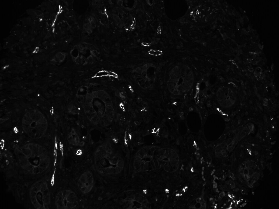
Immunohistochemistry-Paraffin: CD31/PECAM-1 Antibody (C31.3 + C31.7 + C31.10) - Azide and BSA Free [NBP2-47785] - Cholangiocellular carcinoma (liver cancer tissue) stained with ATTO550 conjugated CD31 antibody. Image from verified customer review.![Immunohistochemistry-Paraffin: CD31/PECAM-1 Antibody (C31.3 + C31.7 + C31.10) - Azide and BSA Free [NBP2-47785]](https://resources.rndsystems.com/images/products/CD31-PECAM-1-Antibody-C31-3-+-C31-7-+-C31-10-Azide-and-BSA-Free-Immunohistochemistry-Paraffin-NBP2-47785-img0003.jpg "Immunohistochemistry-Paraffin: CD31/PECAM-1 Antibody (C31.3 + C31.7 + C31.10) - Azide and BSA Free [NBP2-47785]")
Immunohistochemistry-Paraffin: CD31/PECAM-1 Antibody (C31.3 + C31.7 + C31.10) - Azide and BSA Free [NBP2-47785]
Immunohistochemistry-Paraffin: CD31/PECAM-1 Antibody (C31.3 + C31.7 + C31.10) - Azide and BSA Free [NBP2-47785] - Formalin-fixed, paraffin-embedded Angiosarcoma stained with CD31/PECAM-1 Antibody (C31.3 + C31.7 + C31.10) - Azide and BSA Free.Applications for CD31/PECAM-1 Antibody (C31.3 + C31.7 + C31.10) - Azide and BSA Free
Application
Recommended Usage
Immunocytochemistry/ Immunofluorescence
Optimal dilutions of this antibody should be experimentally determined.
Immunohistochemistry
Optimal dilutions of this antibody should be experimentally determined.
Immunohistochemistry-Paraffin
Optimal dilutions of this antibody should be experimentally determined.
Western Blot
Optimal dilutions of this antibody should be experimentally determined.
Application Notes
Use in ICC/IF reported in scientific literature (PMID:10.1016/j.cmet.2021.05.013)
Reviewed Applications
Read 1 review rated 5 using NBP2-47785 in the following applications:
Formulation, Preparation, and Storage
Purification
Protein A or G purified
Formulation
10 mM PBS
Format
Azide and BSA Free
Preservative
No Preservative
Concentration
1.0 mg/ml
Shipping
The product is shipped with polar packs. Upon receipt, store it immediately at the temperature recommended below.
Stability & Storage
Store at -20 to -80C. Avoid freeze-thaw cycles.
Background: CD31/PECAM-1
PECAM's intracellular cytoplasmic domain consists of a sequence of 118 amino acids and contains serine and tyrosine (also referred to as immunoreceptor tyrosine-based inhibitory motifs-ITIMs) residues, which may be phosphorylated upon cellular stimulation (3). ITIMs are phosphorylated by Src-family kinases and non-Src family kinases (e.g., Csk), leading to a conformational change which supports interactions with Src homology 2 (SH2) domain containing proteins such as protein-tyrosine phosphatase, SHP-2 (1,2). Formation of SHP-2/PECAM-1 complexes induces endothelial cell migration through the dephosphorylation of focal adhesion kinase and regulation of RhoA activity (1). Signaling downstream of ITIM tyrosine phosphorylations also plays a role in PECAM's anti-apoptotic activity, a function which is independent of its interaction with SHP-2. In platelets and leukocytes, phosphorylation of PECAM's cytosolic domain is inhibitory, preventing their activation.
References
1. Lertkiatmongkol, P., Liao, D., Mei, H., Hu, Y., & Newman, P. J. (2016). Endothelial functions of PECAM-1 (CD31). Current Opinion in Hematology. https://doi.org/10.1097/MOH.0000000000000239.Endothelial
2. Privratsky, J. R., & Newman, P. J. (2014). PECAM-1: Regulator of endothelial junctional integrity. Cell and Tissue Research. https://doi.org/10.1007/s00441-013-1779-3
3. Newman, P. J., & Newman, D. K. (2003). Signal transduction pathways mediated by PECAM-1: New roles for an old molecule in platelet and vascular cell biology. Arteriosclerosis, Thrombosis, and Vascular Biology. https://doi.org/10.1161/01.ATV.0000071347.69358.D9
Long Name
Platelet Endothelial Cell Adhesion Molecule 1
Alternate Names
CD31, EndoCAM, PECA1, PECAM-1, PECAM1
Gene Symbol
PECAM1
Additional CD31/PECAM-1 Products
Product Documents for CD31/PECAM-1 Antibody (C31.3 + C31.7 + C31.10) - Azide and BSA Free
Certificate of Analysis
To download a Certificate of Analysis, please enter a lot or batch number in the search box below.
Product Specific Notices for CD31/PECAM-1 Antibody (C31.3 + C31.7 + C31.10) - Azide and BSA Free
This product is for research use only and is not approved for use in humans or in clinical diagnosis. Primary Antibodies are guaranteed for 1 year from date of receipt.
Related Research Areas
Citations for CD31/PECAM-1 Antibody (C31.3 + C31.7 + C31.10) - Azide and BSA Free
 Powered by Bioz
Customer Reviews for CD31/PECAM-1 Antibody (C31.3 + C31.7 + C31.10) - Azide and BSA Free (1)
5 out of 5
1 Customer Rating
Have you used CD31/PECAM-1 Antibody (C31.3 + C31.7 + C31.10) - Azide and BSA Free?
Submit a review and receive an Amazon gift card!
$25/€18/£15/$25CAN/¥2500 Yen for a review with an image
$10/€7/£6/$10CAN/¥1110 Yen for a review without an image
Submit a review
Customer Images
Showing
1
-
1 of
1 review
Showing All
Filter By:
-
Application: Immunofluorescence - paraffinSample Tested: Liver cancer tissueSpecies: HumanVerified Customer | Posted 11/18/2018Cholangiocellular carcinoma stained with CD31-ATTO550pH 9 antigen retrieval

There are no reviews that match your criteria.
Protocols
Find general support by application which include: protocols, troubleshooting, illustrated assays, videos and webinars.
- Antigen Retrieval Protocol (PIER)
- Antigen Retrieval for Frozen Sections Protocol
- Appropriate Fixation of IHC/ICC Samples
- Cellular Response to Hypoxia Protocols
- Chromogenic IHC Staining of Formalin-Fixed Paraffin-Embedded (FFPE) Tissue Protocol
- Chromogenic Immunohistochemistry Staining of Frozen Tissue
- ClariTSA™ Fluorophore Kits
- Detection & Visualization of Antibody Binding
- Fluorescent IHC Staining of Frozen Tissue Protocol
- Graphic Protocol for Heat-induced Epitope Retrieval
- Graphic Protocol for the Preparation and Fluorescent IHC Staining of Frozen Tissue Sections
- Graphic Protocol for the Preparation and Fluorescent IHC Staining of Paraffin-embedded Tissue Sections
- Graphic Protocol for the Preparation of Gelatin-coated Slides for Histological Tissue Sections
- ICC Cell Smear Protocol for Suspension Cells
- ICC Immunocytochemistry Protocol Videos
- ICC for Adherent Cells
- IHC Sample Preparation (Frozen sections vs Paraffin)
- Immunocytochemistry (ICC) Protocol
- Immunocytochemistry Troubleshooting
- Immunofluorescence of Organoids Embedded in Cultrex Basement Membrane Extract
- Immunofluorescent IHC Staining of Formalin-Fixed Paraffin-Embedded (FFPE) Tissue Protocol
- Immunohistochemistry (IHC) and Immunocytochemistry (ICC) Protocols
- Immunohistochemistry Frozen Troubleshooting
- Immunohistochemistry Paraffin Troubleshooting
- Preparing Samples for IHC/ICC Experiments
- Preventing Non-Specific Staining (Non-Specific Binding)
- Primary Antibody Selection & Optimization
- Protocol for Heat-Induced Epitope Retrieval (HIER)
- Protocol for Making a 4% Formaldehyde Solution in PBS
- Protocol for VisUCyte™ HRP Polymer Detection Reagent
- Protocol for the Fluorescent ICC Staining of Cell Smears - Graphic
- Protocol for the Fluorescent ICC Staining of Cultured Cells on Coverslips - Graphic
- Protocol for the Preparation & Fixation of Cells on Coverslips
- Protocol for the Preparation and Chromogenic IHC Staining of Frozen Tissue Sections
- Protocol for the Preparation and Chromogenic IHC Staining of Frozen Tissue Sections - Graphic
- Protocol for the Preparation and Chromogenic IHC Staining of Paraffin-embedded Tissue Sections
- Protocol for the Preparation and Chromogenic IHC Staining of Paraffin-embedded Tissue Sections - Graphic
- Protocol for the Preparation and Fluorescent ICC Staining of Cells on Coverslips
- Protocol for the Preparation and Fluorescent ICC Staining of Non-adherent Cells
- Protocol for the Preparation and Fluorescent ICC Staining of Stem Cells on Coverslips
- Protocol for the Preparation and Fluorescent IHC Staining of Frozen Tissue Sections
- Protocol for the Preparation and Fluorescent IHC Staining of Paraffin-embedded Tissue Sections
- Protocol for the Preparation of Gelatin-coated Slides for Histological Tissue Sections
- Protocol for the Preparation of a Cell Smear for Non-adherent Cell ICC - Graphic
- R&D Systems Quality Control Western Blot Protocol
- TUNEL and Active Caspase-3 Detection by IHC/ICC Protocol
- The Importance of IHC/ICC Controls
- Troubleshooting Guide: Immunohistochemistry
- Troubleshooting Guide: Western Blot Figures
- Western Blot Conditions
- Western Blot Protocol
- Western Blot Protocol for Cell Lysates
- Western Blot Troubleshooting
- Western Blot Troubleshooting Guide
- View all Protocols, Troubleshooting, Illustrated assays and Webinars
Loading...