CD31/PECAM-1 Antibody (MEC13.3) - BSA Free
Novus Biologicals | Catalog # NB600-1475
Key Product Details
Species Reactivity
Validated:
Human, Mouse
Cited:
Human, Mouse
Applications
Validated:
Immunohistochemistry, Immunohistochemistry-Frozen, Flow Cytometry, Flow (Cell Surface), Immunocytochemistry/ Immunofluorescence, Immunoprecipitation, In vitro assay, In vivo assay, CyTOF-ready
Cited:
Immunohistochemistry, Immunohistochemistry-Paraffin, Flow Cytometry, Immunocytochemistry/ Immunofluorescence, In vivo assay, IF/IHC, In-vitro
Label
Unconjugated
Antibody Source
Monoclonal Rat IgG2a Kappa Clone # MEC13.3
Format
BSA Free
Loading...
Product Specifications
Immunogen
This CD31/PECAM-1 Antibody (MEC13.3) was developed against mouse endothelial cell line T-end.
Reactivity Notes
Mouse (PMID: 7956830) and Human (PMID: 30626719) reactivity reported in scientific literature.
Localization
Membrane; Single-pass type I membrane protein. Cell junction.
Clonality
Monoclonal
Host
Rat
Isotype
IgG2a Kappa
Theoretical MW
82.5 kDa.
Disclaimer note: The observed molecular weight of the protein may vary from the listed predicted molecular weight due to post translational modifications, post translation cleavages, relative charges, and other experimental factors.
Disclaimer note: The observed molecular weight of the protein may vary from the listed predicted molecular weight due to post translational modifications, post translation cleavages, relative charges, and other experimental factors.
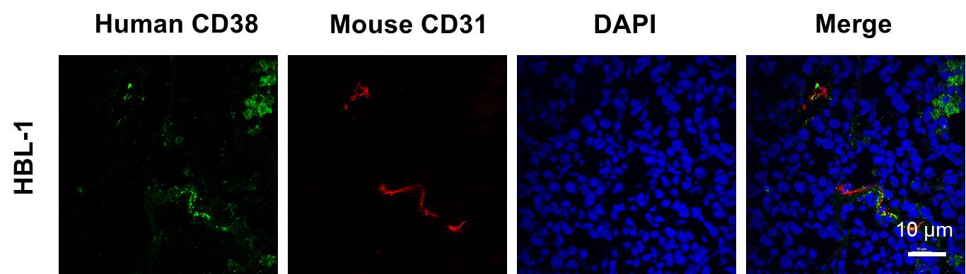
Scientific Data Images for CD31/PECAM-1 Antibody (MEC13.3) - BSA Free
![Immunocytochemistry/ Immunofluorescence: CD31/PECAM-1 Antibody (MEC13.3) - BSA Free [NB600-1475]](https://resources.rndsystems.com/images/products/CD31-PECAM-1-Antibody-MEC13-3-Immunocytochemistry-Immunofluorescence-NB600-1475-img0019.jpg "Immunocytochemistry/ Immunofluorescence: CD31/PECAM-1 Antibody (MEC13.3) - BSA Free [NB600-1475]")
Immunocytochemistry/ Immunofluorescence: CD31/PECAM-1 Antibody (MEC13.3) - BSA Free [NB600-1475]
Immunocytochemistry/Immunofluorescence: CD31/PECAM-1 Antibody (MEC13.3) [NB600-1475] - Mouse MS1 cells were fixed in 4% paraformaldehyde for 10 minutes and permeabilized in 0.05% Triton X-100 in PBS for 5 minutes. The cells were incubated with CD31/PECAM-1 Antibody [MEC13.3] (NB600-1475) at 1ug/ml overnight at 4C and detected with an anti-rat DyLight 488 (Green) at a 1:1000 dilution for 60 minutes. Nuclei were counterstained with DAPI (Blue). Cells were imaged using a 100X objective and digitally deconvolved.![Immunohistochemistry-Frozen: CD31/PECAM-1 Antibody (MEC13.3) - BSA Free [NB600-1475]](https://resources.rndsystems.com/images/products/CD31-PECAM-1-Antibody-MEC13-3-Immunohistochemistry-Frozen-NB600-1475-img0006.jpg "Immunohistochemistry-Frozen: CD31/PECAM-1 Antibody (MEC13.3) - BSA Free [NB600-1475]")
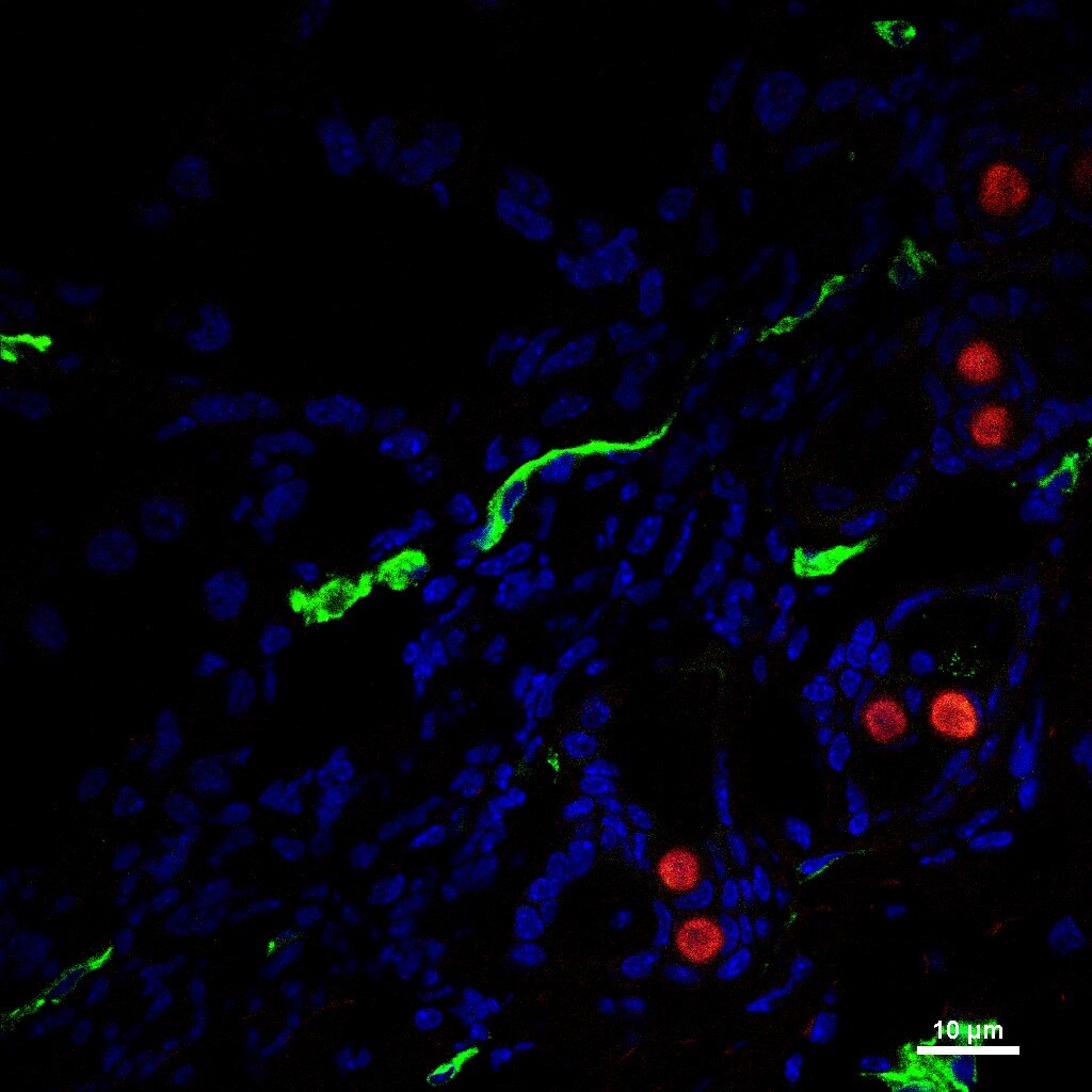
Immunohistochemistry-Frozen: CD31/PECAM-1 Antibody (MEC13.3) - BSA Free [NB600-1475]
Immunohistochemistry-Frozen: CD31/PECAM-1 Antibody (MEC13.3) [NB600-1475] - 4T1 tumor sections were stained for CD31 (green) and CD105 (red).![Flow (Cell Surface): CD31/PECAM-1 Antibody (MEC13.3) - BSA Free [NB600-1475]](https://resources.rndsystems.com/images/products/CD31-PECAM-1-Antibody-MEC13-3-Flow-Cell-Surface-NB600-1475-img0009.jpg "Flow (Cell Surface): CD31/PECAM-1 Antibody (MEC13.3) - BSA Free [NB600-1475]")
Flow (Cell Surface): CD31/PECAM-1 Antibody (MEC13.3) - BSA Free [NB600-1475]
Flow (Cell Surface): CD31/PECAM-1 Antibody (MEC13.3) [NB600-1475] - A surface stain was performed on WEHI-3 Cells with CD31/PECAM-1 Antibody (MEC13.3) (NB600-1475, blue) and a matched isotype control (orange). Cells were incubated in an antibody dilution of 2.5 ug/mL for 20 minutes at room temperature, followed by rat F(ab)2 IgG (H+L) APC-conjugated secondary antibody (F0113, R&D Systems).![Flow Cytometry: CD31/PECAM-1 Antibody (MEC13.3) - BSA Free [NB600-1475]](https://resources.rndsystems.com/images/products/CD31-PECAM-1-Antibody-MEC13-3-Flow-Cytometry-NB600-1475-img0010.jpg "Flow Cytometry: CD31/PECAM-1 Antibody (MEC13.3) - BSA Free [NB600-1475]")
Flow Cytometry: CD31/PECAM-1 Antibody (MEC13.3) - BSA Free [NB600-1475]
Flow Cytometry: CD31/PECAM-1 Antibody (MEC13.3) [NB600-1475] - C57BL/6 mouse splenocytes stained with purified CD31/PECAM-1 Antibody (MEC13.3), followed by anti-rat IgG FITC![Immunocytochemistry/ Immunofluorescence: CD31/PECAM-1 Antibody (MEC13.3) - BSA Free [NB600-1475]](https://resources.rndsystems.com/images/products/CD31-PECAM-1-Antibody-MEC13-3-Immunocytochemistry-Immunofluorescence-NB600-1475-img0017.jpg "Immunocytochemistry/ Immunofluorescence: CD31/PECAM-1 Antibody (MEC13.3) - BSA Free [NB600-1475]")
Immunocytochemistry/ Immunofluorescence: CD31/PECAM-1 Antibody (MEC13.3) - BSA Free [NB600-1475]
Immunocytochemistry/Immunofluorescence: CD31/PECAM-1 Antibody (MEC13.3) [NB600-1475] - Mouse MS1 cells were fixed in 4% paraformaldehyde for 10 minutes and permeabilized in 0.05% Triton X-100 in PBS for 5 minutes. The cells were incubated with CD31/PECAM-1 Antibody [MEC13.3] conjugated to DyLight 488 (NB600-1475G) at 5 ug/ml for 1 hour at room temperature. Nuclei were counterstained with DAPI (Blue). Cells were imaged using a 100X objective and digitally deconvolved.![Immunocytochemistry/ Immunofluorescence: CD31/PECAM-1 Antibody (MEC13.3) - BSA Free [NB600-1475]](https://resources.rndsystems.com/images/products/CD31-PECAM-1-Antibody-MEC13-3-Immunocytochemistry-Immunofluorescence-NB600-1475-img0007.jpg "Immunocytochemistry/ Immunofluorescence: CD31/PECAM-1 Antibody (MEC13.3) - BSA Free [NB600-1475]")
Immunocytochemistry/ Immunofluorescence: CD31/PECAM-1 Antibody (MEC13.3) - BSA Free [NB600-1475]
Immunocytochemistry/Immunofluorescence: CD31/PECAM-1 Antibody (MEC13.3) [NB600-1475] - MS1 cells were fixed for 10 minutes using 10% formalin and then permeabilized for 5 minutes using 1X TBS + 0.5% Triton-X100. The cells were incubated with at 5.0 ug/ml overnight at 4C and detected with an anti-mouse Dylight 488 (Green) at a 1:500 dilution. Actin was detected with Phalloidin 568 (Red) at a 1:200 dilution. Nuclei were counterstained with DAPI (Blue). Cells were imaged using a 40X objective.![Immunocytochemistry/ Immunofluorescence: CD31/PECAM-1 Antibody (MEC13.3) - BSA Free [NB600-1475]](https://resources.rndsystems.com/images/products/CD31-PECAM-1-Antibody-MEC13-3-Immunocytochemistry-Immunofluorescence-NB600-1475-img0011.jpg "Immunocytochemistry/ Immunofluorescence: CD31/PECAM-1 Antibody (MEC13.3) - BSA Free [NB600-1475]")
Immunocytochemistry/ Immunofluorescence: CD31/PECAM-1 Antibody (MEC13.3) - BSA Free [NB600-1475]
Immunocytochemistry/Immunofluorescence: CD31/PECAM-1 Antibody (MEC13.3) [NB600-1475] - was tested in Wehi-3 cells with Dylight 488 (green). Nuclei and beta-tubulin were counterstained with DAPI (blue) and Dylight 550 (red).![Immunohistochemistry-Frozen: CD31/PECAM-1 Antibody (MEC13.3) - BSA Free [NB600-1475]](https://resources.rndsystems.com/images/products/CD31-PECAM-1-Antibody-MEC13-3-Immunohistochemistry-Paraffin-NB600-1475-img0015.jpg "Immunohistochemistry-Frozen: CD31/PECAM-1 Antibody (MEC13.3) - BSA Free [NB600-1475]")
Immunohistochemistry-Frozen: CD31/PECAM-1 Antibody (MEC13.3) - BSA Free [NB600-1475]
CD31-PECAM-1-Antibody-MEC13-3-Immunohistochemistry-Paraffin-NB600-1475-img0015.jpg![Flow Cytometry: CD31/PECAM-1 Antibody (MEC13.3)BSA Free [NB600-1475]](https://resources.rndsystems.com/images/products/CD31-PECAM-1-Antibody-MEC13-3-Flow-Cytometry-NB600-1475-img0022.jpg "Flow Cytometry: CD31/PECAM-1 Antibody (MEC13.3)BSA Free [NB600-1475]")
Flow Cytometry: CD31/PECAM-1 Antibody (MEC13.3)BSA Free [NB600-1475]
Flow Cytometry: CD31/PECAM-1 Antibody (MEC13.3) [NB600-1475] - A surface stain was performed on MS1 cells with CD31/PECAM-1 [MEC13.3] Antibody NB600-1475R (blue) and a matched isotype control (orange). Cells were incubated in an antibody dilution of 2.5 ug/mL for 30 minutes at room temperature. Both antibodies were conjugated to DyLight 550.![Immunocytochemistry/ Immunofluorescence: CD31/PECAM-1 Antibody (MEC13.3) - BSA Free [NB600-1475]](https://resources.rndsystems.com/images/products/CD31-PECAM-1-Antibody-MEC13-3-Immunocytochemistry-Immunofluorescence-NB600-1475-img0016.jpg "Immunocytochemistry/ Immunofluorescence: CD31/PECAM-1 Antibody (MEC13.3) - BSA Free [NB600-1475]")
Immunocytochemistry/ Immunofluorescence: CD31/PECAM-1 Antibody (MEC13.3) - BSA Free [NB600-1475]
Immunocytochemistry/Immunofluorescence: CD31/PECAM-1 Antibody (MEC13.3) [NB600-1475] - Mouse MS1 cells were fixed in 4% paraformaldehyde for 10 minutes and permeabilized in 0.05% Triton X-100 in PBS for 5 minutes. The cells were incubated with CD31/PECAM-1 Antibody [MEC13.3] conjugated to Alexa Fluor 647 (NB600-1475AF647) at 5 ug/ml for 1 hour at room temperature. Nuclei were counterstained with DAPI (Blue). Cells were imaged using a 100X objective and digitally deconvolved.![Immunocytochemistry/ Immunofluorescence: CD31/PECAM-1 Antibody (MEC13.3) - BSA Free [NB600-1475]](https://resources.rndsystems.com/images/products/CD31-PECAM-1-Antibody-MEC13-3-Immunocytochemistry-Immunofluorescence-NB600-1475-img0018.jpg "Immunocytochemistry/ Immunofluorescence: CD31/PECAM-1 Antibody (MEC13.3) - BSA Free [NB600-1475]")
Immunocytochemistry/ Immunofluorescence: CD31/PECAM-1 Antibody (MEC13.3) - BSA Free [NB600-1475]
Immunocytochemistry/Immunofluorescence: CD31/PECAM-1 Antibody (MEC13.3) [NB600-1475] - Mouse MS1 cells were fixed in 4% paraformaldehyde for 10 minutes and permeabilized in 0.05% Triton X-100 in PBS for 5 minutes. The cells were incubated with CD31/PECAM-1 Antibody [MEC13.3] conjugated to DyLight 550 (NB600-1475R) at 5 ug/ml for 1 hour at room temperature. Nuclei were counterstained with DAPI (Blue). Cells were imaged using a 100X objective and digitally deconvolved.![Immunohistochemistry-Frozen: CD31/PECAM-1 Antibody (MEC13.3) - BSA Free [NB600-1475]](https://resources.rndsystems.com/images/products/CD31-PECAM-1-Antibody-MEC13-3-Immunohistochemistry-Frozen-NB600-1475-img0012.jpg "Immunohistochemistry-Frozen: CD31/PECAM-1 Antibody (MEC13.3) - BSA Free [NB600-1475]")
Immunohistochemistry-Frozen: CD31/PECAM-1 Antibody (MEC13.3) - BSA Free [NB600-1475]
Immunohistochemistry-Frozen: CD31/PECAM-1 Antibody (MEC13.3) [NB600-1475] - Staining of mouse CD31 in non-small cell lung cancer tumor sections from a human NSCLC tumor-bearing mouse. This image was submitted via customer Review.![Flow (Cell Surface): CD31/PECAM-1 Antibody (MEC13.3) - BSA Free [NB600-1475]](https://resources.rndsystems.com/images/products/CD31-PECAM-1-Antibody-MEC13-3-Flow-Cell-Surface-NB600-1475-img0013.jpg "Flow (Cell Surface): CD31/PECAM-1 Antibody (MEC13.3) - BSA Free [NB600-1475]")
Flow (Cell Surface): CD31/PECAM-1 Antibody (MEC13.3) - BSA Free [NB600-1475]
Flow (Cell Surface): CD31/PECAM-1 Antibody (MEC13.3) [NB600-1475] - A surface stain was performed on WEHI-3 Cells with CD31 (MEC13.3) antibody NB600-1475 (blue) and a matched isotype control (orange). Cells were incubated in an antibody dilution of 2.5 ug/mL for 20 minutes at room temperature, followed by rat F(ab)2 IgG (H+L) APC-conjugated secondary antibody (F0113, R&D Systems).![Flow Cytometry: CD31/PECAM-1 Antibody (MEC13.3) - BSA Free [NB600-1475]](https://resources.rndsystems.com/images/products/CD31-PECAM-1-Antibody-MEC13-3-Flow-Cytometry-NB600-1475-img0014.jpg "Flow Cytometry: CD31/PECAM-1 Antibody (MEC13.3) - BSA Free [NB600-1475]")
Flow Cytometry: CD31/PECAM-1 Antibody (MEC13.3) - BSA Free [NB600-1475]
Flow Cytometry: CD31/PECAM-1 Antibody (MEC13.3) [NB600-1475] - A surface stain was performed on MS-1 cells with CD31/PECAM-1 Antibody (MEC13.3) (NB600-1475AF647, blue) and a matched isotype control (orange). Cells were incubated in an antibody dilution of 5 ug/mL for 20 minutes at room temperature. Both antibodies were conjugated to Alexa Fluor 647.![Flow Cytometry: CD31/PECAM-1 Antibody (MEC13.3)BSA Free [NB600-1475]](https://resources.rndsystems.com/images/products/CD31-PECAM-1-Antibody-MEC13-3-Flow-Cytometry-NB600-1475-img0020.jpg "Flow Cytometry: CD31/PECAM-1 Antibody (MEC13.3)BSA Free [NB600-1475]")
Flow Cytometry: CD31/PECAM-1 Antibody (MEC13.3)BSA Free [NB600-1475]
Flow Cytometry: CD31/PECAM-1 Antibody (MEC13.3) [NB600-1475] - A surface stain was performed on MS1 cells with CD31/PECAM-1 [MEC13.3] Antibody NB600-1475DL594 (blue) and a matched isotype control (orange). Cells were incubated in an antibody dilution of 2.5 ug/mL for 30 minutes at room temperature. Both antibodies were conjugated to DyLight 594.![Flow Cytometry: CD31/PECAM-1 Antibody (MEC13.3)BSA Free [NB600-1475]](https://resources.rndsystems.com/images/products/CD31-PECAM-1-Antibody-MEC13-3-Flow-Cytometry-NB600-1475-img0021.jpg "Flow Cytometry: CD31/PECAM-1 Antibody (MEC13.3)BSA Free [NB600-1475]")
Flow Cytometry: CD31/PECAM-1 Antibody (MEC13.3)BSA Free [NB600-1475]
Flow Cytometry: CD31/PECAM-1 Antibody (MEC13.3) [NB600-1475] - A surface stain was performed on MS1 cells with CD31/PECAM-1 [MEC13.3] Antibody NB600-1475G (blue) and a matched isotype control (orange). Cells were incubated in an antibody dilution of 5 ug/mL for 30 minutes at room temperature. Both antibodies were conjugated to DyLight 488. - BSA Free [NB600-1475] -")
Immunocytochemistry/ Immunofluorescence: CD31/PECAM-1 Antibody (MEC13.3) - BSA Free [NB600-1475] -
Immunocytochemistry/ Immunofluorescence: CD31/PECAM-1 Antibody (MEC13.3) - BSA Free [NB600-1475] - (A) Representative Western blots depicting phosphorylated JAK3 (pJAK3), total JAK3, & corresponding beta -actin levels in the ipsilateral (CXI) versus the contralateral (CXC) cortex at 24 h in sham & mice subjected to permanent middle cerebral artery occlusion (pMCAO). (B,C) Quantified pJAK3 & JAK3 values normalized to beta -actin in the cortex of sham & mice subjected to stroke. Both pJAK3 & JAK3 are significantly increased in the ipsilateral cortex of stroked animals (*P < 0.05 & **P < 0.01 with respect to sham ipsilateral/contralateral & stroke contralateral; sham, n = 5; stroke, n = 13). Data were analyzed using a two-way ANOVA (ipsilateral/contralateral or sham/stroke) with Bonferroni post-test. (D) Diagram showing peri-infarct brain region from which images were taken (n = 3). Scale bar is 10 µm, & magnification is 60×. (E) JAK3 colocalized with microglia/macrophages labeled with Iba-1. (F) JAK3 colocalized with neurons labeled with NeuN. (G) JAK3 colocalized with endothelial cells labeled with CD31. (H) JAK3 is not colocalized with astrocytes labeled with GFAP. Image collected & cropped by CiteAb from the following publication (https://pubmed.ncbi.nlm.nih.gov/28790974), licensed under a CC-BY license. Not internally tested by Novus Biologicals. in MS1 Mouse Cell Line.")
CD31/PECAM-1 (MEC13.3) in MS1 Mouse Cell Line.
CD31/PECAM-1 (MEC13.3) was detected in immersion fixed MS1 mouse pancreas/Islet of Langerhans endothelial cell line using Rat anti-CD31/PECAM-1 (MEC13.3) Protein-G purified Monoclonal Antibody conjugated to DyLight 550 (Catalog # NB600-1475R) (red) at 2 µg/mL overnight at 4C. Cells were counterstained with DAPI (blue). Cells were imaged using a 100X objective and digitally deconvolved.Applications for CD31/PECAM-1 Antibody (MEC13.3) - BSA Free
Application
Recommended Usage
Flow (Cell Surface)
2.5 ug/ml
Flow Cytometry
2.5 ug/ml
Immunocytochemistry/ Immunofluorescence
1:500 - 1:1000
Immunohistochemistry
1:100-1:500
Immunohistochemistry-Frozen
1:100
Immunoprecipitation
1:10-1:500
In vitro assay
reported in scientific literature (PMID 24647208)
In vivo assay
reported in scientific literature
Application Notes
This CD31/PECAM1 Antibody (MEC13.3) is useful for in vitro and in vivo blocking of CD31-mediated cell-cell interactions. Please Note: NB600-1475 works well on acetone-fixed frozen sections as well as zinc-fixed paraffin-embedded sections. However, inconsistent results were observed for formalin-fixed paraffin-embedded sections. This antibody is CyTOF ready.
Reviewed Applications
Read 2 reviews rated 5 using NB600-1475 in the following applications:
Flow Cytometry Panel Builder
Bio-Techne Knows Flow Cytometry
Save time and reduce costly mistakes by quickly finding compatible reagents using the Panel Builder Tool.
Advanced Features
- Spectra Viewer - Custom analysis of spectra from multiple fluorochromes
- Spillover Popups - Visualize the spectra of individual fluorochromes
- Antigen Density Selector - Match fluorochrome brightness with antigen density

Formulation, Preparation, and Storage
Purification
Protein G purified
Formulation
PBS
Format
BSA Free
Preservative
0.02% Sodium Azide
Concentration
1.0 mg/ml
Shipping
The product is shipped with polar packs. Upon receipt, store it immediately at the temperature recommended below.
Stability & Storage
Store at 4C short term. Aliquot and store at -20C long term. Avoid freeze-thaw cycles.
Background: CD31/PECAM-1
PECAM's intracellular cytoplasmic domain consists of a sequence of 118 amino acids and contains serine and tyrosine (also referred to as immunoreceptor tyrosine-based inhibitory motifs-ITIMs) residues, which may be phosphorylated upon cellular stimulation (3). ITIMs are phosphorylated by Src-family kinases and non-Src family kinases (e.g., Csk), leading to a conformational change which supports interactions with Src homology 2 (SH2) domain containing proteins such as protein-tyrosine phosphatase, SHP-2 (1,2). Formation of SHP-2/PECAM-1 complexes induces endothelial cell migration through the dephosphorylation of focal adhesion kinase and regulation of RhoA activity (1). Signaling downstream of ITIM tyrosine phosphorylations also plays a role in PECAM's anti-apoptotic activity, a function which is independent of its interaction with SHP-2. In platelets and leukocytes, phosphorylation of PECAM's cytosolic domain is inhibitory, preventing their activation.
References
1. Lertkiatmongkol, P., Liao, D., Mei, H., Hu, Y., & Newman, P. J. (2016). Endothelial functions of PECAM-1 (CD31). Current Opinion in Hematology. https://doi.org/10.1097/MOH.0000000000000239.Endothelial
2. Privratsky, J. R., & Newman, P. J. (2014). PECAM-1: Regulator of endothelial junctional integrity. Cell and Tissue Research. https://doi.org/10.1007/s00441-013-1779-3
3. Newman, P. J., & Newman, D. K. (2003). Signal transduction pathways mediated by PECAM-1: New roles for an old molecule in platelet and vascular cell biology. Arteriosclerosis, Thrombosis, and Vascular Biology. https://doi.org/10.1161/01.ATV.0000071347.69358.D9
Long Name
Platelet Endothelial Cell Adhesion Molecule 1
Alternate Names
CD31, EndoCAM, PECA1, PECAM-1, PECAM1, MEC 13.3 CD31, MEC 13.3 Clone
Gene Symbol
PECAM1
Additional CD31/PECAM-1 Products
Product Documents for CD31/PECAM-1 Antibody (MEC13.3) - BSA Free
Certificate of Analysis
To download a Certificate of Analysis, please enter a lot or batch number in the search box below.
Product Specific Notices for CD31/PECAM-1 Antibody (MEC13.3) - BSA Free
This product is for research use only and is not approved for use in humans or in clinical diagnosis. Primary Antibodies are guaranteed for 1 year from date of receipt.
Related Research Areas
Citations for CD31/PECAM-1 Antibody (MEC13.3) - BSA Free
 Powered by Bioz
Customer Reviews for CD31/PECAM-1 Antibody (MEC13.3) - BSA Free (2)
5 out of 5
2 Customer Ratings
Have you used CD31/PECAM-1 Antibody (MEC13.3) - BSA Free?
Submit a review and receive an Amazon gift card!
$25/€18/£15/$25CAN/¥2500 Yen for a review with an image
$10/€7/£6/$10CAN/¥1110 Yen for a review without an image
Submit a review
Customer Images
Showing
1
-
2 of
2 reviews
Showing All
Filter By:
-
Application: Immunohistochemistry-FrozenSample Tested: Non-small cell lung cancerSpecies: HumanVerified Customer | Posted 08/29/2017IHC staining of mouse CD31 in tumor sections from a human NSCLC tumor-bearing mouse

-
Application: Immunohistochemistry-FrozenSample Tested: 4T1 tissueSpecies: MouseVerified Customer | Posted 02/24/20174T1 tumor sections were stained for CD31 (green) and CD105 (red).Green is the CD31, which looked perfect!
There are no reviews that match your criteria.
Protocols
View specific protocols for CD31/PECAM-1 Antibody (MEC13.3) - BSA Free (NB600-1475):
Protocol for Flow Cytometry Cell Surface Staining
Sample Preparation.
1. Grow cells to 60-85% confluency. Flow cytometry requires between 2 x 10⁵ and 1 x 10⁶ cells for optimal performance.
2. If cells are adherent, harvest gently by washing once with staining buffer and then scraping. Avoid using trypsin as this can disrupt certain epitopes of interest. If enzymatic harvest is required, use Accutase, Collagenase, or TrypLE Express for a less damaging option.
3. Reserve 100 uL for counting, then transfer cell volume into a 15 mL conical tube and centrifuge for 4 minutes at 400 RCF.
a. Count cells using a hemocytometer and a 1:1 trypan blue exclusion stain to determine cell viability before starting the flow protocol. If cells appear blue, do not proceed.
4. Re-suspend cells to a concentration of 1 x 10⁶ cells/mL in staining buffer (NBP2-26247).
5. Aliquot out 100 uL samples in accordance with your experimental samples.
Tip: When cell surface and intracellular staining are required in the same sample, it is advisable that the cell surface staining be performed first since the fixation and permeablization steps might reduce the availability of surface antigens.
Cell surface staining
1. Recommended: Block non-specific interactions using 0.5-1 ug of a species specific Fc-blocking reagent such as an anti-mouse CD16/CD32 antibody (NBP1-27946).
2. Add appropriate amount of each antibody (eg. 1 test or 1 ug per sample, as experimentally determined) to 100 uL of staining buffer (NBP2-26247) per sample (eg. use 1 mL of staining buffer for 10 samples).
3. Mix well and incubate at room temperature in dark for 20 minutes.
4. Add 1-2 mL of staining buffer and centrifuge at 400 RCF for 1 minute and discard supernatant.
5. Wash twice by re-suspending cells in staining buffer (2 mL for tubes or 200 uL for wells) and centrifuging at 400 RCF for 5 minutes. Discard supernatant.
6. Add appropriate amount of secondary antibody (as experimentally determined) to each sample.
7. Incubate at room temperature in dark for 20 minutes.
8. Add 1-2 mL of staining buffer and centrifuge at 400 RCF for 1 minute and discard supernatant.
9. Wash twice by re-suspending cells in staining buffer (2 mL for tubes or 200 uL for wells) and centrifuging at 400 RCF for 5 minutes. Discard supernatant.
10. Resuspend in an appropriate volume of staining buffer (usually 500 uL per sample) and proceed with analysis on your flow cytometer.
Immunocytochemistry Protocol
Culture cells to appropriate density in 35 mm culture dishes or 6-well plates.
1. Remove culture medium and wash the cells briefly in PBS. Add 10% formalin to the dish and fix at room temperature for 10 minutes.
2. Remove the formalin and wash the cells in PBS.
3. Permeablize the cells with 0.1% Triton X100 or other suitable detergent for 10 min.
4. Remove the permeablization buffer and wash three times for 10 minutes each in PBS. Be sure to not let the specimen dry out.
5. To block nonspecific antibody binding, incubate in 10% normal goat serum from 1 hour to overnight at room temperature.
6. Add primary antibody at appropriate dilution and incubate overnight at 4C.
7. Remove primary antibody and replace with PBS. Wash three times for 10 minutes each.
8. Add secondary antibody at appropriate dilution. Incubate for 1 hour at room temperature.
9. Remove secondary antibody and replace with PBS. Wash three times for 10 minutes each.
10. Counter stain DNA with DAPi if required.
Find general support by application which include: protocols, troubleshooting, illustrated assays, videos and webinars.
- 7-Amino Actinomycin D (7-AAD) Cell Viability Flow Cytometry Protocol
- Antigen Retrieval Protocol (PIER)
- Antigen Retrieval for Frozen Sections Protocol
- Appropriate Fixation of IHC/ICC Samples
- Cellular Response to Hypoxia Protocols
- Chromogenic IHC Staining of Formalin-Fixed Paraffin-Embedded (FFPE) Tissue Protocol
- Chromogenic Immunohistochemistry Staining of Frozen Tissue
- ClariTSA™ Fluorophore Kits
- Detection & Visualization of Antibody Binding
- Extracellular Membrane Flow Cytometry Protocol
- Flow Cytometry Protocol for Cell Surface Markers
- Flow Cytometry Protocol for Staining Membrane Associated Proteins
- Flow Cytometry Staining Protocols
- Flow Cytometry Troubleshooting Guide
- Fluorescent IHC Staining of Frozen Tissue Protocol
- Graphic Protocol for Heat-induced Epitope Retrieval
- Graphic Protocol for the Preparation and Fluorescent IHC Staining of Frozen Tissue Sections
- Graphic Protocol for the Preparation and Fluorescent IHC Staining of Paraffin-embedded Tissue Sections
- Graphic Protocol for the Preparation of Gelatin-coated Slides for Histological Tissue Sections
- ICC Cell Smear Protocol for Suspension Cells
- ICC Immunocytochemistry Protocol Videos
- ICC for Adherent Cells
- IHC Sample Preparation (Frozen sections vs Paraffin)
- Immunocytochemistry (ICC) Protocol
- Immunocytochemistry Troubleshooting
- Immunofluorescence of Organoids Embedded in Cultrex Basement Membrane Extract
- Immunofluorescent IHC Staining of Formalin-Fixed Paraffin-Embedded (FFPE) Tissue Protocol
- Immunohistochemistry (IHC) and Immunocytochemistry (ICC) Protocols
- Immunohistochemistry Frozen Troubleshooting
- Immunohistochemistry Paraffin Troubleshooting
- Immunoprecipitation Protocol
- Intracellular Flow Cytometry Protocol Using Alcohol (Methanol)
- Intracellular Flow Cytometry Protocol Using Detergents
- Intracellular Nuclear Staining Flow Cytometry Protocol Using Detergents
- Intracellular Staining Flow Cytometry Protocol Using Alcohol Permeabilization
- Intracellular Staining Flow Cytometry Protocol Using Detergents to Permeabilize Cells
- Preparing Samples for IHC/ICC Experiments
- Preventing Non-Specific Staining (Non-Specific Binding)
- Primary Antibody Selection & Optimization
- Propidium Iodide Cell Viability Flow Cytometry Protocol
- Protocol for Heat-Induced Epitope Retrieval (HIER)
- Protocol for Liperfluo
- Protocol for Making a 4% Formaldehyde Solution in PBS
- Protocol for VisUCyte™ HRP Polymer Detection Reagent
- Protocol for the Characterization of Human Th22 Cells
- Protocol for the Characterization of Human Th9 Cells
- Protocol for the Fluorescent ICC Staining of Cell Smears - Graphic
- Protocol for the Fluorescent ICC Staining of Cultured Cells on Coverslips - Graphic
- Protocol for the Preparation & Fixation of Cells on Coverslips
- Protocol for the Preparation and Chromogenic IHC Staining of Frozen Tissue Sections
- Protocol for the Preparation and Chromogenic IHC Staining of Frozen Tissue Sections - Graphic
- Protocol for the Preparation and Chromogenic IHC Staining of Paraffin-embedded Tissue Sections
- Protocol for the Preparation and Chromogenic IHC Staining of Paraffin-embedded Tissue Sections - Graphic
- Protocol for the Preparation and Fluorescent ICC Staining of Cells on Coverslips
- Protocol for the Preparation and Fluorescent ICC Staining of Non-adherent Cells
- Protocol for the Preparation and Fluorescent ICC Staining of Stem Cells on Coverslips
- Protocol for the Preparation and Fluorescent IHC Staining of Frozen Tissue Sections
- Protocol for the Preparation and Fluorescent IHC Staining of Paraffin-embedded Tissue Sections
- Protocol for the Preparation of Gelatin-coated Slides for Histological Tissue Sections
- Protocol for the Preparation of a Cell Smear for Non-adherent Cell ICC - Graphic
- Protocol: Annexin V and PI Staining by Flow Cytometry
- Protocol: Annexin V and PI Staining for Apoptosis by Flow Cytometry
- TUNEL and Active Caspase-3 Detection by IHC/ICC Protocol
- The Importance of IHC/ICC Controls
- Troubleshooting Guide: Fluorokine Flow Cytometry Kits
- Troubleshooting Guide: Immunohistochemistry
- View all Protocols, Troubleshooting, Illustrated assays and Webinars
FAQs for CD31/PECAM-1 Antibody (MEC13.3) - BSA Free
Showing
1
-
2 of
2 FAQs
Showing All
-
Q: CD31/PECAM-1 Antibody (MEC13.3) [NB600-1475] was left out at room temperature for 3 days and is not working. Do you think the antibody is degraded?
A: This antibody is supplied in PBS with 0.05% Sodium Azide and we suggest storing this antibody at 4C while small aliqots can be stored at -20C also. When in lyophillized form, the antibodies can stay stable to some extent at room temperature but in liquid form, they often degrade and thats probably what happened in your case also. We suggest ordering a new stock of antibody so you do not have to waste your valuable time & resources by trying the non-working or degraded antibody.
-
Q: I like to know that CD31/PECAM1 Antibody (MEC13.3) (NB600-1475), will work with NBF fixed paraffin embedded sample, not zinc fixed.
A: It looks like NB600-1475 can be used in IHC of acetone-fixed frozen sections and zinc-fixed paraffin embedded sections. However, it is not recommended for formalin-fixed paraffin-embedded sections due to inconsistent results.
-
Q: CD31/PECAM-1 Antibody (MEC13.3) [NB600-1475] was left out at room temperature for 3 days and is not working. Do you think the antibody is degraded?
A: This antibody is supplied in PBS with 0.05% Sodium Azide and we suggest storing this antibody at 4C while small aliqots can be stored at -20C also. When in lyophillized form, the antibodies can stay stable to some extent at room temperature but in liquid form, they often degrade and thats probably what happened in your case also. We suggest ordering a new stock of antibody so you do not have to waste your valuable time & resources by trying the non-working or degraded antibody.
-
Q: I like to know that CD31/PECAM1 Antibody (MEC13.3) (NB600-1475), will work with NBF fixed paraffin embedded sample, not zinc fixed.
A: It looks like NB600-1475 can be used in IHC of acetone-fixed frozen sections and zinc-fixed paraffin embedded sections. However, it is not recommended for formalin-fixed paraffin-embedded sections due to inconsistent results.
Loading...