Best Seller
CD31/PECAM-1 Antibody
Novus Biologicals | Catalog # NB100-2284

Loading...
Key Product Details
Species Reactivity
Validated:
Human, Mouse, Rat, Porcine, Canine
Cited:
Human, Mouse, Rat, Mammal
Applications
Validated:
Immunohistochemistry, Immunohistochemistry-Paraffin, Immunohistochemistry-Frozen, Western Blot, Immunocytochemistry/ Immunofluorescence
Cited:
Immunohistochemistry, Immunohistochemistry-Paraffin, Immunohistochemistry-Frozen, Western Blot, Immunofluorescence, Immunocytochemistry/ Immunofluorescence, IF/IHC
Label
Unconjugated
Antibody Source
Polyclonal Rabbit IgG
Loading...
Product Specifications
Immunogen
The immunogen recognized by this CD31/PECAM-1 Antibody maps to a region between residue 700 and the C-terminus (residue 738) of human CD31 using the numbering given in entry NP_000433.2 (Gene ID 5175).
Reactivity Notes
Mouse reactivity reported in scientific literature (PMID: 23317813). Rat reactivity reported in scientific literature (PMID: 29960821). Porcine reactivity reported from a verified customer review. Canine reactivity reported from a verified customer review.
Clonality
Polyclonal
Host
Rabbit
Isotype
IgG
Theoretical MW
82.5 kDa.
Disclaimer note: The observed molecular weight of the protein may vary from the listed predicted molecular weight due to post translational modifications, post translation cleavages, relative charges, and other experimental factors.
Disclaimer note: The observed molecular weight of the protein may vary from the listed predicted molecular weight due to post translational modifications, post translation cleavages, relative charges, and other experimental factors.
Scientific Data Images for CD31/PECAM-1 Antibody
Immunohistological Staining of CD31/PECAM-1 in Paraffin Embedded Human Breast Carcinoma
FFPE section of human breast carcinoma. Antibody: Affinity purified rabbit CD31/PECAM-1 Antibody (NB100-2284) used at a dilution of 1:100. Detection: Red-fluorescent goat anti-rabbit IgG-Hilyte PlusTM 555
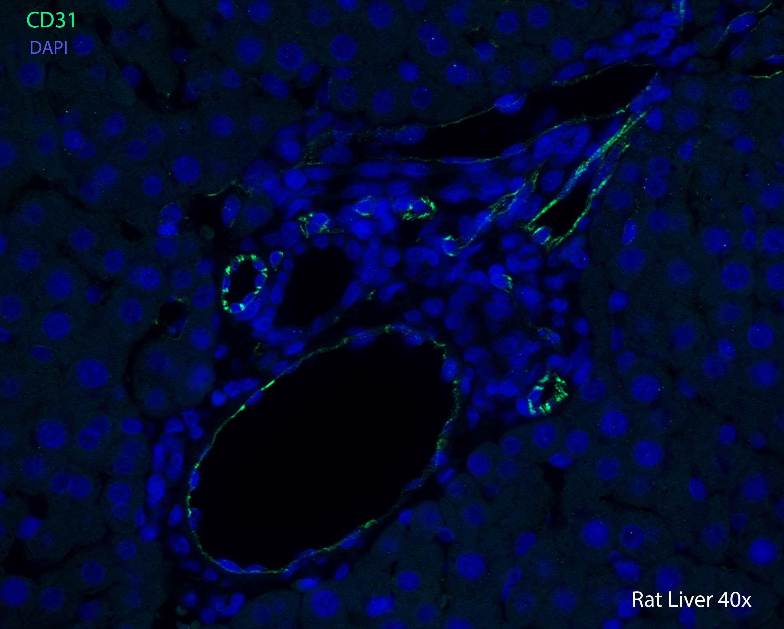
Immunohistological Detection of CD31/PECAM-1 in Paraffin Embedded Rat Liver
Staining of CD31/PECAM-1 in Rat Liver sample. IHC-P image submitted by a verified customer review.
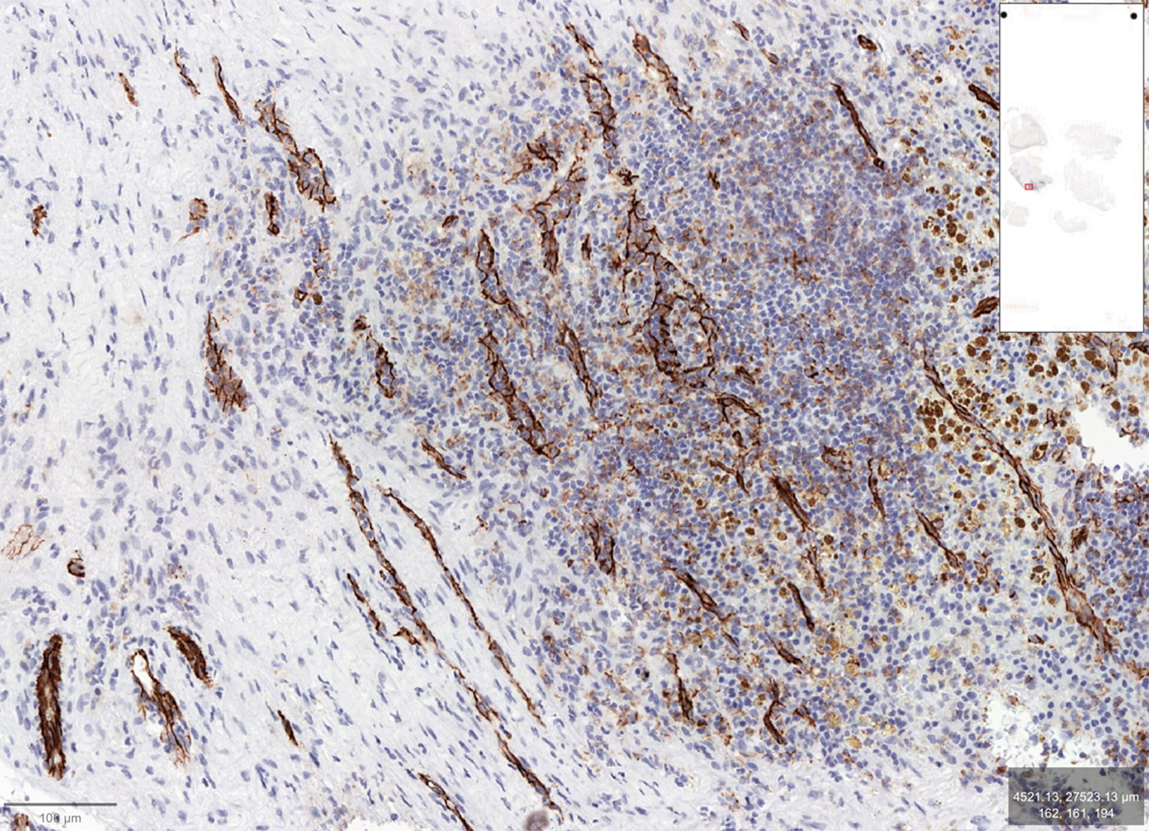
Staining of CD31/PECAM-1 in Paraffin Embedded Human Lung Adenocarcinoma
Human lung adenocarcinoma using. Antibody at 1:100 with citrate epitope retrieval at pH 6.0.
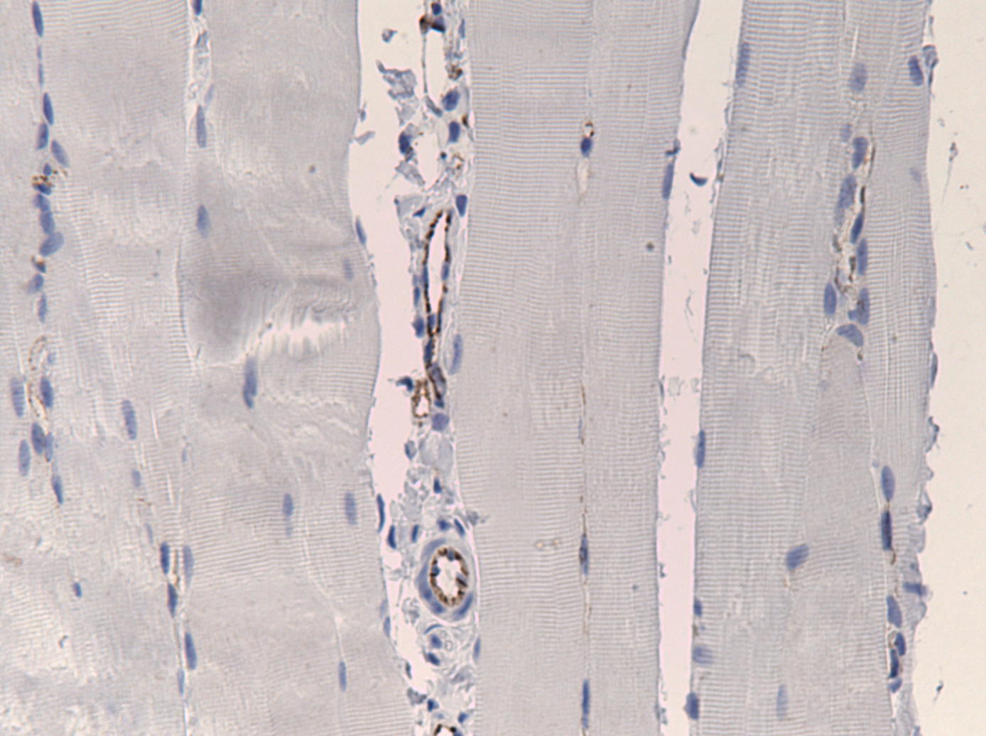
Immunohistological Detection of CD31/PECAM-1 in Paraffin Embedded Rat Muscle
Analysis in rat muscle blood vessels using. IHC-P image submitted by a verifed customer review.
Immunohistological Analysis of HIF-1 alpha in Mouse Tissue Implanted with Different Matrices
Matrix-HA supplemented with strontium was implanted subcutaneously in mice, where immunochemistry analysis and histology occurred of the newly formed tissues. (C) Within each matrix: Matrix-HA, Matrix-8Sr-HA and Matrix-50Sr-HA, were indicated by immunostaining of CD31 for the newly formed tissues. (D) Number of vessels within each tissue was quantified using NDP view software. Unit is square mm. Immunostaining analysis of slides occurred for 2 samples per condition and 3 sections were analyzed per sample and per group of matrix. Citation: Ehret C, Aid-Launais R, Sagardoy T, Siadous R, Bareille R, Rey S, et al. (2017) Strontium-doped hydroxyapatite polysaccharide materials effect on ectopic bone formation. PLoS ONE 12(9): e0184663. https://doi.org/10.1371/journal.pone.0184663
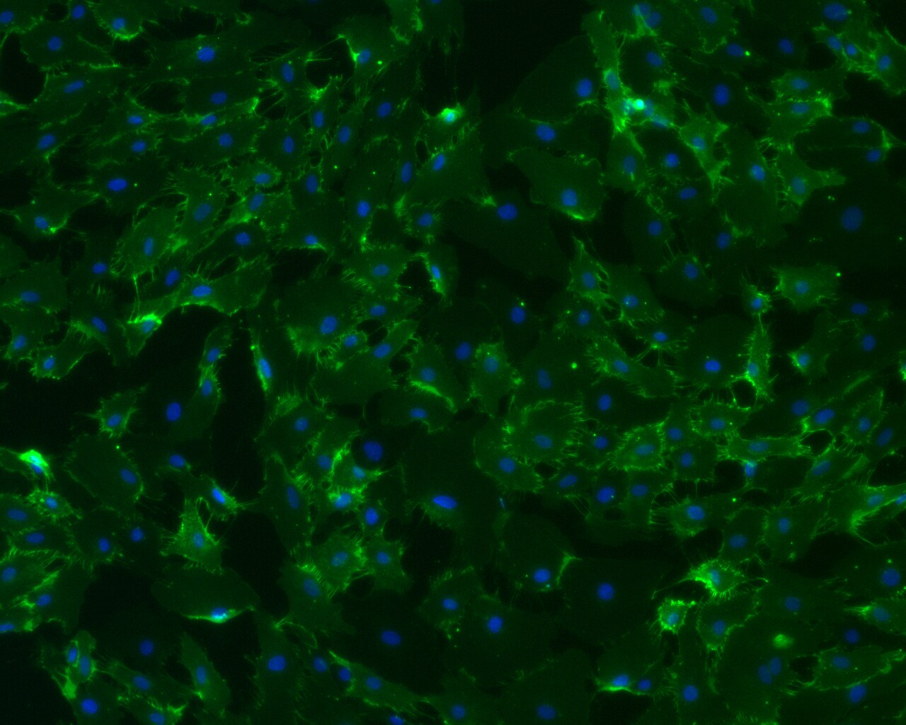
Human Umbilical Vein Endothelial Cells Stained for CD31/PECAM-1
Imaging of HUVEC monolayer on glass substrate. Fixation with 4% PFA. Blocking with Goat Serum at 1:100. Incubation 2 hours at room temperature. ICC/IF image submitted by a verified customer review.
Staining of CD31/PECAM-1 in Paraffin Embedded Canine Bladder Tissue
Analysis of canine kidney tissue using CD31/PECAM-1 antibody. Antibody was used at a 1:50 concentration in Casein in PBS and left at 4C overnight on paraffin embedded canine bladder tissue. HIER was performed in Tris/EDTA buffer, pH 9 for two hours at 75C. Image from verified customer review.
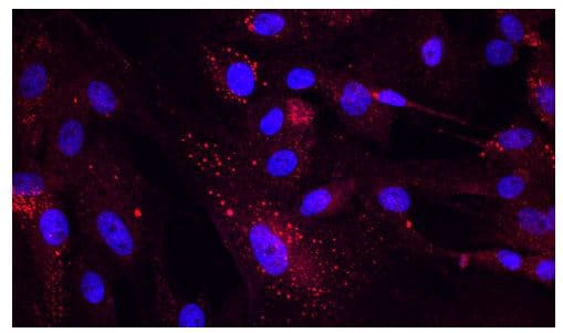
Immunocytochemistry/Immunofluorescence Detection of CD31/PECAM-1 in Porcine SC Cells
Analysis in pig SC cells using CD31/PECAM-1 Antibody (red). Blue color showing nucleus labeling. ICC/IF image submitted by a verified customer review.
Detection of CD31/PECAM-1 in 3 Groups of Rat Tissue Implanted with Matrices
CD31-PECAM-1-Antibody-Immunohistochemistry-NB100-2284-img0021.jpg
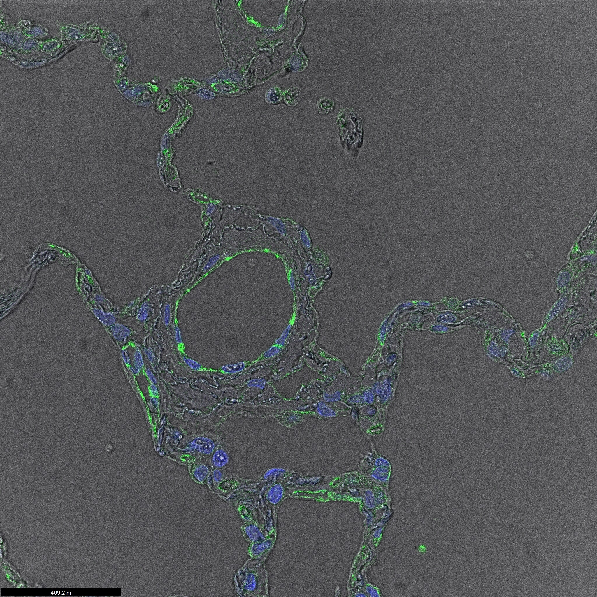
Immunohistochemical Detection of CD31/PECAM-1 in Frozen Mouse Pharyngeal Mesoderm
CD31 staining on E11.5 mouse Pharyngeal Mesoderm. Fixed with 4% PFA overnight. Blocked with 1% BSA. Primary antibody at 1:100. Secondary antibody at 1:1000, conjugated to Alexa Fluor 488. IHC-Fr image submitted by a verfied customer review.
Immunohistochemistry: CD31/PECAM-1 Antibody [NB100-2284] -
Immunohistochemistry evaluation of CD31 expression. (a) Representative images of CD31 staining of vessels in the three groups of implanted matrices (scale bar = 100 µm). (b) Quantification of vessel density inside the implanted matrices at 5 weeks post implantation (n = 6; Average ± SD). NS and **denote Non Significant and p < 0.01, respectively.
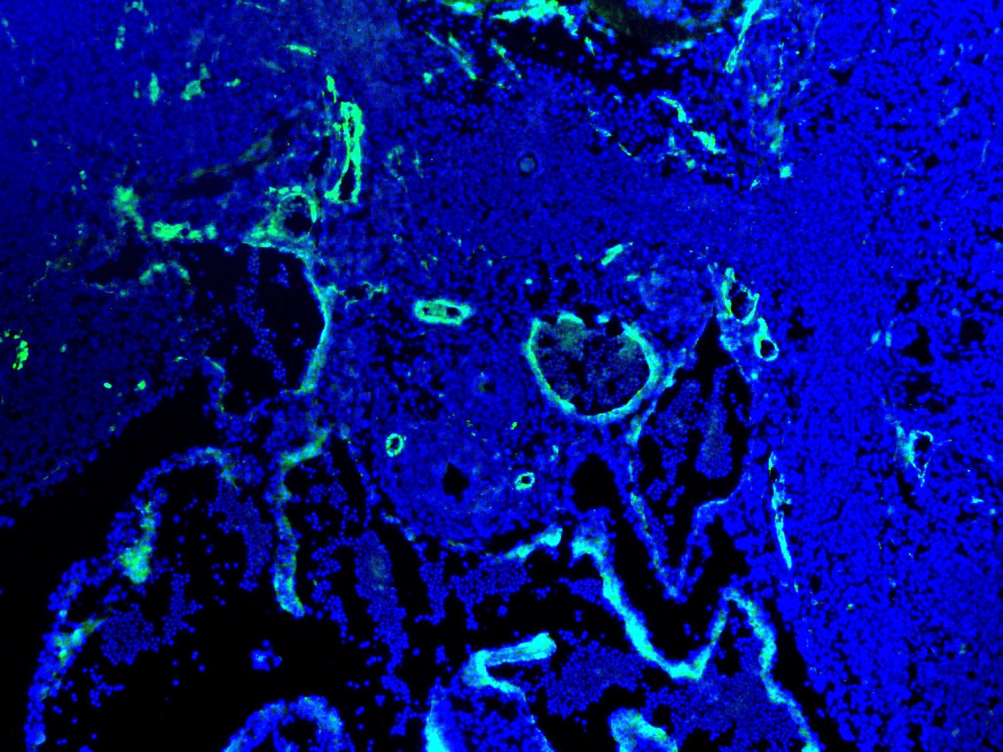
Immunohistochemistry: Rabbit Polyclonal CD31/PECAM-1 Antibody [NB100-2284]
Immunohistochemistry: Rabbit Polyclonal CD31/PECAM-1 Antibody [NB100-2284] - Staining of CD31 in human lung tissue. Nuclei in blue and CD31 (vessel) in green. Mouse anti-CD31 (1:100, overnight incubation at +4°C), followed by donkey anti-mouse 488 (1:400, 3h at RT). Image from a verified customer review.
Immunohistochemistry: CD31/PECAM-1 Antibody [NB100-2284] -
Immunohistochemistry: CD31/PECAM-1 Antibody [NB100-2284] - Antibodies selected for characterization of stromal cells & vasculature in FFPE specimens are species-specific. (A, B) Labeling of a patient colon tumor (A) & Colo205 human cell line xenograft (B) with anti-human mitochondrial antibody (single arrow- tumor cells; double arrows- stroma). (C, D) Labeling of a patient pancreatic tumor (C) & Colo205 human cell line xenograft (D) with anti-human CD34. (E, F) Labeling of a patient lung tumor (E) & Colo205 xenograft (F) with anti-human CD31. (G, H) Labeling of a patient lung tumor (G) & Colo205 xenograft (H) with anti-mouse CD34. (bars = 50 μ). Image collected & cropped by CiteAb from the following publication (https://translational-medicine.biomedcentral.com/articles/10.1186/1479-…), licensed under a CC-BY license. Not internally tested by Novus Biologicals.
Immunohistochemistry: CD31/PECAM-1 Antibody [NB100-2284] -
Immunohistochemistry: CD31/PECAM-1 Antibody [NB100-2284] - At the time of second passage, vessels in successfully growing xenografts of four different tumor types surveyed were of murine origin: A-C Colon; D-F Lung; G-I Pancreatic; J-L Renal Cell Carcinoma. For each tumor type, a representative section of an original patient specimen labeled with anti-huCD31 is shown (A, D, G, J). For each tumor, sections of the first passage xenografted tumor when it was resected are also shown; while no huCD31(+) vessels were identified in the xenografts (B, E, H, K), msCD34(+) vessels were abundant (C, F, I, L). (bars = 50 μ). Image collected & cropped by CiteAb from the following publication (https://translational-medicine.biomedcentral.com/articles/10.1186/1479-…), licensed under a CC-BY license. Not internally tested by Novus Biologicals.
Immunohistochemistry: CD31/PECAM-1 Antibody [NB100-2284] -
Immunohistochemistry: CD31/PECAM-1 Antibody [NB100-2284] - Vascularization of an engrafted patient colon tumor. A patient colon tumor was implanted in a cohort of mice & vessel development was analyzed over 8 weeks. Vessels in the original patient specimen labeled strongly for huCD31 (A) & not for msCD34 (D). Representative sections showing loss of huCD31(+) vessels (B- 4 weeks, C- 7 weeks) & presence of msCD34(+) vessels (E- 4 weeks, F- 7 weeks) are shown. The graph (G) summarizes this process; huCD31(+) vessels were rapidly lost, & by one week, mouse vessels were the predominate vessels present in the colon tumors (no data for msCD34 at 2 weeks; bars = 100 μ). Image collected & cropped by CiteAb from the following publication (https://translational-medicine.biomedcentral.com/articles/10.1186/1479-…), licensed under a CC-BY license. Not internally tested by Novus Biologicals.
Western Blot: CD31/PECAM-1 Antibody [NB100-2284] -
Western Blot: CD31/PECAM-1 Antibody [NB100-2284] - KCl pulse decreased membrane, but increased nuclear, detection of NHE1 in b.End3 endothelial cells.(A) Representative immunofluorescence images of bEnd.3 cells at two time-points after KCl or aCSF pulse. (B) Representative immunoblots of NHE1, alpha -tubulin, PECAM, & lamin B in cytosol, membrane, & nuclear fractions of bEnd.3 cells harvested at 5 min after KCl or aCSF pulse. (C = cytosol, M = membrane, N = nuclear) Values represent the mean ratio of NHE1 detection ± SEM (n = 11–12). (C) Representative immunoblots indicating NHE1 & alpha -tubulin as a loading control in whole cell lysate of bEnd.3 cells harvested at 5 min after KCl or aCSF pulse. Values represent the % of aCSF-treated relative expression ± SEM (n = 6). (D) Representative immunoblots of NHE1 & alpha -tubulin as a loading control in whole lysate of microvessels harvested at 90 min after cortical injection of KCl or aCSF. Values represent the % of naive relative expression ± SEM (n = 6). # denotes significantly different vs naïve (p<0.01), as assessed by one-way ANOVA (E) Intracellular pH during aCSF or KCl pulse. All data represent mean ± SEM (n = 45). (F) Extracellular pH during aCSF or KCl pulse. All data represent mean ± SEM (n = 6) in triplicate. Image collected & cropped by CiteAb from the following publication (https://pubmed.ncbi.nlm.nih.gov/32469979), licensed under a CC-BY license. Not internally tested by Novus Biologicals.
Immunocytochemistry/ Immunofluorescence: CD31/PECAM-1 Antibody [NB100-2284] -
Immunocytochemistry/ Immunofluorescence: CD31/PECAM-1 Antibody [NB100-2284] - Impact of T‐AuNPs on endothelial cells in vivo. Immunofluorescent staining for endothelial cells using an anti‐CD 31 antibody revealed an intact endothelial cell monolayer in the uninjured control carotid artery (N = 3). As expected post balloon angioplasty, there is no endothelial cell monolayer of the injured left carotid arteries in the injury alone, injury + T‐AuNP, & injury + S‐AuNP treatment conditions. Green indicates autofluorescence of the elastic lamina. Magenta indicates endothelial cells. Images obtained using 25× magnification with exposure time of 400 msec. TUNEL staining for apoptosis revealed no evidence of endothelial cell apoptosis in the uninjured control carotid arteries, with a prominent endothelial cell monolayer (white arrowhead). Mild apoptosis was noted in the media of balloon‐injured left carotid arteries, as expected. Black arrows indicate apoptotic cells. Representative images obtained using 40× magnification. Image collected & cropped by CiteAb from the following publication (https://pubmed.ncbi.nlm.nih.gov/28242820), licensed under a CC-BY license. Not internally tested by Novus Biologicals.
Immunohistochemistry: CD31/PECAM-1 Antibody [NB100-2284] -
Immunohistochemistry: CD31/PECAM-1 Antibody [NB100-2284] - The growth of a patient mesothelioma xenograft was supported by development of a murine vascular network. A patient mesothelioma was implanted in a cohort of mice & monitored for tumor growth. Once tumors began to actively grow, representative tumors were resected at weekly intervals & analyzed for vessel content. Staining for huCD31 was prominent in the original patient specimen (A), much reduced at 4 weeks (B) & negligible at 9 weeks (C). In contrast, patient specimens were not stained for msCD34 (D), whereas at 4 weeks large numbers of vessels stained for msCD34 (E) & by 9 weeks, msCD34 labeled vessels were predominant (F). The graph (G) summarizes the loss of detectable human vessels & acquisition of murine vessels over a 9 week period. (bars = 100 μ). Image collected & cropped by CiteAb from the following publication (https://translational-medicine.biomedcentral.com/articles/10.1186/1479-…), licensed under a CC-BY license. Not internally tested by Novus Biologicals.
Immunocytochemistry/ Immunofluorescence: CD31/PECAM-1 Antibody [NB100-2284] -
Immunocytochemistry/ Immunofluorescence: CD31/PECAM-1 Antibody [NB100-2284] - TGF‐ beta 1 released by MSCs contributes to induced myofibroblast differentiation & granulation tissue formationA–C2.5 × 105 of TGF‐ beta 1 siRNA or control siRNA‐transfected AT‐MSCs were intradermally injected around each of CD18−/− murine wound. PBS mock injection served as negative control. Wound tissue was harvested at day 2, 5, & 7 post‐wounding for quantification of human TGF‐ beta 1 mRNA (A) at day 2 by qPCR, total TGF‐ beta 1 (B), & active TGF‐ beta 1 (C) protein at day 5 by ELISA. Data are expressed as mean ± SEM, n = 3 wounds per group, **P < 0.01, by one‐way ANOVA with Tukey's test.D–GExpression of alpha ‐SMA (D & E) & CD31 (F & G) at days 5 & 7 by immunostaining on tissue sections. The dashed lines indicate the border of the wound bed & epidermis or eschar. e, epidermis; es, eschar; wb, wound bed. Scale bars: 100 μm. Quantification data are expressed as mean ± SEM, n = 3 wounds per group, *P < 0.05, ***P < 0.001, by one‐way ANOVA with Tukey's test.Source data are available online for this figure. Image collected & cropped by CiteAb from the following publication (https://pubmed.ncbi.nlm.nih.gov/32080965), licensed under a CC-BY license. Not internally tested by Novus Biologicals.
Immunohistochemistry-Paraffin: Rabbit Polyclonal CD31/PECAM-1 Antibody [NB100-2284]
Immunohistochemistry-Paraffin: Rabbit Polyclonal CD31/PECAM-1 Antibody [NB100-2284] - CD31 positive vessels in a canine splenic hemangiosarcoma.Tissue slides were loaded into automated research stainer, dewaxed and pretreated with EDTA-based epitope retrieval ER2 solution for 20 mins at 100°C. After a peroxide incubation for 5 min, the rabbit antibody against CD31 (NB100-2284) (1:100) was incubated for 60 mins at room temperature followed by Bond Polymer (anti-rabbit HRP) incubation for 8 min at room temperature. Mixed DAB reagent (Polymer Refine Detection Kit) was incubated for 10 mins, and Hematoxylin (Refine Detection Kit) counterstaining for 10 mins. After staining, sample slides were washed in water, dehydrated using ethanol gradient (70%, 90%, 100%), washed three times, and mounted in mounting medium. Image from a verified customer review.Applications for CD31/PECAM-1 Antibody
Application
Recommended Usage
Immunocytochemistry/ Immunofluorescence
1:50 - 1:500
Immunohistochemistry
1:100 - 1:500
Immunohistochemistry-Frozen
1:10 - 1:500
Immunohistochemistry-Paraffin
1:100 - 1:500
Western Blot
1:100 - 1:2000
Application Notes
For IHC-P: Epitope exposure is recommended, with citrate buffer will enhance staining. In some cases, the antibody may be diluted further than indicated. IHC-Fr, WB reactivity reported in scientific literature (PMID:23317813). ICC/IF reactivity reported in scientific literature (PMID: 27328066).
Reviewed Applications
Read 8 reviews rated 4.8 using NB100-2284 in the following applications:
Formulation, Preparation, and Storage
Purification
Immunogen affinity purified
Formulation
TBS, 0.1% BSA
Preservative
0.09% Sodium Azide
Concentration
0.1 mg/ml
Shipping
The product is shipped with polar packs. Upon receipt, store it immediately at the temperature recommended below.
Stability & Storage
Store at 4C. Do not freeze.
Background: CD31/PECAM-1
PECAM's intracellular cytoplasmic domain consists of a sequence of 118 amino acids and contains serine and tyrosine (also referred to as immunoreceptor tyrosine-based inhibitory motifs-ITIMs) residues, which may be phosphorylated upon cellular stimulation (3). ITIMs are phosphorylated by Src-family kinases and non-Src family kinases (e.g., Csk), leading to a conformational change which supports interactions with Src homology 2 (SH2) domain containing proteins such as protein-tyrosine phosphatase, SHP-2 (1,2). Formation of SHP-2/PECAM-1 complexes induces endothelial cell migration through the dephosphorylation of focal adhesion kinase and regulation of RhoA activity (1). Signaling downstream of ITIM tyrosine phosphorylations also plays a role in PECAM's anti-apoptotic activity, a function which is independent of its interaction with SHP-2. In platelets and leukocytes, phosphorylation of PECAM's cytosolic domain is inhibitory, preventing their activation.
References
1. Lertkiatmongkol, P., Liao, D., Mei, H., Hu, Y., & Newman, P. J. (2016). Endothelial functions of PECAM-1 (CD31). Current Opinion in Hematology. https://doi.org/10.1097/MOH.0000000000000239.Endothelial
2. Privratsky, J. R., & Newman, P. J. (2014). PECAM-1: Regulator of endothelial junctional integrity. Cell and Tissue Research. https://doi.org/10.1007/s00441-013-1779-3
3. Newman, P. J., & Newman, D. K. (2003). Signal transduction pathways mediated by PECAM-1: New roles for an old molecule in platelet and vascular cell biology. Arteriosclerosis, Thrombosis, and Vascular Biology. https://doi.org/10.1161/01.ATV.0000071347.69358.D9
Long Name
Platelet Endothelial Cell Adhesion Molecule 1
Alternate Names
CD31, EndoCAM, PECA1, PECAM-1, PECAM1
Entrez Gene IDs
5175 (Human)
Gene Symbol
PECAM1
UniProt
Additional CD31/PECAM-1 Products
Product Documents for CD31/PECAM-1 Antibody
Certificate of Analysis
To download a Certificate of Analysis, please enter a lot or batch number in the search box below.
Product Specific Notices for CD31/PECAM-1 Antibody
This product is for research use only and is not approved for use in humans or in clinical diagnosis. Primary Antibodies are guaranteed for 1 year from date of receipt.
Related Research Areas
Citations for CD31/PECAM-1 Antibody
 Powered by Bioz
Customer Reviews for CD31/PECAM-1 Antibody (8)
4.8 out of 5
8 Customer Ratings
Have you used CD31/PECAM-1 Antibody?
Submit a review and receive an Amazon gift card!
$25/€18/£15/$25CAN/¥2500 Yen for a review with an image
$10/€7/£6/$10CAN/¥1110 Yen for a review without an image
Submit a review
Customer Images
Showing
1
-
5 of
8 reviews
Showing All
Filter By:
-
Application: Immunohistochemistry-ParaffinSample Tested: spleen and Splenic hemangiosarcomaSpecies: CanineVerified Customer | Posted 03/13/2025CD31 positive vessels in a canine splenic hemangiosarcomaTissue slides were loaded into automated research stainer, dewaxed and pretreated with EDTA-based epitope retrieval ER2 solution for 20 mins at 100°C. After a peroxide incubation for 5 min, the rabbit antibody against CD31 (NB100-2284) (1:100) was incubated for 60 mins at room temperature followed by Bond Polymer (anti-rabbit HRP) incubation for 8 min at room temperature. Mixed DAB reagent (Polymer Refine Detection Kit) was incubated for 10 mins, and Hematoxylin (Refine Detection Kit) counterstaining for 10 mins. After staining, sample slides were washed in water, dehydrated using ethanol gradient (70%, 90%, 100%), washed three times, and mounted in mounting medium.

-
Application: ImmunofluorescenceSample Tested: Lung tissueSpecies: HumanVerified Customer | Posted 08/28/2024Nuclei in blue CD31 (vessel) in greenmouse anti-CD31 (1:100, overnight incubation at +4°C), followed by donkey anti-mouse 488 (1:400, 3h at RT)
-
Application: Immunohistochemistry-ParaffinSample Tested: Adult kidneySpecies: CanineVerified Customer | Posted 04/28/2022Analysis of canine kidney tissue using PECAM-1 antibody.Antibody was used at a 1:50 concentration in Casein in PBS and left at 4C overnight on paraffin embedded canine bladder tissue. HIER was performed in Tris/EDTA buffer, pH 9 for two hours at 75C.
-
Application: Immunohistochemistry-FrozenSample Tested: E11.5 mouse embryo fixed in 4% PFASpecies: MouseVerified Customer | Posted 01/07/2021CD31 staining on E11.5 mouse Pharyngeal MesodermFixed 4% PFA overnight. Blocked with 1% BSA Primary antibody dilution - 1:100 Secondary antibody - Invitrogen Alexa Fluor 488 Secondary antibody dilution - 1:1000
-
Application: Immunohistochemistry-ParaffinSample Tested: Rat liverSpecies: RatVerified Customer | Posted 02/01/2018Novus CD31/PECAM-1 [NB100-2284] Rat LiverFormalin fixed Paraffin embedded rat liver tissue Antigen retrieval pH6.0 citrate buffer. Sea Block blocking solution 30min. NB100-2284 diluted 1/100 in Sea Block and incubated overnight at 4degC Alexa Fluor® 488 Goat anti Rabbit 1/300 in Sea Block for 1hr at 37degC. Antifade blocking mounting media with DAPI Axio Observer 7 Apotome Imaging 40x
-
Application: ImmunocytochemistrySample Tested: HUVEC (Human Umbilical Vein Endothelial Cells)Species: HumanVerified Customer | Posted 08/29/2017HUVEC monolayer on glass substrateFixation 4% PFA Blocking Goat Serum Dilution 1:100 Incubation 2h @ RT
-
Application: ImmunocytochemistrySample Tested: SC cellsSpecies: OtherVerified Customer | Posted 01/07/2016Pig SC cells stained with CD31 antibody
-
Application: Immunohistochemistry-ParaffinSample Tested: rat muscle FFPESpecies: RatVerified Customer | Posted 01/27/2015CD31 in rat muscle blood vessels
There are no reviews that match your criteria.
Protocols
Find general support by application which include: protocols, troubleshooting, illustrated assays, videos and webinars.
- Antigen Retrieval Protocol (PIER)
- Antigen Retrieval for Frozen Sections Protocol
- Appropriate Fixation of IHC/ICC Samples
- Cellular Response to Hypoxia Protocols
- Chromogenic IHC Staining of Formalin-Fixed Paraffin-Embedded (FFPE) Tissue Protocol
- Chromogenic Immunohistochemistry Staining of Frozen Tissue
- ClariTSA™ Fluorophore Kits
- Detection & Visualization of Antibody Binding
- Fluorescent IHC Staining of Frozen Tissue Protocol
- Graphic Protocol for Heat-induced Epitope Retrieval
- Graphic Protocol for the Preparation and Fluorescent IHC Staining of Frozen Tissue Sections
- Graphic Protocol for the Preparation and Fluorescent IHC Staining of Paraffin-embedded Tissue Sections
- Graphic Protocol for the Preparation of Gelatin-coated Slides for Histological Tissue Sections
- ICC Cell Smear Protocol for Suspension Cells
- ICC Immunocytochemistry Protocol Videos
- ICC for Adherent Cells
- IHC Sample Preparation (Frozen sections vs Paraffin)
- Immunocytochemistry (ICC) Protocol
- Immunocytochemistry Troubleshooting
- Immunofluorescence of Organoids Embedded in Cultrex Basement Membrane Extract
- Immunofluorescent IHC Staining of Formalin-Fixed Paraffin-Embedded (FFPE) Tissue Protocol
- Immunohistochemistry (IHC) and Immunocytochemistry (ICC) Protocols
- Immunohistochemistry Frozen Troubleshooting
- Immunohistochemistry Paraffin Troubleshooting
- Preparing Samples for IHC/ICC Experiments
- Preventing Non-Specific Staining (Non-Specific Binding)
- Primary Antibody Selection & Optimization
- Protocol for Heat-Induced Epitope Retrieval (HIER)
- Protocol for Making a 4% Formaldehyde Solution in PBS
- Protocol for VisUCyte™ HRP Polymer Detection Reagent
- Protocol for the Fluorescent ICC Staining of Cell Smears - Graphic
- Protocol for the Fluorescent ICC Staining of Cultured Cells on Coverslips - Graphic
- Protocol for the Preparation & Fixation of Cells on Coverslips
- Protocol for the Preparation and Chromogenic IHC Staining of Frozen Tissue Sections
- Protocol for the Preparation and Chromogenic IHC Staining of Frozen Tissue Sections - Graphic
- Protocol for the Preparation and Chromogenic IHC Staining of Paraffin-embedded Tissue Sections
- Protocol for the Preparation and Chromogenic IHC Staining of Paraffin-embedded Tissue Sections - Graphic
- Protocol for the Preparation and Fluorescent ICC Staining of Cells on Coverslips
- Protocol for the Preparation and Fluorescent ICC Staining of Non-adherent Cells
- Protocol for the Preparation and Fluorescent ICC Staining of Stem Cells on Coverslips
- Protocol for the Preparation and Fluorescent IHC Staining of Frozen Tissue Sections
- Protocol for the Preparation and Fluorescent IHC Staining of Paraffin-embedded Tissue Sections
- Protocol for the Preparation of Gelatin-coated Slides for Histological Tissue Sections
- Protocol for the Preparation of a Cell Smear for Non-adherent Cell ICC - Graphic
- R&D Systems Quality Control Western Blot Protocol
- TUNEL and Active Caspase-3 Detection by IHC/ICC Protocol
- The Importance of IHC/ICC Controls
- Troubleshooting Guide: Immunohistochemistry
- Troubleshooting Guide: Western Blot Figures
- Western Blot Conditions
- Western Blot Protocol
- Western Blot Protocol for Cell Lysates
- Western Blot Troubleshooting
- Western Blot Troubleshooting Guide
- View all Protocols, Troubleshooting, Illustrated assays and Webinars
FAQs for CD31/PECAM-1 Antibody
Showing
1
-
3 of
3 FAQs
Showing All
-
Q: I am looking for references concerning the use of NBP2-15797, NBP1-47913 and NB100-2284 in IHC on PFA- fixed section.
A: Among your mentioned catalog numbers (NBP2-15797, NBP1-47913 and NB100-2284), we have published references for NB100-2284 only. Hylander et al. 2013 (Journal of Translational Medicine 2013, 11:110) have cited the IHC-P use of NB100-2284 and this paper has also explained a nice detailed IHC-P protocol that you may follow if you want to. NBP1-71663 is our bestselling catalog number among all CD31/PECAM1 antibodies and it has been cited in two publications, both explaining its use in IHC-P assays (Pratheeshkumar et al. 2012 PLoS One. 2012;7(10):e47516; and Pratheeshkumar et al. 2012 PLoS One. 2012;7(12):e52279)
-
Q: I'm interested in your antibody CD31/PECAM1 ref: NB100-2284. I'd like to stain human endothelial cells injected in mouse muscle. Do you know if your antibody cross-reacts with mouse? Is this antibody working on Immunohistochemistry frozen sections?
A:
As is listed on our datasheet, NB100-2284 reacts with human and mouse in WB, immunocytochemistry and IHC on both frozen and paraffin-embedded tissues. Novus also offers sample sizes of several products, which may be identified by a "SS" at the end of the catalog number (ie. NB100-105SS is the sample size of NB100-105). We also offer Super Novus packs to many common antigens. These kits enable customers to test several different antibodies to the target at a discounted rate.
-
Q: I'm interested in your antibody CD31/PECAM1 ref: NB100-2284. I'd like to stain human endothelial cells injected in mouse muscle. Do you know if your antibody cross-reacts with mouse? Is this antibody working on Immunohistochemistry frozen sections?
A: NB100-2284 reacts with human and mouse in WB, immunocytochemistry and IHC on both frozen and paraffin-embedded tissues. Novus also offers sample sizes of several products, which may be identified by a SS at the end of the catalog number (ie. NB100-105SS is the sample size of NB100-105). We also offer Super Novus packs to many common antigens. These kits enable customers to test several different antibodies to the target at a discounted rate.
-
Q: I am looking for references concerning the use of NBP2-15797, NBP1-47913 and NB100-2284 in IHC on PFA- fixed section.
A: Among your mentioned catalog numbers (NBP2-15797, NBP1-47913 and NB100-2284), we have published references for NB100-2284 only. Hylander et al. 2013 (Journal of Translational Medicine 2013, 11:110) have cited the IHC-P use of NB100-2284 and this paper has also explained a nice detailed IHC-P protocol that you may follow if you want to. NBP1-71663 is our bestselling catalog number among all CD31/PECAM1 antibodies and it has been cited in two publications, both explaining its use in IHC-P assays (Pratheeshkumar et al. 2012 PLoS One. 2012;7(10):e47516; and Pratheeshkumar et al. 2012 PLoS One. 2012;7(12):e52279)
-
Q: I'm interested in your antibody CD31/PECAM1 ref: NB100-2284. I'd like to stain human endothelial cells injected in mouse muscle. Do you know if your antibody cross-reacts with mouse? Is this antibody working on Immunohistochemistry frozen sections?
A:
As is listed on our datasheet, NB100-2284 reacts with human and mouse in WB, immunocytochemistry and IHC on both frozen and paraffin-embedded tissues. Novus also offers sample sizes of several products, which may be identified by a "SS" at the end of the catalog number (ie. NB100-105SS is the sample size of NB100-105). We also offer Super Novus packs to many common antigens. These kits enable customers to test several different antibodies to the target at a discounted rate.
-
Q: I'm interested in your antibody CD31/PECAM1 ref: NB100-2284. I'd like to stain human endothelial cells injected in mouse muscle. Do you know if your antibody cross-reacts with mouse? Is this antibody working on Immunohistochemistry frozen sections?
A: NB100-2284 reacts with human and mouse in WB, immunocytochemistry and IHC on both frozen and paraffin-embedded tissues. Novus also offers sample sizes of several products, which may be identified by a SS at the end of the catalog number (ie. NB100-105SS is the sample size of NB100-105). We also offer Super Novus packs to many common antigens. These kits enable customers to test several different antibodies to the target at a discounted rate.
Loading...