CD63 Antibody (H5C6) - BSA Free
Novus Biologicals | Catalog # NBP2-42225
Clone H5C6 was used by HLDA to establish CD designation.
Key Product Details
Species Reactivity
Validated:
Human, Canine
Cited:
Human, Mouse, Rat, Canine
Applications
Validated:
Immunohistochemistry Whole-Mount, Western Blot, ELISA, Block/Neutralize, Flow Cytometry, Flow (Intracellular), Immunocytochemistry/ Immunofluorescence, Immunoprecipitation, Dot Blot, Functional, In vitro assay, Electron Microscopy
Cited:
Western Blot, ELISA, Immunoassay, Block/Neutralize, Flow Cytometry, Immunocytochemistry/ Immunofluorescence, Immunoprecipitation, Cytometric Bead Assay Standard, Dot Blot, In vitro assay, Functional Assay, Electron Microscopy
Label
Unconjugated
Antibody Source
Monoclonal Mouse IgG1 kappa Clone # H5C6
Format
BSA Free
Loading...
Product Specifications
Immunogen
Human splenic adherent cells.
Marker
Exosome Marker
Clonality
Monoclonal
Host
Mouse
Isotype
IgG1 kappa
Scientific Data Images for CD63 Antibody (H5C6) - BSA Free
![Western Blot: CD63 Antibody (H5C6)BSA Free [NBP2-42225]](https://resources.rndsystems.com/images/products/CD63-Antibody-H5C6-Western-Blot-NBP2-42225-img0002.jpg "Western Blot: CD63 Antibody (H5C6)BSA Free [NBP2-42225]")
Western Blot: CD63 Antibody (H5C6)BSA Free [NBP2-42225]
Western Blot: CD63 Antibody (H5C6) [NBP2-42225] - THP1 whole cell protein was separated by SDS-PAGE on a 12% gel and transferred to PVDF membrane. The membrane was probed with anti-CD63 antibody at 2 ug/mL and detected with an anti-mouse HRP secondary antibody using chemiluminescence.![Immunocytochemistry/ Immunofluorescence: CD63 Antibody (H5C6) - BSA Free [NBP2-42225]](https://resources.rndsystems.com/images/products/CD63-Antibody-H5C6-Immunocytochemistry-Immunofluorescence-NBP2-42225-img0001.jpg "Immunocytochemistry/ Immunofluorescence: CD63 Antibody (H5C6) - BSA Free [NBP2-42225]")
Immunocytochemistry/ Immunofluorescence: CD63 Antibody (H5C6) - BSA Free [NBP2-42225]
Immunocytochemistry/Immunofluorescence: CD63 Antibody (H5C6) [NBP2-42225] - The CD63 (H5C6) antibody was tested in HeLa cells at a 1:50 dilution against DyLight 488 (Green). Actin and nuclei were counterstained against Phalloidin 568 (Red) and DAPI (Blue), respectively.![Immunocytochemistry/ Immunofluorescence: CD63 Antibody (H5C6) - BSA Free [NBP2-42225]](https://resources.rndsystems.com/images/products/CD63-Antibody-H5C6-Immunocytochemistry-Immunofluorescence-NBP2-42225-img0014.jpg "Immunocytochemistry/ Immunofluorescence: CD63 Antibody (H5C6) - BSA Free [NBP2-42225]")
Immunocytochemistry/ Immunofluorescence: CD63 Antibody (H5C6) - BSA Free [NBP2-42225]
Immunocytochemistry/Immunofluorescence: CD63 Antibody (H5C6) [NBP2-42225] - HeLa cells were fixed in 4% paraformaldehyde for 10 minutes and permeabilized in 0.05% Triton X-100 in PBS for 5 minutes. The cells were incubated with anti-CD63 Antibody [H5C6] conjugated to DyLight 550 (NBP2-42225R) at 5 ug/ml for 1 hour at room temperature. Nuclei were counterstained with DAPI (Blue). Cells were imaged using a 100X objective and digitally deconvolved.![Flow Cytometry: CD63 Antibody (H5C6) - BSA Free [NBP2-42225]](https://resources.rndsystems.com/images/products/CD63-Antibody-H5C6-Flow-Cytometry-NBP2-42225-img0013.jpg "Flow Cytometry: CD63 Antibody (H5C6) - BSA Free [NBP2-42225]")
Flow Cytometry: CD63 Antibody (H5C6) - BSA Free [NBP2-42225]
Flow Cytometry: CD63 Antibody (H5C6) [NBP2-42225] - An intracellular stain was performed on SK-MEL-28 cells with CD63 (H5C6) Antibody NBP2-42225R (blue) and a matched isotype control (orange). Cells were fixed with 4% PFA and then permeabilized with 0.1% saponin. Cells were incubated in an antibody dilution of 5 ug/mL for 30 minutes at room temperature. Both antibodies were conjugated to DyLight 550.![Immunocytochemistry/ Immunofluorescence: CD63 Antibody (H5C6) - BSA Free [NBP2-42225]](https://resources.rndsystems.com/images/products/CD63-Antibody-H5C6-Immunocytochemistry-Immunofluorescence-NBP2-42225-img0006.jpg "Immunocytochemistry/ Immunofluorescence: CD63 Antibody (H5C6) - BSA Free [NBP2-42225]")
Immunocytochemistry/ Immunofluorescence: CD63 Antibody (H5C6) - BSA Free [NBP2-42225]
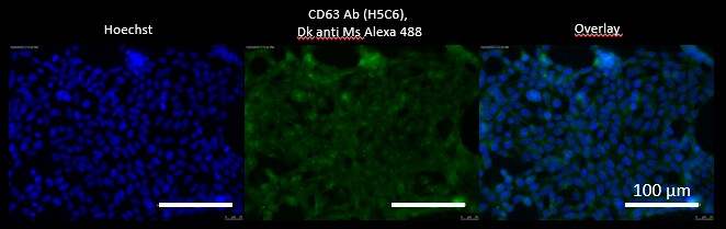
Immunocytochemistry/Immunofluorescence: CD63 Antibody (H5C6) [NBP2-42225] - MDCK cells stained with CD63 antibody at a dilution of 1:50 followed by Donkey anti-mouse secondary antibody conjugated with Alexa Fluor 488 (1:500). Nuclei were stained with Hoechst 33342. ICC/IF image submitted by a verified customer review.![Immunocytochemistry/ Immunofluorescence: CD63 Antibody (H5C6) - BSA Free [NBP2-42225]](https://resources.rndsystems.com/images/products/CD63-Antibody-H5C6-Immunocytochemistry-Immunofluorescence-NBP2-42225-img0015.jpg "Immunocytochemistry/ Immunofluorescence: CD63 Antibody (H5C6) - BSA Free [NBP2-42225]")
Immunocytochemistry/ Immunofluorescence: CD63 Antibody (H5C6) - BSA Free [NBP2-42225]
Immunocytochemistry/Immunofluorescence: CD63 Antibody (H5C6) [NBP2-42225] - U2OS cells were fixed in 4% paraformaldehyde for 10 minutes and permeabilized in 0.05% Triton X-100 in PBS for 5 minutes. The cells were incubated with anti-CD63 Antibody [H5C6] conjugated to Alexa Fluor 488 (NBP2-42225AF488) at 5 ug/ml for 1 hour at room temperature. Nuclei were counterstained with DAPI (Blue). Cells were imaged using a 100X objective and digitally deconvolved.![Immunocytochemistry/ Immunofluorescence: CD63 Antibody (H5C6) - BSA Free [NBP2-42225]](https://resources.rndsystems.com/images/products/CD63-Antibody-H5C6-Immunocytochemistry-Immunofluorescence-NBP2-42225-img0017.jpg "Immunocytochemistry/ Immunofluorescence: CD63 Antibody (H5C6) - BSA Free [NBP2-42225]")
Immunocytochemistry/ Immunofluorescence: CD63 Antibody (H5C6) - BSA Free [NBP2-42225]
Immunocytochemistry/Immunofluorescence: CD63 Antibody (H5C6) [NBP2-42225] - A431 cells were fixed in 4% paraformaldehyde for 10 minutes and permeabilized in 0.05% Triton X-100 in PBS for 5 minutes. The cells were incubated with anti-CD63 Antibody [H5C6] conjugated to Biotin (NBP2-42225B) at 5 ug/ml for 60 minutes at room temperature and detected with Streptavidin Protein conjugated to DyLight 488 at a 1:500 dilution for 60 minutes. Nuclei were counterstained with DAPI (Blue). Cells were imaged using a 100X objective and digitally deconvolved.![Immunocytochemistry/ Immunofluorescence: CD63 Antibody (H5C6) - BSA Free [NBP2-42225]](https://resources.rndsystems.com/images/products/CD63-Antibody-H5C6-Immunocytochemistry-Immunofluorescence-NBP2-42225-img0016.jpg "Immunocytochemistry/ Immunofluorescence: CD63 Antibody (H5C6) - BSA Free [NBP2-42225]")
Immunocytochemistry/ Immunofluorescence: CD63 Antibody (H5C6) - BSA Free [NBP2-42225]
Immunocytochemistry/Immunofluorescence: CD63 Antibody (H5C6) [NBP2-42225] - HeLa cells were fixed in 4% paraformaldehyde for 10 minutes and permeabilized in 0.05% Triton X-100 in PBS for 5 minutes. The cells were incubated with anti-CD63 Antibody [H5C6] conjugated to Biotin (NBP2-42225B) at 5 ug/ml for 1 hour at room temperature and detected with Streptavidin conjugated to DyLight 488. Nuclei were counterstained with DAPI (Blue). Cells were imaged using a 100X objective and digitally deconvolved.![Immunocytochemistry/ Immunofluorescence: CD63 Antibody (H5C6) - BSA Free [NBP2-42225]](https://resources.rndsystems.com/images/products/CD63-Antibody-H5C6-Immunocytochemistry-Immunofluorescence-NBP2-42225-img0008.jpg "Immunocytochemistry/ Immunofluorescence: CD63 Antibody (H5C6) - BSA Free [NBP2-42225]")
Immunocytochemistry/ Immunofluorescence: CD63 Antibody (H5C6) - BSA Free [NBP2-42225]
Immunocytochemistry/Immunofluorescence: CD63 Antibody (H5C6) [NBP2-42225] - HeLa cells were fixed for 10 minutes using 10% formalin and then permeabilized for 5 minutes using 1X PBS + 0.05% Triton X-100. The cells were incubated with anti-CD63 [H5C6] conjugated to Alexa Fluor 488 [NBP2-42225AF488] at 10ug/mL for 1 hour at room temperature. Nuclei were counterstained with DAPI (Blue). Cells were imaged using a 40X objective.![Western Blot: CD63 Antibody (H5C6)BSA Free [NBP2-42225]](https://resources.rndsystems.com/images/products/CD63-Antibody-H5C6-BSA-Free-Western-Blot-NBP2-42225-img0019.jpg "Western Blot: CD63 Antibody (H5C6)BSA Free [NBP2-42225]")
Western Blot: CD63 Antibody (H5C6)BSA Free [NBP2-42225]
CD63-Antibody-H5C6-BSA-Free-Western-Blot-NBP2-42225-img0019.jpg![Flow Cytometry: CD63 Antibody (H5C6) - BSA Free [NBP2-42225]](https://resources.rndsystems.com/images/products/CD63-Antibody-H5C6-Flow-Cytometry-NBP2-42225-img0018.jpg "Flow Cytometry: CD63 Antibody (H5C6) - BSA Free [NBP2-42225]")
Flow Cytometry: CD63 Antibody (H5C6) - BSA Free [NBP2-42225]
Flow Cytometry: CD63 Antibody (H5C6) [NBP2-42225] - An intracellular stain was performed on HeLa cells with CD63 [H5C6] Antibody NBP2-42225AF594 (blue) and a matched isotype control (orange). Cells were fixed with 4% PFA and then permeabilized with 0.1% saponin. Cells were incubated in an antibody dilution of 2.5 ug/mL for 30 minutes at room temperature. Both antibodies were conjugated to Alexa Fluor 594.![Western Blot: CD63 Antibody (H5C6)BSA Free [NBP2-42225]](https://resources.rndsystems.com/images/products/CD63-Antibody-H5C6-Western-Blot-NBP2-42225-img0007.jpg "Western Blot: CD63 Antibody (H5C6)BSA Free [NBP2-42225]")
Western Blot: CD63 Antibody (H5C6)BSA Free [NBP2-42225]
Western Blot: CD63 Antibody (H5C6) [NBP2-42225] - The same samples and volumes were run under reducing and non-reducing conditions. All procedures were performed in parallel for both conditions. L- ladder +- exosomes. Image visualized with HRP linked to secondary antibody. Western blot image submitted by a verified customer review.![Immunocytochemistry/ Immunofluorescence: CD63 Antibody (H5C6) - BSA Free [NBP2-42225]](https://resources.rndsystems.com/images/products/CD63-Antibody-H5C6-Immunocytochemistry-Immunofluorescence-NBP2-42225-img0005.jpg "Immunocytochemistry/ Immunofluorescence: CD63 Antibody (H5C6) - BSA Free [NBP2-42225]")
Immunocytochemistry/ Immunofluorescence: CD63 Antibody (H5C6) - BSA Free [NBP2-42225]
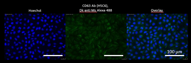
Immunocytochemistry/Immunofluorescence: CD63 Antibody (H5C6) [NBP2-42225] - HEK cells stained with CD63 antibody at a dilution of 1:50 followed by Donkey anti-mouse secondary antibody conjugated with Alexa Fluor 488 (1:500). Nuclei were stained with Hoechst 33342. ICC/IF image submitted by a verified customer review.![Flow Cytometry: CD63 Antibody (H5C6) - BSA Free [NBP2-42225]](https://resources.rndsystems.com/images/products/CD63-Antibody-H5C6-Flow-Cytometry-NBP2-42225-img0003.jpg "Flow Cytometry: CD63 Antibody (H5C6) - BSA Free [NBP2-42225]")
Flow Cytometry: CD63 Antibody (H5C6) - BSA Free [NBP2-42225]
Flow Cytometry: CD63 Antibody (H5C6) [NBP2-42225] - Human peripheral blood cells were stained (2 x 10^6 cells/mL) using the anti-CD63 antibody (Blue) at a dilution of 1:1000. Signal was detected using a Gt x Ms DyLight 488 Secondary and gated to the monocyte/granulocyte cell populations. Isotype was Mouse IgG1 kappa (orange). Data collected on BD FACS Calibur flow cytometer.![Flow Cytometry: CD63 Antibody (H5C6) - BSA Free [NBP2-42225]](https://resources.rndsystems.com/images/products/CD63-Antibody-H5C6-Flow-Cytometry-NBP2-42225-img0004.jpg "Flow Cytometry: CD63 Antibody (H5C6) - BSA Free [NBP2-42225]")
Flow Cytometry: CD63 Antibody (H5C6) - BSA Free [NBP2-42225]
Flow Cytometry: CD63 Antibody (H5C6) [NBP2-42225] - A431 cells were stained (1 x10^6 cells/mL) using the anti-CD63 antibody at a 1:1000 dilution (blue). Signal was detected with Gt x Ms DyLight 488 secondary. Isotype control (orange).![Flow (Intracellular): CD63 Antibody (H5C6) - BSA Free [NBP2-42225]](https://resources.rndsystems.com/images/products/CD63-Antibody-H5C6-Flow-Intracellular-NBP2-42225-img0009.jpg "Flow (Intracellular): CD63 Antibody (H5C6) - BSA Free [NBP2-42225]")
Flow (Intracellular): CD63 Antibody (H5C6) - BSA Free [NBP2-42225]
Flow (Intracellular): CD63 Antibody (H5C6) [NBP2-42225] - An intracellular stain was performed on HepG2 cells with CD63 Antibody (H5C6) NBP2-42225AF488 (blue) and a matched isotype control (orange). Cells were fixed with 4% PFA and then permeabilized with 0.1% saponin. Cells were incubated in an antibody dilution of 10 ug/mL for 30 minutes at room temperature. Both antibodies were conjugated to Alexa Fluor 488.![Flow Cytometry: CD63 Antibody (H5C6) - BSA Free [NBP2-42225]](https://resources.rndsystems.com/images/products/CD63-Antibody-H5C6-Flow-Cytometry-NBP2-42225-img0010.jpg "Flow Cytometry: CD63 Antibody (H5C6) - BSA Free [NBP2-42225]")
Flow Cytometry: CD63 Antibody (H5C6) - BSA Free [NBP2-42225]
Flow Cytometry: CD63 Antibody (H5C6) [NBP2-42225] - An intracellular stain was performed on HeLa cells with CD63 Antibody (H5C6) NBP2-42225APC (blue) and a matched isotype control (orange). Cells were fixed with 4% PFA and then permeabilized with 0.1% saponin. Cells were incubated in an antibody dilution of 1 ug/mL for 30 minutes at room temperature. Both antibodies were conjugated to Allophycocyanin.![Flow Cytometry: CD63 Antibody (H5C6) - BSA Free [NBP2-42225]](https://resources.rndsystems.com/images/products/CD63-Antibody-H5C6-Flow-Cytometry-NBP2-42225-img0011.jpg "Flow Cytometry: CD63 Antibody (H5C6) - BSA Free [NBP2-42225]")
Flow Cytometry: CD63 Antibody (H5C6) - BSA Free [NBP2-42225]
Flow Cytometry: CD63 Antibody (H5C6) [NBP2-42225] - An intracellular stain was performed on SK-MEL-28 cells with CD63 Antibody (H5C6) NBP2-42225AF488 (blue) and a matched isotype control (orange). Cells were fixed with 4% PFA and then permeabilized with 0.1% saponin. Cells were incubated in an antibody dilution of 5 ug/mL for 30 minutes at room temperature. Both antibodies were conjugated to Alexa Fluor 488.![Flow Cytometry: CD63 Antibody (H5C6) - BSA Free [NBP2-42225]](https://resources.rndsystems.com/images/products/CD63-Antibody-H5C6-Flow-Cytometry-NBP2-42225-img0012.jpg "Flow Cytometry: CD63 Antibody (H5C6) - BSA Free [NBP2-42225]")
Flow Cytometry: CD63 Antibody (H5C6) - BSA Free [NBP2-42225]
Flow Cytometry: CD63 Antibody (H5C6) [NBP2-42225] - An intracellular stain was performed on HeLa cells with CD63 [H5C6] Antibody NBP2-42225AF488 (blue) and a matched isotype control (orange). Cells were fixed with 4% PFA and then permeabilized with 0.1% saponin. Cells were incubated in an antibody dilution of 5 ug/mL for 30 minutes at room temperature. Both antibodies were conjugated to Alexa Fluor 488. in MCF7 Human Cell Line -")
CD63 (H5C6) in MCF7 Human Cell Line -
CD63 (H5C6) was detected in immersion fixed MCF7 human breast cancer cell line using Mouse anti-CD63 (H5C6) Protein G Purified Monoclonal Antibody conjugated to Alexa Fluor® 488 (Catalog # NBP2-42225AF488) (green) at 5 µg/mL overnight at 4C. Cells were counterstained with DAPI (blue). Cells were imaged using a 100X objective and digitally deconvolved. in A431 Human Cell Line -")
CD63 (H5C6) in A431 Human Cell Line -
CD63 (H5C6) was detected in immersion fixed A431 human skin carcinoma cell line using Mouse anti-CD63 (H5C6) Protein-G purified Monoclonal Antibody conjugated to DyLight 550 (Catalog # NBP2-42225R) (red) at 5 µg/mL overnight at 4C. Cells were counterstained with DAPI (blue). Cells were imaged using a 100X objective and digitally deconvolved. in A431 Human Cell Line by Flow Cytometry.")
Detection of CD63 (H5C6) in A431 Human Cell Line by Flow Cytometry.
An intracellular stain was performed on A431 human skin carcinoma cell line using Mouse anti- CD63 (H5C6) Protein-G purified Monoclonal Antibody conjugated to DyLight 550 (Catalog # NBP2-42225R, blue histogram) or matched control antibody (orange histogram) at 2.5 µg/mL for 30 minutes at RT. in A431 Human Cell Line.")
CD63 (H5C6) in A431 Human Cell Line.
CD63 (H5C6) was detected in immersion fixed A431 human skin carcinoma cell line using Mouse anti-CD63 (H5C6) Protein-G purified Monoclonal Antibody conjugated to Alexa Fluor® 647 (Catalog # NBP2-42225AF647) (light blue) at 2 µg/mL overnight at 4C. Cells were counterstained with DAPI (blue). Cells were imaged using a 100X objective and digitally deconvolved. in A431 Human Cell Line by Flow Cytometry.")
Detection of CD63 (H5C6) in A431 Human Cell Line by Flow Cytometry.
An intracellular stain was performed on A431 human skin carcinoma cell line using Mouse anti- CD63 (H5C6) Protein-G purified Monoclonal Antibody conjugated to Alexa Fluor® 647 (Catalog # NBP2-42225AF647, blue histogram) or matched control antibody (orange histogram) at 2.5 µg/mL for 30 minutes at RT. - BSA Free [NBP2-42225] -")
Western Blot: CD63 Antibody (H5C6) - BSA Free [NBP2-42225] -
Western Blot: CD63 Antibody (H5C6) - BSA Free [NBP2-42225] - NCAM as a neuronal exosome marker in plasma. (A–C) Quantitative analysis of plasma EVs using flow cytometry. Dot plots of fluorescent intensity for plasma EVs stained with PE-CY7-labeled CD63 antibody. EVs were unstained (A) & stained with either isotype control (B) or CD63 antibody (C). (D) A18945 iPSC-derived neurons cultured for 6 weeks & immunostaining with mature neuronal markers (DAPI, MAP2, NeuN, & merged image). Images were taken using Zeiss LSM confocal microscope at ×63 magnification. Scale bar, 20 μm. (E) Representative images of cortical organoids at day 90 of differentiation. Scale bar, 1 mm. (F) The organoids express cortical layer marker BRN2 (also called POU3F2). The nuclei were stained with DAPI. Scale bar, 100 μm. (Gi) Markers for proliferating neural progenitors (SOX2) & cortical neuron marker CTIP2 (also known as BCL11B) with nuclei DAPI staining (Gii). (H) Western blot analysis of NCAM & CD63 from EVs released from iPSCs, iPSC-derived cortical neurons, & iPSC-derived brain organoids. EVs were isolated using SEC from the cell culture media of each sample, & equal EV particle numbers (6 × 10⁸) were subject to immunoblotting with NCAM & CD63 antibodies. (I–L) Flow cytometry dot plots of fluorescent intensity for plasma EVs double-stained with DiI & NCAM antibody. EV samples were unstained (I), single stained with DiI (J), & double-stained with DiI & NCAM antibody in the absence (K) & presence (L) of 0.1% Triton X-100. Image collected & cropped by CiteAb from the following publication (https://pubmed.ncbi.nlm.nih.gov/35655952), licensed under a CC-BY license. Not internally tested by Novus Biologicals. in U-2 OS Human Cell Line.")
CD63 (H5C6) in U-2 OS Human Cell Line.
CD63 (H5C6) was detected in immersion fixed U-2 OS human osteosarcoma cell line using Mouse anti- CD63 (H5C6) Protein-G purified Monoclonal Antibody conjugated to Biotin (Catalog # NBP2-42225B) at 5 µg/mL overnight at 4C. Cells were stained using Streptavidin conjugated to DyLight 550 (red) and counterstained with DAPI (blue). Cells were imaged using a 100X objective and digitally deconvolved.Applications for CD63 Antibody (H5C6) - BSA Free
Application
Recommended Usage
Block/Neutralize
reported in scientific literature (PMID 35602933)
Electron Microscopy
reported in scientific literature (PMID 16735575)
Flow Cytometry
1:1000
Functional
reported in scientific literature (PMID 9811687)
Immunocytochemistry/ Immunofluorescence
1:50-1:100
Immunohistochemistry Whole-Mount
reported in scientific literature (PMID 21464080)
In vitro assay
reported in scientific literature (PMID 21464080)
Reviewed Applications
Read 7 reviews rated 3.4 using NBP2-42225 in the following applications:
Flow Cytometry Panel Builder
Bio-Techne Knows Flow Cytometry
Save time and reduce costly mistakes by quickly finding compatible reagents using the Panel Builder Tool.
Advanced Features
- Spectra Viewer - Custom analysis of spectra from multiple fluorochromes
- Spillover Popups - Visualize the spectra of individual fluorochromes
- Antigen Density Selector - Match fluorochrome brightness with antigen density

Formulation, Preparation, and Storage
Purification
Protein G purified
Formulation
PBS
Format
BSA Free
Preservative
0.05% Sodium Azide
Concentration
1.0 mg/ml
Shipping
The product is shipped with polar packs. Upon receipt, store it immediately at the temperature recommended below.
Stability & Storage
Store at 4C short term. Aliquot and store at -20C long term. Avoid freeze-thaw cycles.
Background: CD63
References
1. Pols, M. S., & Klumperman, J. (2009). Trafficking and function of the tetraspanin CD63. Experimental cell research. https://doi.org/10.1016/j.yexcr.2008.09.020
2. Metzelaar, M. J., Wijngaard, P. L., Peters, P. J., Sixma, J. J., Nieuwenhuis, H. K., & Clevers, H. C. (1991). CD63 antigen. A novel lysosomal membrane glycoprotein, cloned by a screening procedure for intracellular antigens in eukaryotic cells. The Journal of biological chemistry.
3. Horejsi, V., & Vlcek, C. (1991). Novel structurally distinct family of leucocyte surface glycoproteins including CD9, CD37, CD53 and CD63. FEBS letters. https://doi.org/10.1016/0014-5793(91)80988-f
4. Eckfeld, C., HauBler, D., Schoeps, B., Hermann, C. D., & Kruger, A. (2019). Functional disparities within the TIMP family in cancer: hints from molecular divergence. Cancer metastasis reviews. https://doi.org/10.1007/s10555-019-09812-6
5. Hoffmann, H. J., Santos, A. F., Mayorga, C., Nopp, A., Eberlein, B., Ferrer, M., Rouzaire, P., Ebo, D. G., Sabato, V., Sanz, M. L., Pecaric-Petkovic, T., Patil, S. U., Hausmann, O. V., Shreffler, W. G., Korosec, P., & Knol, E. F. (2015). The clinical utility of basophil activation testing in diagnosis and monitoring of allergic disease. Allergy. https://doi.org/10.1111/all.12698
6. Dell'Angelica, E. C., Shotelersuk, V., Aguilar, R. C., Gahl, W. A., & Bonifacino, J. S. (1999). Altered trafficking of lysosomal proteins in Hermansky-Pudlak syndrome due to mutations in the beta 3A subunit of the AP-3 adaptor. Molecular cell. https://doi.org/10.1016/s1097-2765(00)80170-7
Alternate Names
CD63, Granulophysin, Lamp-3, ME491, OMA81H, Tspan30
Entrez Gene IDs
967 (Human)
Gene Symbol
CD63
UniProt
Additional CD63 Products
Product Documents for CD63 Antibody (H5C6) - BSA Free
Certificate of Analysis
To download a Certificate of Analysis, please enter a lot or batch number in the search box below.
Product Specific Notices for CD63 Antibody (H5C6) - BSA Free
This product is for research use only and is not approved for use in humans or in clinical diagnosis. Primary Antibodies are guaranteed for 1 year from date of receipt.
Related Research Areas
Citations for CD63 Antibody (H5C6) - BSA Free
 Powered by Bioz
Customer Reviews for CD63 Antibody (H5C6) - BSA Free (7)
3.4 out of 5
7 Customer Ratings
Have you used CD63 Antibody (H5C6) - BSA Free?
Submit a review and receive an Amazon gift card!
$25/€18/£15/$25CAN/¥2500 Yen for a review with an image
$10/€7/£6/$10CAN/¥1110 Yen for a review without an image
Submit a review
Customer Images
Showing
1
-
5 of
7 reviews
Showing All
Filter By:
-
Application: Flow CytometrySample Tested: BALSpecies: MouseVerified Customer | Posted 10/30/2025Bio-Techne ResponseThis review reflects a new species or application tested on a primary antibody.
-
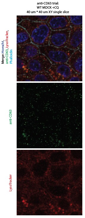
Application: ImmunocytochemistrySample Tested: MDCK canine kidney epithelial cell lineSpecies: CanineVerified Customer | Posted 09/01/2022IF staining of fixed, permeabilized MDCK cells in which acidic organelles were stained with LysoTracker. Anti-CD63 Ab gave some labeling of late endosomes/lysosomes, but was less sensitive than LysoTracker, and also had nonspecific puncta.

-
Application: Dot BlotSample Tested: cell lysate canine umbilical cord-derived mesenchymal stromal cellsSpecies: CanineVerified Customer | Posted 10/09/2020Dot blot of cell lysate from canine umbilical cord-derived mesenchymal stromal cells. Water as negative control. Preliminary data only.
-
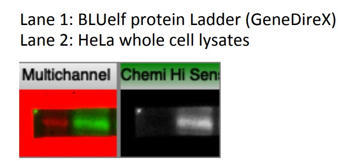
Application: Western BlotSample Tested: Hela whole cell lysateSpecies: HumanVerified Customer | Posted 10/02/2019
-
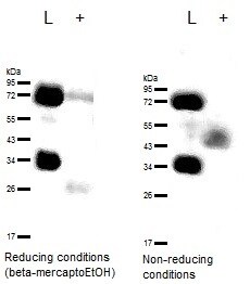
Application: Western BlotSample Tested: Blood plasma and UrineSpecies: HumanVerified Customer | Posted 09/18/2017Western blots with and without reducting conditions. The same samples and sample volumes were used for both. All procedure was performed parallely for both conditions. L- ladder +- exosomesAntibody labelled with HRP
-
Application: Immunocytochemistry/ImmunofluorescenceSample Tested: HEK cellsSpecies: HumanVerified Customer | Posted 11/01/2016HEK cells stained with CD63 antibody at a dilution of 1:50 followed by Donkey anti-mouse secondary antibody conjugated with Alexa Fluor 488 (1:500). Nuclei were stained with Hoechst 33342.
-
Application: Immunocytochemistry/ImmunofluorescenceSample Tested: Madin-Darby Canine Kidney Epithelial Cells (MDCK Line)Species: CanineVerified Customer | Posted 11/01/2016MDCD cells stained with CD63 antibody at a dilution of 1:50 followed by Donkey anti-mouse secondary antibody conjugated with Alexa Fluor 488 (1:500). Nuclei were stained with Hoechst 33342.
There are no reviews that match your criteria.
Protocols
Find general support by application which include: protocols, troubleshooting, illustrated assays, videos and webinars.
- 7-Amino Actinomycin D (7-AAD) Cell Viability Flow Cytometry Protocol
- Antigen Retrieval Protocol (PIER)
- Antigen Retrieval for Frozen Sections Protocol
- Appropriate Fixation of IHC/ICC Samples
- Cellular Response to Hypoxia Protocols
- Chromogenic IHC Staining of Formalin-Fixed Paraffin-Embedded (FFPE) Tissue Protocol
- Chromogenic Immunohistochemistry Staining of Frozen Tissue
- ClariTSA™ Fluorophore Kits
- Detection & Visualization of Antibody Binding
- ELISA Sample Preparation & Collection Guide
- ELISA Troubleshooting Guide
- Extracellular Membrane Flow Cytometry Protocol
- Flow Cytometry Protocol for Cell Surface Markers
- Flow Cytometry Protocol for Staining Membrane Associated Proteins
- Flow Cytometry Staining Protocols
- Flow Cytometry Troubleshooting Guide
- Fluorescent IHC Staining of Frozen Tissue Protocol
- Graphic Protocol for Heat-induced Epitope Retrieval
- Graphic Protocol for the Preparation and Fluorescent IHC Staining of Frozen Tissue Sections
- Graphic Protocol for the Preparation and Fluorescent IHC Staining of Paraffin-embedded Tissue Sections
- Graphic Protocol for the Preparation of Gelatin-coated Slides for Histological Tissue Sections
- How to Run an R&D Systems DuoSet ELISA
- How to Run an R&D Systems Quantikine ELISA
- How to Run an R&D Systems Quantikine™ QuicKit™ ELISA
- ICC Cell Smear Protocol for Suspension Cells
- ICC Immunocytochemistry Protocol Videos
- ICC for Adherent Cells
- IHC Sample Preparation (Frozen sections vs Paraffin)
- Immunocytochemistry (ICC) Protocol
- Immunocytochemistry Troubleshooting
- Immunofluorescence of Organoids Embedded in Cultrex Basement Membrane Extract
- Immunofluorescent IHC Staining of Formalin-Fixed Paraffin-Embedded (FFPE) Tissue Protocol
- Immunohistochemistry (IHC) and Immunocytochemistry (ICC) Protocols
- Immunohistochemistry Frozen Troubleshooting
- Immunohistochemistry Paraffin Troubleshooting
- Immunoprecipitation Protocol
- Intracellular Flow Cytometry Protocol Using Alcohol (Methanol)
- Intracellular Flow Cytometry Protocol Using Detergents
- Intracellular Nuclear Staining Flow Cytometry Protocol Using Detergents
- Intracellular Staining Flow Cytometry Protocol Using Alcohol Permeabilization
- Intracellular Staining Flow Cytometry Protocol Using Detergents to Permeabilize Cells
- Preparing Samples for IHC/ICC Experiments
- Preventing Non-Specific Staining (Non-Specific Binding)
- Primary Antibody Selection & Optimization
- Propidium Iodide Cell Viability Flow Cytometry Protocol
- Protocol for Heat-Induced Epitope Retrieval (HIER)
- Protocol for Liperfluo
- Protocol for Making a 4% Formaldehyde Solution in PBS
- Protocol for VisUCyte™ HRP Polymer Detection Reagent
- Protocol for the Characterization of Human Th22 Cells
- Protocol for the Characterization of Human Th9 Cells
- Protocol for the Fluorescent ICC Staining of Cell Smears - Graphic
- Protocol for the Fluorescent ICC Staining of Cultured Cells on Coverslips - Graphic
- Protocol for the Preparation & Fixation of Cells on Coverslips
- Protocol for the Preparation and Chromogenic IHC Staining of Frozen Tissue Sections
- Protocol for the Preparation and Chromogenic IHC Staining of Frozen Tissue Sections - Graphic
- Protocol for the Preparation and Chromogenic IHC Staining of Paraffin-embedded Tissue Sections
- Protocol for the Preparation and Chromogenic IHC Staining of Paraffin-embedded Tissue Sections - Graphic
- Protocol for the Preparation and Fluorescent ICC Staining of Cells on Coverslips
- Protocol for the Preparation and Fluorescent ICC Staining of Non-adherent Cells
- Protocol for the Preparation and Fluorescent ICC Staining of Stem Cells on Coverslips
- Protocol for the Preparation and Fluorescent IHC Staining of Frozen Tissue Sections
- Protocol for the Preparation and Fluorescent IHC Staining of Paraffin-embedded Tissue Sections
- Protocol for the Preparation of Gelatin-coated Slides for Histological Tissue Sections
- Protocol for the Preparation of a Cell Smear for Non-adherent Cell ICC - Graphic
- Protocol: Annexin V and PI Staining by Flow Cytometry
- Protocol: Annexin V and PI Staining for Apoptosis by Flow Cytometry
- Quantikine HS ELISA Kit Assay Principle, Alkaline Phosphatase
- Quantikine HS ELISA Kit Principle, Streptavidin-HRP Polymer
- R&D Systems Quality Control Western Blot Protocol
- Sandwich ELISA (Colorimetric) – Biotin/Streptavidin Detection Protocol
- Sandwich ELISA (Colorimetric) – Direct Detection Protocol
- TUNEL and Active Caspase-3 Detection by IHC/ICC Protocol
- The Importance of IHC/ICC Controls
- Troubleshooting Guide: ELISA
- Troubleshooting Guide: Fluorokine Flow Cytometry Kits
- Troubleshooting Guide: Immunohistochemistry
- Troubleshooting Guide: Western Blot Figures
- Western Blot Conditions
- Western Blot Protocol
- Western Blot Protocol for Cell Lysates
- Western Blot Troubleshooting
- Western Blot Troubleshooting Guide
- View all Protocols, Troubleshooting, Illustrated assays and Webinars
Loading...