![Western Blot: CD69 Antibody (8B6)BSA Free [NBP1-51607]](https://resources.rndsystems.com/images/products/CD69-Antibody-8B6-Western-Blot-NBP1-51607-img0006.jpg "Western Blot: CD69 Antibody (8B6)BSA Free [NBP1-51607]")
Loading...
Key Product Details
Species Reactivity
Validated:
Human
Cited:
Human
Applications
Validated:
Immunohistochemistry, Immunohistochemistry-Paraffin, Western Blot, ELISA, Flow Cytometry, Imaging Mass Cytometry
Cited:
Western Blot
Label
Unconjugated
Antibody Source
Monoclonal Mouse IgG1 Clone # 8B6
Loading...
Product Specifications
Immunogen
Purified recombinant fragment of human CD69 expressed in E. Coli.
Clonality
Monoclonal
Host
Mouse
Isotype
IgG1
Theoretical MW
22.5 kDa.
Disclaimer note: The observed molecular weight of the protein may vary from the listed predicted molecular weight due to post translational modifications, post translation cleavages, relative charges, and other experimental factors.
Disclaimer note: The observed molecular weight of the protein may vary from the listed predicted molecular weight due to post translational modifications, post translation cleavages, relative charges, and other experimental factors.
Scientific Data Images for CD69 Antibody (8B6)
Western Blot: CD69 Antibody (8B6)BSA Free [NBP1-51607]
Western Blot: CD69 Antibody (8B6) [NBP1-51607] - Analysis using CD69 mouse mAb against, Jurkat (1), L1210 (2) and TPH-1 (3) cell lysate.![Immunohistochemistry-Paraffin: CD69 Antibody (8B6) - BSA Free [NBP1-51607]](https://resources.rndsystems.com/images/products/CD69-Antibody-8B6-Immunohistochemistry-Paraffin-NBP1-51607-img0008.jpg "Immunohistochemistry-Paraffin: CD69 Antibody (8B6) - BSA Free [NBP1-51607]")
Immunohistochemistry-Paraffin: CD69 Antibody (8B6) - BSA Free [NBP1-51607]
Immunohistochemistry-Paraffin: CD69 Antibody (8B6) [NBP1-51607] - Human tonsil tissue stained with CD69 antibody NBP1-51607. IHC-P image submitted by a verified customer review.![Flow Cytometry: CD69 Antibody (8B6) - BSA Free [NBP1-51607]](https://resources.rndsystems.com/images/products/CD69-Antibody-8B6-Flow-Cytometry-NBP1-51607-img0007.jpg "Flow Cytometry: CD69 Antibody (8B6) - BSA Free [NBP1-51607]")
Flow Cytometry: CD69 Antibody (8B6) - BSA Free [NBP1-51607]
Flow Cytometry: CD69 Antibody (8B6) [NBP1-51607] - Analysis of Jurkat cells using CD69 mouse mAb (green) and negative control (purple).![Immunohistochemistry-Paraffin: CD69 Antibody (8B6) - BSA Free [NBP1-51607]](https://resources.rndsystems.com/images/products/CD69-Antibody-8B6-Immunohistochemistry-Paraffin-NBP1-51607-img0005.jpg "Immunohistochemistry-Paraffin: CD69 Antibody (8B6) - BSA Free [NBP1-51607]")
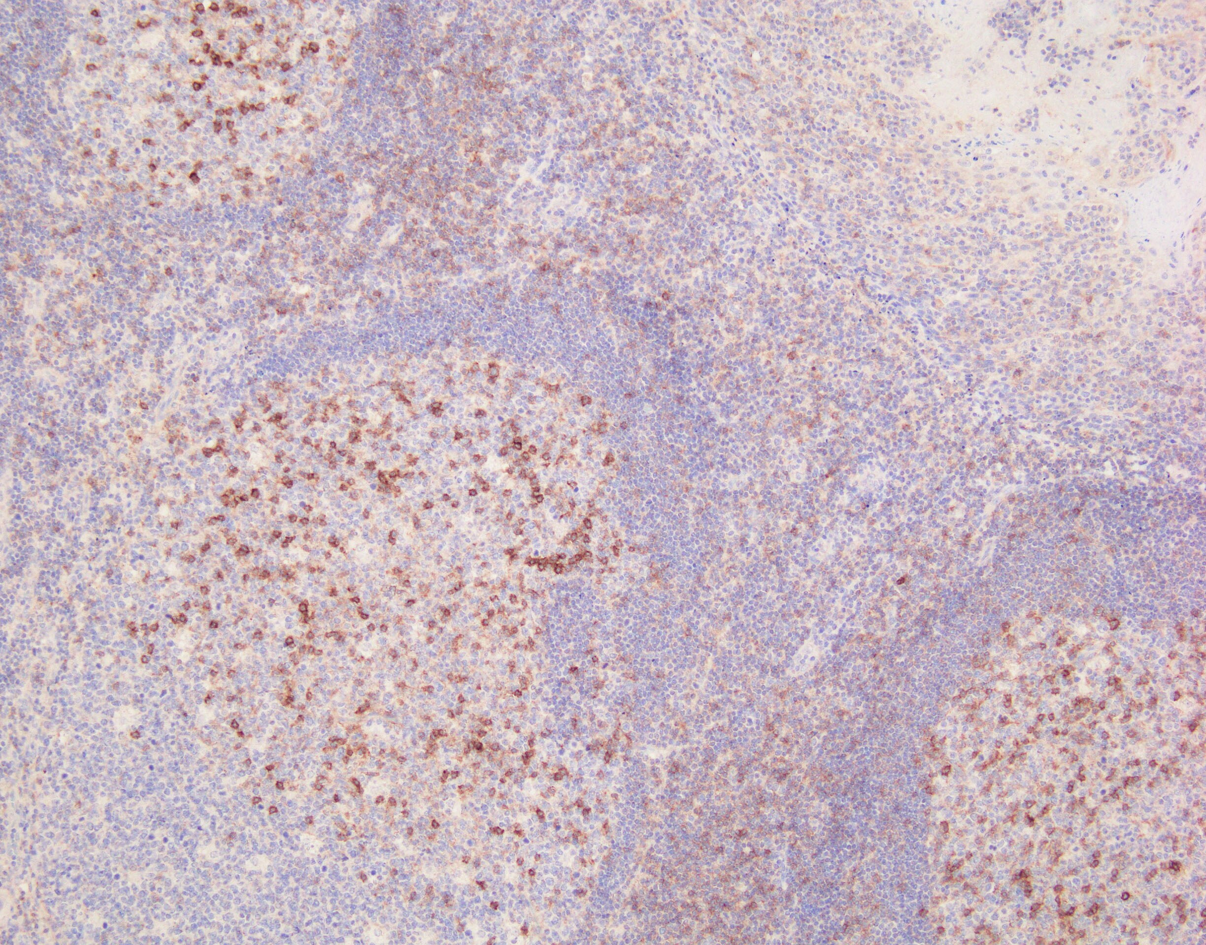
Immunohistochemistry-Paraffin: CD69 Antibody (8B6) - BSA Free [NBP1-51607]
Immunohistochemistry-Paraffin: CD69 Antibody (8B6) [NBP1-51607] - Analysis of human Tonsil tissues using anti-CD69 mouse mAb![Imaging Mass Cytometry: CD69 Antibody (8B6) - BSA Free [NBP1-51607]](https://resources.rndsystems.com/images/products/CD69-Antibody-8B6-Imaging-Mass-Cytometry-NBP1-51607-img0009.jpg "Imaging Mass Cytometry: CD69 Antibody (8B6) - BSA Free [NBP1-51607]")
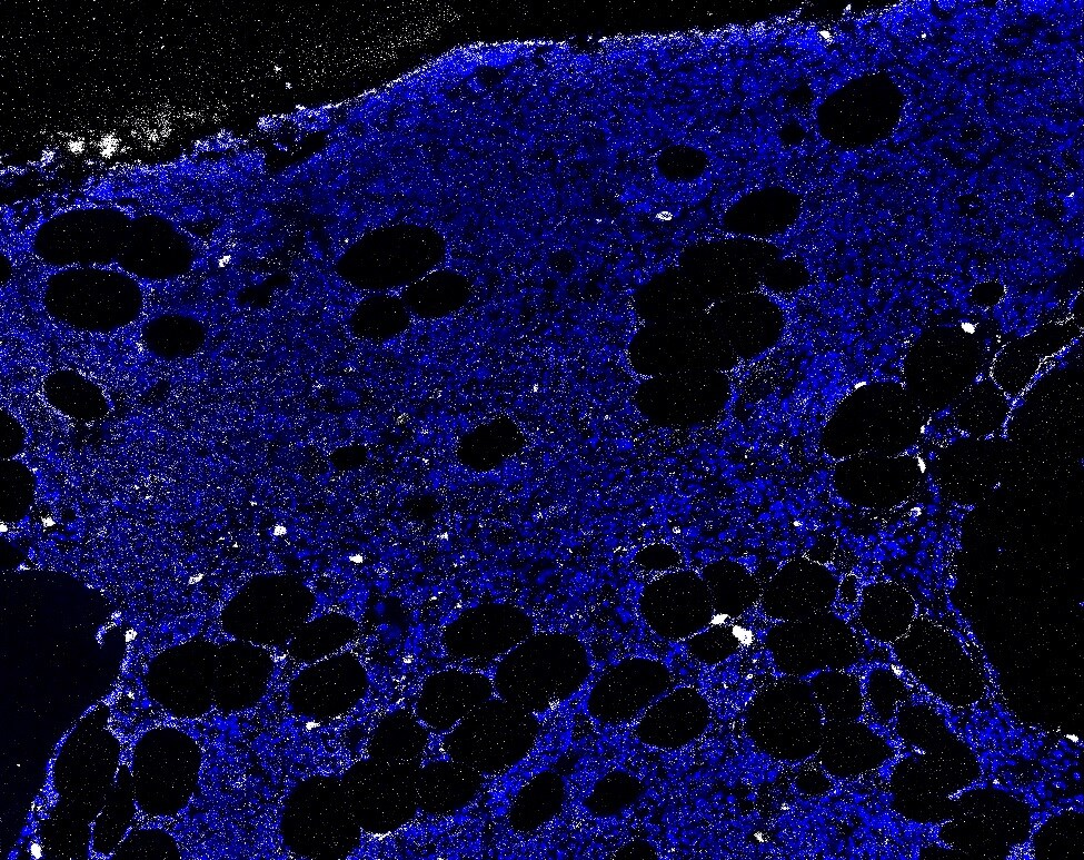
Imaging Mass Cytometry: CD69 Antibody (8B6) - BSA Free [NBP1-51607]
Imaging Mass Cytometry: CD69 Antibody (8B6) [NBP1-51607] - Human bone marrow FFPE tissue section. DNA stained in blue, CD69 stained in white. IMC image submitted by a verified customer review.Applications for CD69 Antibody (8B6)
Application
Recommended Usage
ELISA
1:10000
Flow Cytometry
1:200 - 1:400
Immunohistochemistry
1:200 - 1:1000
Immunohistochemistry-Paraffin
1:200 - 1:1000
Western Blot
1:500 - 1:2000
Application Notes
CD69 antibody validated for IHC-P and Imaging Mass Cytometry from verified customer reviews.
Reviewed Applications
Read 2 reviews rated 4 using NBP1-51607 in the following applications:
Flow Cytometry Panel Builder
Bio-Techne Knows Flow Cytometry
Save time and reduce costly mistakes by quickly finding compatible reagents using the Panel Builder Tool.
Advanced Features
- Spectra Viewer - Custom analysis of spectra from multiple fluorochromes
- Spillover Popups - Visualize the spectra of individual fluorochromes
- Antigen Density Selector - Match fluorochrome brightness with antigen density

Formulation, Preparation, and Storage
Purification
Ascites
Formulation
Ascites
Preservative
0.03% Sodium Azide
Concentration
This product is unpurified. The exact concentration of antibody is not quantifiable.
Shipping
The product is shipped with polar packs. Upon receipt, store it immediately at the temperature recommended below.
Stability & Storage
Store at 4C short term. Aliquot and store at -20C long term. Avoid freeze-thaw cycles.
Background: CD69
Alternate Names
CD69, EA-1, Leu23, MLR-3, p60
Gene Symbol
CD69
Additional CD69 Products
Product Documents for CD69 Antibody (8B6)
Certificate of Analysis
To download a Certificate of Analysis, please enter a lot or batch number in the search box below.
Product Specific Notices for CD69 Antibody (8B6)
This product is for research use only and is not approved for use in humans or in clinical diagnosis. Primary Antibodies are guaranteed for 1 year from date of receipt.
Citations for CD69 Antibody (8B6)
 Powered by Bioz
Customer Reviews for CD69 Antibody (8B6) (2)
4 out of 5
2 Customer Ratings
Have you used CD69 Antibody (8B6)?
Submit a review and receive an Amazon gift card!
$25/€18/£15/$25CAN/¥2500 Yen for a review with an image
$10/€7/£6/$10CAN/¥1110 Yen for a review without an image
Submit a review
Customer Images
Showing
1
-
2 of
2 reviews
Showing All
Filter By:
-
Application: Imaging Mass CytometrySample Tested: bone marrowSpecies: HumanVerified Customer | Posted 11/12/2021IMC on human bone marrow FFPE slides. DNA-blue CD69-white
 Bio-Techne ResponseThis review was submitted through the legacy Novus Innovators Program, reflecting a new species or application tested on a primary antibody.
 -
Application: Immunohistochemistry-ParaffinSample Tested: human tonsilSpecies: HumanVerified Customer | Posted 09/11/20208B6 was reacted to activated lymphocytesPretreatment: HIER, ER2(Leica) Primary Ab: 1:1000 Detection system: BOND polymer refine detection kit (Leica)
There are no reviews that match your criteria.
Protocols
Find general support by application which include: protocols, troubleshooting, illustrated assays, videos and webinars.
- 7-Amino Actinomycin D (7-AAD) Cell Viability Flow Cytometry Protocol
- Antigen Retrieval Protocol (PIER)
- Antigen Retrieval for Frozen Sections Protocol
- Appropriate Fixation of IHC/ICC Samples
- Cellular Response to Hypoxia Protocols
- Chromogenic IHC Staining of Formalin-Fixed Paraffin-Embedded (FFPE) Tissue Protocol
- Chromogenic Immunohistochemistry Staining of Frozen Tissue
- ClariTSA™ Fluorophore Kits
- Detection & Visualization of Antibody Binding
- ELISA Sample Preparation & Collection Guide
- ELISA Troubleshooting Guide
- Extracellular Membrane Flow Cytometry Protocol
- Flow Cytometry Protocol for Cell Surface Markers
- Flow Cytometry Protocol for Staining Membrane Associated Proteins
- Flow Cytometry Staining Protocols
- Flow Cytometry Troubleshooting Guide
- Fluorescent IHC Staining of Frozen Tissue Protocol
- Graphic Protocol for Heat-induced Epitope Retrieval
- Graphic Protocol for the Preparation and Fluorescent IHC Staining of Frozen Tissue Sections
- Graphic Protocol for the Preparation and Fluorescent IHC Staining of Paraffin-embedded Tissue Sections
- Graphic Protocol for the Preparation of Gelatin-coated Slides for Histological Tissue Sections
- How to Run an R&D Systems DuoSet ELISA
- How to Run an R&D Systems Quantikine ELISA
- How to Run an R&D Systems Quantikine™ QuicKit™ ELISA
- IHC Sample Preparation (Frozen sections vs Paraffin)
- Immunofluorescent IHC Staining of Formalin-Fixed Paraffin-Embedded (FFPE) Tissue Protocol
- Immunohistochemistry (IHC) and Immunocytochemistry (ICC) Protocols
- Immunohistochemistry Frozen Troubleshooting
- Immunohistochemistry Paraffin Troubleshooting
- Intracellular Flow Cytometry Protocol Using Alcohol (Methanol)
- Intracellular Flow Cytometry Protocol Using Detergents
- Intracellular Nuclear Staining Flow Cytometry Protocol Using Detergents
- Intracellular Staining Flow Cytometry Protocol Using Alcohol Permeabilization
- Intracellular Staining Flow Cytometry Protocol Using Detergents to Permeabilize Cells
- Preparing Samples for IHC/ICC Experiments
- Preventing Non-Specific Staining (Non-Specific Binding)
- Primary Antibody Selection & Optimization
- Propidium Iodide Cell Viability Flow Cytometry Protocol
- Protocol for Heat-Induced Epitope Retrieval (HIER)
- Protocol for Liperfluo
- Protocol for Making a 4% Formaldehyde Solution in PBS
- Protocol for VisUCyte™ HRP Polymer Detection Reagent
- Protocol for the Characterization of Human Th22 Cells
- Protocol for the Characterization of Human Th9 Cells
- Protocol for the Preparation & Fixation of Cells on Coverslips
- Protocol for the Preparation and Chromogenic IHC Staining of Frozen Tissue Sections
- Protocol for the Preparation and Chromogenic IHC Staining of Frozen Tissue Sections - Graphic
- Protocol for the Preparation and Chromogenic IHC Staining of Paraffin-embedded Tissue Sections
- Protocol for the Preparation and Chromogenic IHC Staining of Paraffin-embedded Tissue Sections - Graphic
- Protocol for the Preparation and Fluorescent IHC Staining of Frozen Tissue Sections
- Protocol for the Preparation and Fluorescent IHC Staining of Paraffin-embedded Tissue Sections
- Protocol for the Preparation of Gelatin-coated Slides for Histological Tissue Sections
- Protocol: Annexin V and PI Staining by Flow Cytometry
- Protocol: Annexin V and PI Staining for Apoptosis by Flow Cytometry
- Quantikine HS ELISA Kit Assay Principle, Alkaline Phosphatase
- Quantikine HS ELISA Kit Principle, Streptavidin-HRP Polymer
- R&D Systems Quality Control Western Blot Protocol
- Sandwich ELISA (Colorimetric) – Biotin/Streptavidin Detection Protocol
- Sandwich ELISA (Colorimetric) – Direct Detection Protocol
- TUNEL and Active Caspase-3 Detection by IHC/ICC Protocol
- The Importance of IHC/ICC Controls
- Troubleshooting Guide: ELISA
- Troubleshooting Guide: Fluorokine Flow Cytometry Kits
- Troubleshooting Guide: Immunohistochemistry
- Troubleshooting Guide: Western Blot Figures
- Western Blot Conditions
- Western Blot Protocol
- Western Blot Protocol for Cell Lysates
- Western Blot Troubleshooting
- Western Blot Troubleshooting Guide
- View all Protocols, Troubleshooting, Illustrated assays and Webinars
Loading...
Associated Pathways
