COX4 Antibody - BSA Free
Novus Biologicals | Catalog # NB110-39115
![Western Blot: COX4 AntibodyBSA Free [NB110-39115]](https://resources.rndsystems.com/images/products/COX4-Antibody---BSA-Free-Western-Blot-NB110-39115-img0023.jpg "Western Blot: COX4 AntibodyBSA Free [NB110-39115]")
Key Product Details
Validated by
Biological Validation
Species Reactivity
Validated:
Human, Mouse, Rat, Porcine, Bovine, Drosophila, Insect, Opossum, Primate
Cited:
Human, Mouse, Rat, Insect, Opossum
Applications
Validated:
Immunohistochemistry, Immunohistochemistry-Paraffin, Western Blot, Immunocytochemistry/ Immunofluorescence, Simple Western, Chromatin Immunoprecipitation (ChIP), Knockdown Validated
Cited:
Immunohistochemistry-Paraffin, Western Blot, IF/IHC, Knockdown Validated
Label
Unconjugated
Antibody Source
Polyclonal Rabbit IgG
Format
BSA Free
Loading...
Product Specifications
Immunogen
A synthetic peptide made to an internal region of human COX IV isoform 1 (within residues 1-100). [Swiss-Prot# P13073]
Reactivity Notes
Opossum reactivity reported in scientific literature (PMID: 28720662).
Localization
Mitochondrion, mito inner membrane
Marker
Mitochondria Marker
Clonality
Polyclonal
Host
Rabbit
Isotype
IgG
Scientific Data Images for COX4 Antibody - BSA Free
Western Blot: COX4 AntibodyBSA Free [NB110-39115]
COX4-Antibody---BSA-Free-Western-Blot-NB110-39115-img0023.jpg![Immunocytochemistry/ Immunofluorescence: COX4 Antibody - BSA Free [NB110-39115]](https://resources.rndsystems.com/images/products/COX4-Antibody---BSA-Free-Immunocytochemistry-Immunofluorescence-NB110-39115-img0021.jpg "Immunocytochemistry/ Immunofluorescence: COX4 Antibody - BSA Free [NB110-39115]")
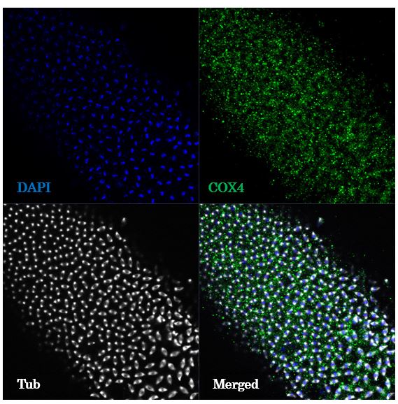
Immunocytochemistry/ Immunofluorescence: COX4 Antibody - BSA Free [NB110-39115]
Immunocytochemistry/Immunofluorescence: COX4 Antibody - BSA Free [NB110-39115] - NIH3T3 cells were fixed for 10 minutes using 10% formalin and then permeabilized for 5 minutes using 1X PBS + 0.05% Triton X-100. The cells were incubated with anti-COX4 at 10 ug/mL overnight at 4C and detected with an anti-rabbit DyLight 488 (Green) at a 1:500 dilution. Nuclei were counterstained with DAPI (Blue). Cells were imaged using a 40X objective.![Immunohistochemistry: COX4 Antibody - BSA Free [NB110-39115]](https://resources.rndsystems.com/images/products/COX4-Antibody---BSA-Free-Immunohistochemistry-NB110-39115-img0015.jpg "Immunohistochemistry: COX4 Antibody - BSA Free [NB110-39115]")
Immunohistochemistry: COX4 Antibody - BSA Free [NB110-39115]
Immunohistochemistry: COX4 Antibody - BSA Free [NB110-39115] - Analysis of COX4 in human breast cancer using DAB with hematoxylin counterstain.![Western Blot: COX4 AntibodyBSA Free [NB110-39115]](https://resources.rndsystems.com/images/products/COX4-Antibody---BSA-Free-Western-Blot-NB110-39115-img0012.jpg "Western Blot: COX4 AntibodyBSA Free [NB110-39115]")
Western Blot: COX4 AntibodyBSA Free [NB110-39115]
Western Blot: COX4 Antibody - BSA Free [NB110-39115] - Analysis of COX4 in the following cell lysates: 1. HeLa, 2. Ntera, 3. A431, 4. HepG2, 5. MCF7 and 6. 3T3.![Western Blot: COX4 AntibodyBSA Free [NB110-39115]](https://resources.rndsystems.com/images/products/COX4-Antibody---BSA-Free-Western-Blot-NB110-39115-img0016.jpg "Western Blot: COX4 AntibodyBSA Free [NB110-39115]")
Western Blot: COX4 AntibodyBSA Free [NB110-39115]
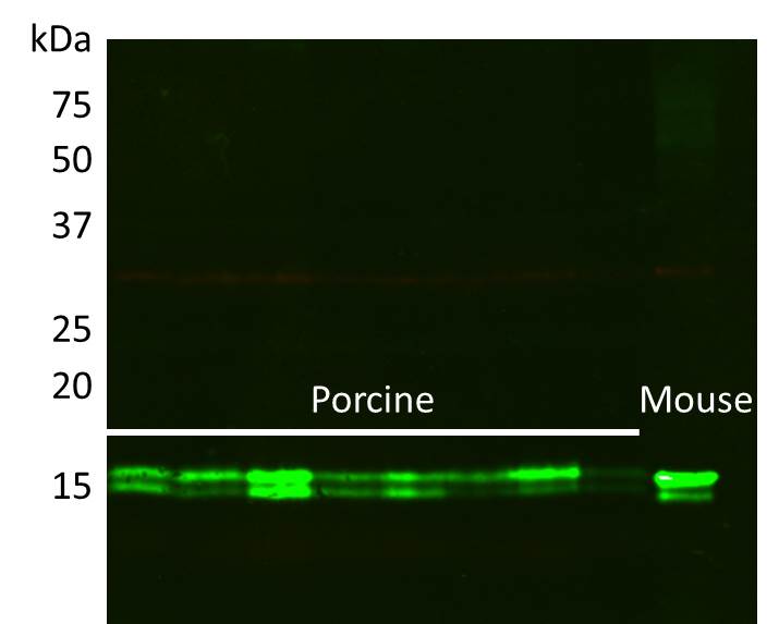
Western Blot: COX4 Antibody - BSA Free [NB110-39115] - Analysis of COX4 in porcine whole skeletal muscle lysate using anti-COX4 antibody. Image from verified customer review.![Western Blot: COX4 AntibodyBSA Free [NB110-39115]](https://resources.rndsystems.com/images/products/COX4-Antibody---BSA-Free-Western-Blot-NB110-39115-img0017.jpg "Western Blot: COX4 AntibodyBSA Free [NB110-39115]")
Western Blot: COX4 AntibodyBSA Free [NB110-39115]
Western Blot: COX4 Antibody - BSA Free [NB110-39115] - Detection of COX4-1 isoform using COX4 antibody NB110-39115. Lane 1: HEK 293 with empty vector, Lane 2: HEK 293 with flag-tagged human COX4-1, Lane 3: HEK 293 with flag-tagged human COX4-2. Photo courtesy of Ryo Fukuda, G. Semenza lab. Johns Hopkins, SOM![Western Blot: COX4 AntibodyBSA Free [NB110-39115]](https://resources.rndsystems.com/images/products/COX4-Antibody---BSA-Free-Western-Blot-NB110-39115-img0020.jpg "Western Blot: COX4 AntibodyBSA Free [NB110-39115]")
Western Blot: COX4 AntibodyBSA Free [NB110-39115]
Western Blot: COX4 Antibody - BSA Free [NB110-39115] - Total protein from HeLa, 3T3 and PC12 was separated on a 4-20% gel by SDS-PAGE, transferred to PVDF membrane and blocked in 5% non-fat milk in TBST. The membrane was probed with 0.5 ug/mL anti-COX4 in 5% block buffer and detected with an anti-rabbit HRP secondary antibody using chemiluminescence.![Immunocytochemistry/ Immunofluorescence: COX4 Antibody - BSA Free [NB110-39115]](https://resources.rndsystems.com/images/products/COX4-Antibody---BSA-Free-Immunocytochemistry-Immunofluorescence-NB110-39115-img0011.jpg "Immunocytochemistry/ Immunofluorescence: COX4 Antibody - BSA Free [NB110-39115]")
Immunocytochemistry/ Immunofluorescence: COX4 Antibody - BSA Free [NB110-39115]
Immunocytochemistry/Immunofluorescence: COX4 Antibody - BSA Free [NB110-39115] - COX4 antibody was tested in HeLa cells with Dylight 488 (green). Nuclei and alpha-tubulin were counterstained with DAPI (blue) and Dylight 550 (red).![Immunocytochemistry/ Immunofluorescence: COX4 Antibody - BSA Free [NB110-39115]](https://resources.rndsystems.com/images/products/COX4-Antibody---BSA-Free-Immunocytochemistry-Immunofluorescence-NB110-39115-img0014.jpg "Immunocytochemistry/ Immunofluorescence: COX4 Antibody - BSA Free [NB110-39115]")
Immunocytochemistry/ Immunofluorescence: COX4 Antibody - BSA Free [NB110-39115]
Immunocytochemistry/Immunofluorescence: COX4 Antibody - BSA Free [NB110-39115] - Analysis of HeLa cells using COX4 antibody (NB110-39115, 1:5). An Alexa Fluor 488-conjugated Goat to rabbit IgG was used as secondary antibody (green). Actin filaments were labeled with Alexa Fluor 568 phalloidin (red). DAPI was used to stain the cell nuclei (blue).![Immunocytochemistry: COX4 Antibody - BSA Free [NB110-39115]](https://resources.rndsystems.com/images/products/COX4-Antibody---BSA-Free-Immunocytochemistry-NB110-39115-img0019.jpg "Immunocytochemistry: COX4 Antibody - BSA Free [NB110-39115]")
Immunocytochemistry: COX4 Antibody - BSA Free [NB110-39115]
Immunocytochemistry: COX4 Antibody - BSA Free [NB110-39115] - Analysis of methanol fixed drosophila embryo using 1:200 dilution of COX4 antibody. The signal was developed using AF488 conjugated Donkey anti-Rabbit IgG (H+L) secondary antibody and the sections were further counterstained for tubulin and DAPI. The antibody generated a specific staining of COX4 in mitochondria near mitotic spindles at early stage of embryo development. Image from verified customer review.![Simple Western: COX4 AntibodyBSA Free [NB110-39115]](https://resources.rndsystems.com/images/products/COX4-Antibody---BSA-Free-Simple-Western-NB110-39115-img0009.jpg "Simple Western: COX4 AntibodyBSA Free [NB110-39115]")
Simple Western: COX4 AntibodyBSA Free [NB110-39115]
Simple Western: COX4 Antibody - BSA Free [NB110-39115] - Image shows a specific band for COX4 in 1.0 mg/mL of HeLa lysate. This experiment was performed under reducing conditions using the 12-230 kDa separation system.
Western Blot: COX4 Antibody - BSA Free [NB110-39115] -
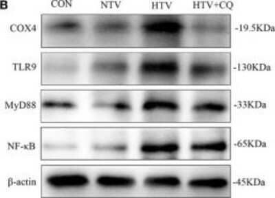
Western Blot: COX4 Antibody - BSA Free [NB110-39115] - The expression levels of microtubule protein light chain 3 (LC3B), PTEN inducing putative kinase 1 (PINK1), Parkin, mitofusin 1 (Mfn1), toll-like receptor (TLR) 9, cytochrome c oxidase 4 (COX4), myeloid differentiation factor 88 (MyD88), & nuclear factor (NF)-kappa B in lung tissues from animals with spontaneous breathing (CON) or mechanical ventilation at high tidal volume (HTV) with saline, antimycin A (AmA) or cyclosporine A (CsA). (A) Levels of PINK1, Parkin, & Mfn1 mRNA. (B) Levels of LC3B, PINK1, Parkin, & Mfn1 protein by Western blot. (C) Relative expression of LC3B-II/LC3B-I & PINK1 protein. (D) Relative expression of Parkin & Mfn1 protein. (E) Levels of TLR9 & COX4 mRNA. (F) Levels of MyD88 & nuclear factor-kappa B (NF-kappa B) mRNA. (G) Levels of TLR9, COX4, MyD88, & NF-kappa B protein by Western blot. (H) Relative expression of TLR9 & COX4 protein. (I) Relative expression of MyD88 & NF-kappa B protein. Fold expression for target genes was normalized to that measured for the beta -actin gene. Both of these experiments were in triplicate. aP < 0.05 vs. CON group;bP < 0.05 vs. HTV group; & cP < 0.05 vs. AmA group. Image collected & cropped by CiteAb from the following publication (https://www.frontiersin.org/article/10.3389/fimmu.2018.01477/full), licensed under a CC-BY license. Not internally tested by Novus Biologicals.Applications for COX4 Antibody - BSA Free
Application
Recommended Usage
Immunocytochemistry/ Immunofluorescence
1:40
Immunohistochemistry
1:100
Immunohistochemistry-Paraffin
1:100
Knockdown Validated
reported in scientific literature (PMID 31655343)
Simple Western
1:25
Western Blot
1:2000
Application Notes
In Western Blot, a band is seen ~19.5 kDa representing COX IV. In ICC/IF, mitochondrion staining was observed in HeLa cells. In IHC-P, staining is observed in the cytoplasm and mitochondria of human breast cancer tissue. Prior to immunostaining paraffin tissues, antigen retrieval with sodium citrate buffer (pH 6.0) is recommended. Higher dilutions may be needed for mitochondrial membrane enriched preparations. In Simple Western only 10 - 15 uL of the recommended dilution is used per data point.
See Simple Western Antibody Database for Simple Western validation: Tested in HeLa lysate 1.0 mg/mL, titrated to saturation using various models; separated by Size; antibody dilution of 1:25, 75 ug/mL; apparent MW was 22 kDa. Separated by Size-Wes, Sally Sue/Peggy Sue.
See Simple Western Antibody Database for Simple Western validation: Tested in HeLa lysate 1.0 mg/mL, titrated to saturation using various models; separated by Size; antibody dilution of 1:25, 75 ug/mL; apparent MW was 22 kDa. Separated by Size-Wes, Sally Sue/Peggy Sue.
Reviewed Applications
Read 4 reviews rated 4.5 using NB110-39115 in the following applications:
Formulation, Preparation, and Storage
Purification
Immunogen affinity purified
Formulation
PBS
Format
BSA Free
Preservative
0.02% Sodium Azide
Concentration
1.0 mg/ml
Shipping
The product is shipped with polar packs. Upon receipt, store it immediately at the temperature recommended below.
Stability & Storage
Aliquot and store at -20C or -80C. Avoid freeze-thaw cycles.
Background: COX4
Long Name
Cytochrome c Oxidase Subunit IV
Alternate Names
COX4I2
Gene Symbol
COX4I1
Additional COX4 Products
Product Documents for COX4 Antibody - BSA Free
Certificate of Analysis
To download a Certificate of Analysis, please enter a lot or batch number in the search box below.
Product Specific Notices for COX4 Antibody - BSA Free
This product is for research use only and is not approved for use in humans or in clinical diagnosis. Primary Antibodies are guaranteed for 1 year from date of receipt.
Related Research Areas
Citations for COX4 Antibody - BSA Free
 Powered by Bioz
Customer Reviews for COX4 Antibody - BSA Free (4)
4.5 out of 5
4 Customer Ratings
Have you used COX4 Antibody - BSA Free?
Submit a review and receive an Amazon gift card!
$25/€18/£15/$25CAN/¥2500 Yen for a review with an image
$10/€7/£6/$10CAN/¥1110 Yen for a review without an image
Submit a review
Customer Images

-(01-ml)_NB110-39115_8326.bmp)
Showing
1
-
4 of
4 reviews
Showing All
Filter By:
-
Application: Western BlotSample Tested: 20 ug whole body lysateSpecies: DrosophilaVerified Customer | Posted 06/26/2017The Cox IV band runs just below the 15 Kda marker20 ug whole body lysate run in a 12% gel. Antibody used 1:4000

-
Application: ImmunocytochemistrySample Tested:Species: OtherVerified Customer | Posted 10/29/2015ICC analysis of COX4 in Mitochondria of drosophila embryo
-
Application: Western BlotSample Tested: Whole Skeletal Muscle LysateSpecies: OtherVerified Customer | Posted 04/23/2015Whole Skeletal Muscle Lysate
-
Application: ImmunocytochemistrySample Tested:Species: HumanVerified Customer | Posted 06/16/2014IF confocal analysis of HeLa cells using COX IV antibody (NB110-39115, 1:5).
There are no reviews that match your criteria.
Protocols
View specific protocols for COX4 Antibody - BSA Free (NB110-39115):
Immunocytochemistry Protocol
Culture cells to appropriate density in 35 mm culture dishes or 6-well plates.
1. Remove culture medium and add 10% formalin to the dish. Fix at room temperature for 30 minutes.
2. Remove the formalin and add ice cold methanol. Incubate for 5-10 minutes.
3. Remove methanol and add washing solution (i.e. PBS). Be sure to not let the specimen dry out. Wash three times for 10 minutes.
4. To block nonspecific antibody binding incubate in 10% normal goat serum from 1 hour to overnight at room temperature.
5. Add primary antibody at appropriate dilution and incubate at room temperature from 2 hours to overnight at room temperature.
6. Remove primary antibody and replace with washing solution. Wash three times for 10 minutes.
7. Add secondary antibody at appropriate dilution. Incubate for 1 hour at room temperature.
8. Remove antibody and replace with wash solution, then wash for 10 minutes. Add Hoechst 33258 to wash solution at 1:25,0000 and incubate for 10 minutes. Wash a third time for 10 minutes.
9. Cells can be viewed directly after washing. The plates can also be stored in PBS containing Azide covered in Parafilm (TM). Cells can also be cover-slipped using Fluoromount, with appropriate sealing.
*The above information is only intended as a guide. The researcher should determine what protocol best meets their needs. Please follow safe laboratory procedures.
Immunohistochemistry-Paraffin Embedded Sections Protocol
Antigen Unmasking:
Bring slides to a boil in 10 mM sodium citrate buffer (pH 6.0) then maintain at a sub-boiling temperature for 10 minutes. Cool slides on bench-top for 30 minutes.
Staining:
1. Wash sections in deionized water three times for 5 minutes each.
2. Wash sections in wash buffer for 5 minutes.
3. Block each section with 100-400 ul blocking solution for 1 hour at room temperature.
4. Remove blocking solution and add 100-400 ul diluted primary antibody. Incubate overnight at 4 C.
5. Remove antibody solution and wash sections in wash buffer three times for 5 minutes each.
6. Add 100-400 ul biotinylated diluted secondary antibody. Incubate 30 minutes at room temperature.
7. Remove secondary antibody solution and wash sections three times with wash buffer for 5 minutes each.
8. Add 100-400 ul Streptavidin-HRP reagent to each section and incubate for 30 minutes at room temperature.
9. Wash sections three times in wash buffer for 5 minutes each.
10. Add 100-400 ul DAB substrate to each section and monitor staining closely.
11. As soon as the sections develop, immerse slides in deionized water.
12. Counterstain sections in hematoxylin.
13. Wash sections in deionized water two times for 5 minutes each.
14. Dehydrate sections.
15. Mount coverslips.
*The above information is only intended as a guide. The researcher should determine what protocol best meets their needs. Please follow safe laboratory procedures.
Western Blot Protocol
1. Perform SDS-PAGE on samples to be analyzed, loading 40 ug of total protein per lane.
2. Transfer proteins to membrane according to the instructions provided by the manufacturer of the membrane and transfer apparatus.
3. Stain according to standard Ponceau S procedure (or similar product) to assess transfer success, and mark molecular weight standards where appropriate.
4. Rinse the blot.
5. Block the membrane using standard blocking buffer for at least 1 hour.
6. Wash the membrane in wash buffer three times for 10 minutes each.
7. Dilute primary antibody in blocking buffer and incubate 1 hour at room temperature.
8. Wash the membrane in wash buffer three times for 10 minutes each.
9. Apply the diluted HRP conjugated secondary antibody in blocking buffer (as per manufacturers instructions) and incubate 1 hour at room temperature.
10. Wash the blot in wash buffer three times for 10 minutes each (this step can be repeated as required to reduce background).
11. Apply the detection reagent of choice in accordance with the manufacturers instructions.
Note: Tween-20 can be added to the blocking or antibody dilution buffer at a final concentration of 0.05-0.2%.
*The above information is only intended as a guide. The researcher should determine what protocol best meets their needs. Please follow safe laboratory procedures.
Find general support by application which include: protocols, troubleshooting, illustrated assays, videos and webinars.
- Antigen Retrieval Protocol (PIER)
- Antigen Retrieval for Frozen Sections Protocol
- Appropriate Fixation of IHC/ICC Samples
- Cellular Response to Hypoxia Protocols
- ChIP Protocol Video
- Chromatin Immunoprecipitation (ChIP) Protocol
- Chromatin Immunoprecipitation Protocol
- Chromogenic IHC Staining of Formalin-Fixed Paraffin-Embedded (FFPE) Tissue Protocol
- Chromogenic Immunohistochemistry Staining of Frozen Tissue
- ClariTSA™ Fluorophore Kits
- Detection & Visualization of Antibody Binding
- Fluorescent IHC Staining of Frozen Tissue Protocol
- Graphic Protocol for Heat-induced Epitope Retrieval
- Graphic Protocol for the Preparation and Fluorescent IHC Staining of Frozen Tissue Sections
- Graphic Protocol for the Preparation and Fluorescent IHC Staining of Paraffin-embedded Tissue Sections
- Graphic Protocol for the Preparation of Gelatin-coated Slides for Histological Tissue Sections
- ICC Cell Smear Protocol for Suspension Cells
- ICC Immunocytochemistry Protocol Videos
- ICC for Adherent Cells
- IHC Sample Preparation (Frozen sections vs Paraffin)
- Immunocytochemistry (ICC) Protocol
- Immunocytochemistry Troubleshooting
- Immunofluorescence of Organoids Embedded in Cultrex Basement Membrane Extract
- Immunofluorescent IHC Staining of Formalin-Fixed Paraffin-Embedded (FFPE) Tissue Protocol
- Immunohistochemistry (IHC) and Immunocytochemistry (ICC) Protocols
- Immunohistochemistry Frozen Troubleshooting
- Immunohistochemistry Paraffin Troubleshooting
- Preparing Samples for IHC/ICC Experiments
- Preventing Non-Specific Staining (Non-Specific Binding)
- Primary Antibody Selection & Optimization
- Protocol for Heat-Induced Epitope Retrieval (HIER)
- Protocol for Making a 4% Formaldehyde Solution in PBS
- Protocol for VisUCyte™ HRP Polymer Detection Reagent
- Protocol for the Fluorescent ICC Staining of Cell Smears - Graphic
- Protocol for the Fluorescent ICC Staining of Cultured Cells on Coverslips - Graphic
- Protocol for the Preparation & Fixation of Cells on Coverslips
- Protocol for the Preparation and Chromogenic IHC Staining of Frozen Tissue Sections
- Protocol for the Preparation and Chromogenic IHC Staining of Frozen Tissue Sections - Graphic
- Protocol for the Preparation and Chromogenic IHC Staining of Paraffin-embedded Tissue Sections
- Protocol for the Preparation and Chromogenic IHC Staining of Paraffin-embedded Tissue Sections - Graphic
- Protocol for the Preparation and Fluorescent ICC Staining of Cells on Coverslips
- Protocol for the Preparation and Fluorescent ICC Staining of Non-adherent Cells
- Protocol for the Preparation and Fluorescent ICC Staining of Stem Cells on Coverslips
- Protocol for the Preparation and Fluorescent IHC Staining of Frozen Tissue Sections
- Protocol for the Preparation and Fluorescent IHC Staining of Paraffin-embedded Tissue Sections
- Protocol for the Preparation of Gelatin-coated Slides for Histological Tissue Sections
- Protocol for the Preparation of a Cell Smear for Non-adherent Cell ICC - Graphic
- R&D Systems Quality Control Western Blot Protocol
- TUNEL and Active Caspase-3 Detection by IHC/ICC Protocol
- The Importance of IHC/ICC Controls
- Troubleshooting Guide: Immunohistochemistry
- Troubleshooting Guide: Western Blot Figures
- Western Blot Conditions
- Western Blot Protocol
- Western Blot Protocol for Cell Lysates
- Western Blot Troubleshooting
- Western Blot Troubleshooting Guide
- View all Protocols, Troubleshooting, Illustrated assays and Webinars
Loading...