CRBN Antibody - BSA Free
Novus Biologicals | Catalog # NBP1-91810
![Immunocytochemistry/ Immunofluorescence: CRBN Antibody [NBP1-91810]](https://resources.rndsystems.com/images/products/CRBN-Antibody-Immunocytochemistry-Immunofluorescence-NBP1-91810-img0027.jpg "Immunocytochemistry/ Immunofluorescence: CRBN Antibody [NBP1-91810]")
Key Product Details
Validated by
Species Reactivity
Validated:
Cited:
Applications
Validated:
Cited:
Label
Antibody Source
Format
Product Specifications
Immunogen
Clonality
Host
Isotype
Scientific Data Images for CRBN Antibody - BSA Free
Immunocytochemistry/ Immunofluorescence: CRBN Antibody [NBP1-91810]
Immunocytochemistry/Immunofluorescence: CRBN Antibody [NBP1-91810] - Staining of human cell line U-251 MG shows localization to nucleoli. Antibody staining is shown in green.![Western Blot: CRBN Antibody [NBP1-91810]](https://resources.rndsystems.com/images/products/CRBN-Antibody-Western-Blot-NBP1-91810-img0025.jpg "Western Blot: CRBN Antibody [NBP1-91810]")
Western Blot: CRBN Antibody [NBP1-91810]
Western Blot: CRBN Antibody [NBP1-91810] - Analysis in mouse cell line NIH-3T3 and rat cell line NBT-II.![Simple Western: CRBN Antibody [NBP1-91810]](https://resources.rndsystems.com/images/products/CRBN-Antibody-Simple-Western-NBP1-91810-img0014.jpg "Simple Western: CRBN Antibody [NBP1-91810]")
Simple Western: CRBN Antibody [NBP1-91810]
Simple Western: CRBN Antibody [NBP1-91810] - Simple Western lane view shows a specific band for CRBN in 0.2 mg/ml of RT4 (left), U-251MG (right) lysate. This experiment was performed under reducing conditions using the 12-230 kDa separation system.![Simple Western: CRBN Antibody [NBP1-91810]](https://resources.rndsystems.com/images/products/CRBN-Antibody-Simple-Western-NBP1-91810-img0015.jpg "Simple Western: CRBN Antibody [NBP1-91810]")
Simple Western: CRBN Antibody [NBP1-91810]
Simple Western: CRBN Antibody [NBP1-91810] - Electropherogram image(s) of corresponding Simple Western lane view. CRBN antibody was used at 1:60 dilution on RT4 and U-251MG lysate(s).![Single Cell Western: CRBN Antibody [NBP1-91810]](https://resources.rndsystems.com/images/products/CRBN-Antibody-Single-Cell-Western-NBP1-91810-img0035.jpg "Single Cell Western: CRBN Antibody [NBP1-91810]")
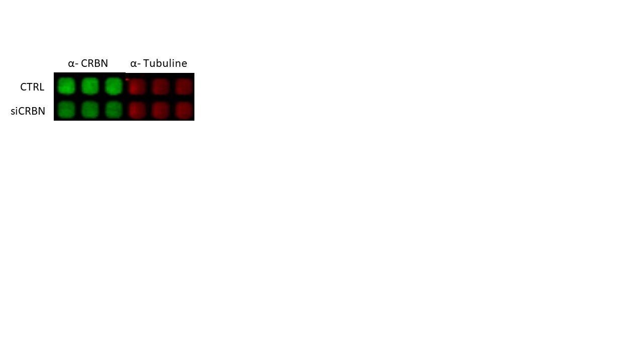
![Knockdown Validated: CRBN Antibody [NBP1-91810]](https://resources.rndsystems.com/images/products/CRBN-Antibody-Western-Blot-NBP1-91810-img0036.jpg "Western Blot: CRBN Antibody [NBP1-91810]")
![CRBN Antibody - BSA Free Western Blot: CRBN Antibody - BSA Free [NBP1-91810]](https://resources.rndsystems.com/images/products/nbp1-91810_rabbit-polyclonal-crbn-antibody-305202510774.jpg "Western Blot: CRBN Antibody - BSA Free [NBP1-91810]")
![CRBN Antibody - BSA Free Western Blot: CRBN Antibody - BSA Free [NBP1-91810]](https://resources.rndsystems.com/images/products/nbp1-91810_rabbit-polyclonal-crbn-antibody-295202516111812.jpg "Western Blot: CRBN Antibody - BSA Free [NBP1-91810]")
Western Blot: CRBN Antibody - BSA Free [NBP1-91810]
Analysis in mouse cell line NIH-3T3 and rat cell line NBT-II.Applications for CRBN Antibody - BSA Free
Immunocytochemistry/ Immunofluorescence
Knockout Validated
Simple Western
Western Blot
See Simple Western Antibody Database for Simple Western validation: Tested in RT4, U-251MG, separated by Size, antibody dilution of 1:60, apparent MW was 60 kDa. Separated by Size-Wes, Sally Sue/Peggy Sue.
Reviewed Applications
Read 1 review rated 4 using NBP1-91810 in the following applications:
Formulation, Preparation, and Storage
Purification
Formulation
Format
Preservative
Concentration
Shipping
Stability & Storage
Background: CRBN
Long Name
Alternate Names
Entrez Gene IDs
Gene Symbol
UniProt
Additional CRBN Products
Product Documents for CRBN Antibody - BSA Free
Certificate of Analysis
To download a Certificate of Analysis, please enter a lot or batch number in the search box below.
Product Specific Notices for CRBN Antibody - BSA Free
This product is for research use only and is not approved for use in humans or in clinical diagnosis. Primary Antibodies are guaranteed for 1 year from date of receipt.
Citations for CRBN Antibody - BSA Free
 Powered by Bioz
Customer Reviews for CRBN Antibody - BSA Free (1)
Have you used CRBN Antibody - BSA Free?
Submit a review and receive an Amazon gift card!
$25/€18/£15/$25CAN/¥2500 Yen for a review with an image
$10/€7/£6/$10CAN/¥1110 Yen for a review without an image
Submit a review
Customer Images
-
Application: In Cell WesternSample Tested: Cancer CellsSpecies: HumanVerified Customer | Posted 11/14/2019CRBN expression after Downregulation by siRNA1/500 dilution

There are no reviews that match your criteria.
Protocols
Find general support by application which include: protocols, troubleshooting, illustrated assays, videos and webinars.
- Appropriate Fixation of IHC/ICC Samples
- Cellular Response to Hypoxia Protocols
- ClariTSA™ Fluorophore Kits
- Detection & Visualization of Antibody Binding
- ICC Cell Smear Protocol for Suspension Cells
- ICC Immunocytochemistry Protocol Videos
- ICC for Adherent Cells
- Immunocytochemistry (ICC) Protocol
- Immunocytochemistry Troubleshooting
- Immunofluorescence of Organoids Embedded in Cultrex Basement Membrane Extract
- Immunohistochemistry (IHC) and Immunocytochemistry (ICC) Protocols
- Preparing Samples for IHC/ICC Experiments
- Preventing Non-Specific Staining (Non-Specific Binding)
- Primary Antibody Selection & Optimization
- Protocol for VisUCyte™ HRP Polymer Detection Reagent
- Protocol for the Fluorescent ICC Staining of Cell Smears - Graphic
- Protocol for the Fluorescent ICC Staining of Cultured Cells on Coverslips - Graphic
- Protocol for the Preparation and Fluorescent ICC Staining of Cells on Coverslips
- Protocol for the Preparation and Fluorescent ICC Staining of Non-adherent Cells
- Protocol for the Preparation and Fluorescent ICC Staining of Stem Cells on Coverslips
- Protocol for the Preparation of a Cell Smear for Non-adherent Cell ICC - Graphic
- R&D Systems Quality Control Western Blot Protocol
- TUNEL and Active Caspase-3 Detection by IHC/ICC Protocol
- The Importance of IHC/ICC Controls
- Troubleshooting Guide: Western Blot Figures
- Western Blot Conditions
- Western Blot Protocol
- Western Blot Protocol for Cell Lysates
- Western Blot Troubleshooting
- Western Blot Troubleshooting Guide
- View all Protocols, Troubleshooting, Illustrated assays and Webinars
FAQs for CRBN Antibody - BSA Free
-
Q: For the picture on your website, you run four cell lines. The lane 2 and 3 all have two bands. Which is the CRBN?
A: Human CRBN has two known isoforms, both of which are approximately the same molecular weight, 50kDa. Given this information, it would be extremely difficult to distinguish the two in a WB. In our testing of the two cell lines, we did get multiple bands very close to each other, while in other lines or tissue lysates, we get one clean band. Unfortunately, we have not yet fully investigated the multiple banding pattern shown in the image. The antibody is polyclonal, so we typically tend to see some non-specific banding that can be cleaned up with further protocol optimization.