CXCR7/RDC-1 is a G protein-coupled receptor (GPCR) member of the CXC subfamily of chemokine receptors. Human CXCR7/RDC-1 is 362 amino acids (aa) in length with a predicted molecular weight of 41 kDa. Mouse and rat CXCR7/RDC-1 share 93% aa sequence identity with the human protein. CXCR7/RDC-1 binds to and acts as a scavenger for CXCL11/I-TAC and CXCL12/SDF-1. CXCR7/RDC-1 can also function as a co-receptor for HIV and SIV. Unlike other chemokine receptors, CXCR7/RDC-1 does not activate G protein signaling, but instead initiates beta-Arrestin-mediated receptor-ligand internalization. Although CXCR7/RDC-1 itself does not activate G protein signaling, the receptor can heterodimerize with CXCR4 to activate G proteins in response to CXCL12/SDF-1 binding. Studies on CXCR7/RDC-1 knockout mice suggest that it is critical for cardiovascular development. While CXCR7/RDC-1 does not appear to signal in normal hematopoietic cells, it is highly expressed in leukemic cells where it activates Akt signaling that promotes cell trafficking and adhesion. CXCR7/RDC-1 also mediates neuronal migration, displays aberrant signaling in astrocytes, and is highly expressed in glioma cells.
CXCR7/RDC-1 Antibody - BSA Free
Novus Biologicals | Catalog # NBP2-24779
![Simple Western: CXCR7/RDC-1 AntibodyBSA Free [NBP2-24779]](https://resources.rndsystems.com/images/products/CXCR7-RDC-1-Antibody-Simple-Western-NBP2-24779-img0003.jpg "Simple Western: CXCR7/RDC-1 AntibodyBSA Free [NBP2-24779]")
Key Product Details
Species Reactivity
Validated:
Human, Mouse, Rat, Sheep
Cited:
Human, Mouse, Rat, Ovine
Predicted:
Bovine (100%), Equine (100%), Primate (100%). Backed by our 100% Guarantee.
Applications
Validated:
Immunohistochemistry, Immunohistochemistry-Paraffin, Western Blot, Immunocytochemistry/ Immunofluorescence, Simple Western, Flow Cytometry (Negative)
Cited:
Immunohistochemistry, Immunohistochemistry-Frozen, Western Blot, Flow Cytometry, Flow (Cell Surface)
Label
Unconjugated
Antibody Source
Polyclonal Rabbit IgG
Format
BSA Free
Loading...
Product Specifications
Immunogen
A synthetic peptide corresponding amino acids 106-129 of human CXCR7/RDC1 was used as the immunogen, GenBank NP_064707.1.
Clonality
Polyclonal
Host
Rabbit
Isotype
IgG
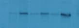
Scientific Data Images for CXCR7/RDC-1 Antibody - BSA Free
Simple Western: CXCR7/RDC-1 AntibodyBSA Free [NBP2-24779]
Simple Western: CXCR7/RDC-1 Antibody [NBP2-24779] - Lane view shows a specific band for CXCR7 in 0.5 mg/mL of Jurkat lysate. This experiment was performed under reducing conditions using the 12-230 kDa separation system.![Immunohistochemistry-Paraffin: CXCR7/RDC-1 Antibody - BSA Free [NBP2-24779]](https://resources.rndsystems.com/images/products/CXCR7-RDC-1-Antibody-Immunohistochemistry-Paraffin-NBP2-24779-img0006.jpg "Immunohistochemistry-Paraffin: CXCR7/RDC-1 Antibody - BSA Free [NBP2-24779]")
Immunohistochemistry-Paraffin: CXCR7/RDC-1 Antibody - BSA Free [NBP2-24779]
Immunohistochemistry-Paraffin: CXCR7/RDC-1 Antibody [NBP2-24779] - Analysis of a FFPE tissue section of human tonsil using 5 ug/mL concentration of CXCR7/RDC-1 antibody. The staining was developed using HRP labeled anti-rabbit secondary antibody and DAB reagent, and nuclei of cells were counter-stained with hematoxylin. This CXCR7 antibody generated a specific membrane cytoplasmic staining in most of the cells, and the signal was highest/very intense in the endothelial cells/blood vessels.![Immunohistochemistry-Paraffin: CXCR7/RDC-1 Antibody - BSA Free [NBP2-24779]](https://resources.rndsystems.com/images/products/CXCR7-RDC-1-Antibody-Immunohistochemistry-Paraffin-NBP2-24779-img0007.jpg "Immunohistochemistry-Paraffin: CXCR7/RDC-1 Antibody - BSA Free [NBP2-24779]")
Immunohistochemistry-Paraffin: CXCR7/RDC-1 Antibody - BSA Free [NBP2-24779]
Immunohistochemistry-Paraffin: CXCR7/RDC-1 Antibody [NBP2-24779] - Analysis of a FFPE tissue section of human tonsil using 5 ug/mL concentration of CXCR7/RDC-1 antibody. The staining was developed using HRP labeled anti-rabbit secondary antibody and DAB reagent, and nuclei of cells were counter-stained with hematoxylin. This CXCR7 antibody generated a specific membrane cytoplasmic staining in most of the cells, and the signal was highest/very intense in the endothelial cells/blood vessels.![Western Blot: CXCR7/RDC-1 AntibodyBSA Free [NBP2-24779]](https://resources.rndsystems.com/images/products/CXCR7-RDC-1-Antibody-Western-Blot-NBP2-24779-img0008.jpg "Western Blot: CXCR7/RDC-1 AntibodyBSA Free [NBP2-24779]")
Western Blot: CXCR7/RDC-1 AntibodyBSA Free [NBP2-24779]
Western Blot: CXCR7/RDC-1 Antibody [NBP2-24779] - Total protein from human spleen, tonsil, lymph node and mouse spleen was separated on a 7.5% gel by SDS-PAGE, transferred to PVDF membrane and blocked in 5% non-fat milk in TBST. The membrane was probed with 2.0 ug/mL anti-CXCR7 in 1% BSA in TBST and detected with an anti-rabbit HRP secondary antibody using chemiluminescence.
Western Blot: CXCR7/RDC-1 Antibody - BSA Free [NBP2-24779] -
CXCR7 expression is upregulated in middle cerebral artery occlusion (MCAO) rat ischemic penumbra brain tissue, in C6 cells under oxygen and glucose deprivation (OGD), and in the peripheral blood of a patient with acute ischemic stroke with thrombectomy. (A) Experimental procedure and timeline. (B) The modified neurological severity score (mNSS) following MCAO establishment. (C) Relative gene expression of CXCR7 in the ischemic penumbra brain tissues of the rats at 6 h, 12 h, 1, 3, and 7 days after MCAO establishment and in the sham group (n = 3). (D) Densitometric quantification of CXCR7/GAPDH. (E) Relative gene expression of CXCR7 in the C6 cells at 0, 2, 4, 6, and 8 h under hypoxia condition and in the sham group (n = 3). (F) Densitometric quantification of CXCR7/GAPDH. (G) Correlation between CXCR7 expression and mNSS scores using Pearson's correlation coefficient. (H) Levels of CXCR7 are increased in the serum of patients with acute ischemic stroke undergoing mechanical thrombectomy (n = 17) compared with that in healthy control participants (n = 10). *p < 0.05 versus the sham group Image collected and cropped by CiteAb from the following open publication (https://pubmed.ncbi.nlm.nih.gov/36523152), licensed under a CC-BY license. Not internally tested by Novus Biologicals.
Western Blot: CXCR7/RDC-1 Antibody - BSA Free [NBP2-24779] -
CXCR7 expression is upregulated in middle cerebral artery occlusion (MCAO) rat ischemic penumbra brain tissue, in C6 cells under oxygen and glucose deprivation (OGD), and in the peripheral blood of a patient with acute ischemic stroke with thrombectomy. (A) Experimental procedure and timeline. (B) The modified neurological severity score (mNSS) following MCAO establishment. (C) Relative gene expression of CXCR7 in the ischemic penumbra brain tissues of the rats at 6 h, 12 h, 1, 3, and 7 days after MCAO establishment and in the sham group (n = 3). (D) Densitometric quantification of CXCR7/GAPDH. (E) Relative gene expression of CXCR7 in the C6 cells at 0, 2, 4, 6, and 8 h under hypoxia condition and in the sham group (n = 3). (F) Densitometric quantification of CXCR7/GAPDH. (G) Correlation between CXCR7 expression and mNSS scores using Pearson's correlation coefficient. (H) Levels of CXCR7 are increased in the serum of patients with acute ischemic stroke undergoing mechanical thrombectomy (n = 17) compared with that in healthy control participants (n = 10). *p < 0.05 versus the sham group Image collected and cropped by CiteAb from the following open publication (https://pubmed.ncbi.nlm.nih.gov/36523152), licensed under a CC-BY license. Not internally tested by Novus Biologicals.Applications for CXCR7/RDC-1 Antibody - BSA Free
Application
Recommended Usage
Immunocytochemistry/ Immunofluorescence
1 - 2 ug/ml
Immunohistochemistry
5 - 10 ug/ml
Immunohistochemistry-Paraffin
5 - 10 ug/mL
Simple Western
1:100
Western Blot
4 - 6 ug/mL
Application Notes
In Simple Western only 10 - 15 uL of the recommended dilution is used per data point.
See Simple Western Antibody Database for Simple Western validation: Tested in Jurkat lysate 0.5 mg/mL, separated by Size, antibody dilution of 1:50, apparent MW was 65 kDa.
See Simple Western Antibody Database for Simple Western validation: Tested in Jurkat lysate 0.5 mg/mL, separated by Size, antibody dilution of 1:50, apparent MW was 65 kDa.
Reviewed Applications
Read 1 review rated 5 using NBP2-24779 in the following applications:
Formulation, Preparation, and Storage
Purification
Peptide affinity purified
Formulation
PBS
Format
BSA Free
Preservative
0.02% Sodium Azide
Concentration
1.0 mg/ml
Shipping
The product is shipped with polar packs. Upon receipt, store it immediately at the temperature recommended below.
Stability & Storage
Store at 4C short term. Aliquot and store at -20C long term. Avoid freeze-thaw cycles.
Background: CXCR7/RDC-1
Additional CXCR7/RDC-1 Products
Product Documents for CXCR7/RDC-1 Antibody - BSA Free
Certificate of Analysis
To download a Certificate of Analysis, please enter a lot or batch number in the search box below.
Product Specific Notices for CXCR7/RDC-1 Antibody - BSA Free
This product is for research use only and is not approved for use in humans or in clinical diagnosis. Primary Antibodies are guaranteed for 1 year from date of receipt.
Citations for CXCR7/RDC-1 Antibody - BSA Free
 Powered by Bioz
Customer Reviews for CXCR7/RDC-1 Antibody - BSA Free (1)
5 out of 5
1 Customer Rating
Have you used CXCR7/RDC-1 Antibody - BSA Free?
Submit a review and receive an Amazon gift card!
$25/€18/£15/$25CAN/¥2500 Yen for a review with an image
$10/€7/£6/$10CAN/¥1110 Yen for a review without an image
Submit a review
Customer Images
Showing
1
-
1 of
1 review
Showing All
Filter By:
-
Application: Western BlotSample Tested: T47D human breast cancer cell lineSpecies: HumanVerified Customer | Posted 03/07/2020

There are no reviews that match your criteria.
Protocols
View specific protocols for CXCR7/RDC-1 Antibody - BSA Free (NBP2-24779):
Immunocytochemistry Protocol
Culture cells to appropriate density in 35 mm culture dishes or 6-well plates.
1. Remove culture medium and wash the cells briefly in PBS. Add 10% formalin to the dish and fix at room temperature for 10 minutes.
2. Remove the formalin and wash the cells in PBS.
3. Permeablize the cells with 0.1% Triton X100 or other suitable detergent for 10 min.
4. Remove the permeablization buffer and wash three times for 10 minutes each in PBS. Be sure to not let the specimen dry out.
5. To block nonspecific antibody binding, incubate in 10% normal goat serum from 1 hour to overnight at room temperature.
6. Add primary antibody at appropriate dilution and incubate overnight at 4C.
7. Remove primary antibody and replace with PBS. Wash three times for 10 minutes each.
8. Add secondary antibody at appropriate dilution. Incubate for 1 hour at room temperature.
9. Remove secondary antibody and replace with PBS. Wash three times for 10 minutes each.
10. Counter stain DNA with DAPi if required.
Western Blot Protocol
1. Perform SDS-PAGE on samples to be analyzed, loading 10-25 ug of total protein per lane.
2. Transfer proteins to PVDF membrane according to the instructions provided by the manufacturer of the membrane and transfer apparatus.
3. Stain the membrane with Ponceau S (or similar product) to assess transfer success, and mark molecular weight standards where appropriate.
4. Rinse the blot TBS -0.05% Tween 20 (TBST).
5. Block the membrane in 5% Non-fat milk in TBST (blocking buffer) for at least 1 hour.
6. Wash the membrane in TBST three times for 10 minutes each.
7. Dilute primary antibody in blocking buffer and incubate overnight at 4C with gentle rocking.
8. Wash the membrane in TBST three times for 10 minutes each.
9. Incubate the membrane in diluted HRP conjugated secondary antibody in blocking buffer (as per manufacturer's instructions) for 1 hour at room temperature.
10. Wash the blot in TBST three times for 10 minutes each (this step can be repeated as required to reduce background).
11. Apply the detection reagent of choice in accordance with the manufacturer's instructions.
Find general support by application which include: protocols, troubleshooting, illustrated assays, videos and webinars.
- Antigen Retrieval Protocol (PIER)
- Antigen Retrieval for Frozen Sections Protocol
- Appropriate Fixation of IHC/ICC Samples
- Cellular Response to Hypoxia Protocols
- Chromogenic IHC Staining of Formalin-Fixed Paraffin-Embedded (FFPE) Tissue Protocol
- Chromogenic Immunohistochemistry Staining of Frozen Tissue
- ClariTSA™ Fluorophore Kits
- Detection & Visualization of Antibody Binding
- Fluorescent IHC Staining of Frozen Tissue Protocol
- Graphic Protocol for Heat-induced Epitope Retrieval
- Graphic Protocol for the Preparation and Fluorescent IHC Staining of Frozen Tissue Sections
- Graphic Protocol for the Preparation and Fluorescent IHC Staining of Paraffin-embedded Tissue Sections
- Graphic Protocol for the Preparation of Gelatin-coated Slides for Histological Tissue Sections
- ICC Cell Smear Protocol for Suspension Cells
- ICC Immunocytochemistry Protocol Videos
- ICC for Adherent Cells
- IHC Sample Preparation (Frozen sections vs Paraffin)
- Immunocytochemistry (ICC) Protocol
- Immunocytochemistry Troubleshooting
- Immunofluorescence of Organoids Embedded in Cultrex Basement Membrane Extract
- Immunofluorescent IHC Staining of Formalin-Fixed Paraffin-Embedded (FFPE) Tissue Protocol
- Immunohistochemistry (IHC) and Immunocytochemistry (ICC) Protocols
- Immunohistochemistry Frozen Troubleshooting
- Immunohistochemistry Paraffin Troubleshooting
- Preparing Samples for IHC/ICC Experiments
- Preventing Non-Specific Staining (Non-Specific Binding)
- Primary Antibody Selection & Optimization
- Protocol for Heat-Induced Epitope Retrieval (HIER)
- Protocol for Making a 4% Formaldehyde Solution in PBS
- Protocol for VisUCyte™ HRP Polymer Detection Reagent
- Protocol for the Fluorescent ICC Staining of Cell Smears - Graphic
- Protocol for the Fluorescent ICC Staining of Cultured Cells on Coverslips - Graphic
- Protocol for the Preparation & Fixation of Cells on Coverslips
- Protocol for the Preparation and Chromogenic IHC Staining of Frozen Tissue Sections
- Protocol for the Preparation and Chromogenic IHC Staining of Frozen Tissue Sections - Graphic
- Protocol for the Preparation and Chromogenic IHC Staining of Paraffin-embedded Tissue Sections
- Protocol for the Preparation and Chromogenic IHC Staining of Paraffin-embedded Tissue Sections - Graphic
- Protocol for the Preparation and Fluorescent ICC Staining of Cells on Coverslips
- Protocol for the Preparation and Fluorescent ICC Staining of Non-adherent Cells
- Protocol for the Preparation and Fluorescent ICC Staining of Stem Cells on Coverslips
- Protocol for the Preparation and Fluorescent IHC Staining of Frozen Tissue Sections
- Protocol for the Preparation and Fluorescent IHC Staining of Paraffin-embedded Tissue Sections
- Protocol for the Preparation of Gelatin-coated Slides for Histological Tissue Sections
- Protocol for the Preparation of a Cell Smear for Non-adherent Cell ICC - Graphic
- R&D Systems Quality Control Western Blot Protocol
- TUNEL and Active Caspase-3 Detection by IHC/ICC Protocol
- The Importance of IHC/ICC Controls
- Troubleshooting Guide: Immunohistochemistry
- Troubleshooting Guide: Western Blot Figures
- Western Blot Conditions
- Western Blot Protocol
- Western Blot Protocol for Cell Lysates
- Western Blot Troubleshooting
- Western Blot Troubleshooting Guide
- View all Protocols, Troubleshooting, Illustrated assays and Webinars
Loading...
Associated Pathways
