Cyclophilin B (SCYLP, CyPB and peptidyl-prolyl cis-trans isomerase B) is a 24 kDa glycoprotein member of the B subfamily of the cyclophilin-type PPIase family of molecules. It is both secreted and retained in the ER. When secreted, Cyclophilin B mediates chemotaxis and T cell adhesion to fibronectin. This is likely due to its prolyl cis/trans isomerase activity. Intracellularly, Cyclophilin B appears to serve as a molecular chaperone for molecules destined for secretion. It does so via stabilization, and facilitating the activity of additional chaperones. The human Cyclophilin B precursor is 216 amino acids (aa) in length. It contains a 25 aa signal sequence plus a 191 aa mature region. There is a partial heparin-binding sequence (aa 27-34), a PPIase domain (aa 47-204) and a C-terminal ER retention motif (aa 213-216). Over aa 34-216, the human and mouse sequences are 95% aa identical.
Cyclophilin B Antibody - BSA Free
Novus Biologicals | Catalog # NBP1-85358
![Immunohistochemistry-Paraffin: Cyclophilin B Antibody [NBP1-85358]](https://resources.rndsystems.com/images/products/Cyclophilin-B-Antibody-Immunohistochemistry-Paraffin-NBP1-85358-img0013.jpg "Immunohistochemistry-Paraffin: Cyclophilin B Antibody [NBP1-85358]")
Loading...
Key Product Details
Validated by
Knockout/Knockdown, Orthogonal Validation
Species Reactivity
Human, Mouse, Rat
Applications
Immunohistochemistry, Immunohistochemistry-Paraffin, Western Blot, Immunocytochemistry/ Immunofluorescence, Simple Western, Knockdown Validated
Label
Unconjugated
Antibody Source
Polyclonal Rabbit IgG
Format
BSA Free
Loading...
Product Specifications
Immunogen
This antibody was developed against Recombinant Protein corresponding to amino acids: LKHYGPGWVSMANAGKDTNGSQFFITTVKTAWLDGKHVVFGKVLEGMEVVRKVESTKTDSRDKPLKDVIIADCGKIEVEKPFAIAKE
Clonality
Polyclonal
Host
Rabbit
Isotype
IgG
Scientific Data Images for Cyclophilin B Antibody - BSA Free
![Western Blot: Cyclophilin B Antibody [NBP1-85358]](https://resources.rndsystems.com/images/products/Cyclophilin-B-Antibody-Western-Blot-NBP1-85358-img0009.jpg "Western Blot: Cyclophilin B Antibody [NBP1-85358]")
![Western Blot: Cyclophilin B Antibody [NBP1-85358]](https://resources.rndsystems.com/images/products/Cyclophilin-B-Antibody-Western-Blot-NBP1-85358-img0014.jpg "Western Blot: Cyclophilin B Antibody [NBP1-85358]")
Western Blot: Cyclophilin B Antibody [NBP1-85358]
Western Blot: Cyclophilin B Antibody [NBP1-85358] - Analysis in mouse cell line NIH-3T3 and rat cell line NBT-II.![Immunocytochemistry/ Immunofluorescence: Cyclophilin B Antibody [NBP1-85358]](https://resources.rndsystems.com/images/products/Cyclophilin-B-Antibody-Immunocytochemistry-Immunofluorescence-NBP1-85358-img0007.jpg "Immunocytochemistry/ Immunofluorescence: Cyclophilin B Antibody [NBP1-85358]")
Immunocytochemistry/ Immunofluorescence: Cyclophilin B Antibody [NBP1-85358]
Immunocytochemistry/Immunofluorescence: Cyclophilin B Antibody [NBP1-85358] - Staining of human cell line HUVEC TERT2 shows localization to endoplasmic reticulum. Antibody staining is shown in green.![Immunohistochemistry-Paraffin: Cyclophilin B Antibody [NBP1-85358]](https://resources.rndsystems.com/images/products/Cyclophilin-B-Antibody-Immunohistochemistry-Paraffin-NBP1-85358-img0017.jpg "Immunohistochemistry-Paraffin: Cyclophilin B Antibody [NBP1-85358]")
Immunohistochemistry-Paraffin: Cyclophilin B Antibody [NBP1-85358]
Immunohistochemistry-Paraffin: Cyclophilin B Antibody [NBP1-85358] - Staining of human placenta shows strong granular cytoplasmic positivity in trophoblastic cells.![Immunohistochemistry-Paraffin: Cyclophilin B Antibody [NBP1-85358]](https://resources.rndsystems.com/images/products/Cyclophilin-B-Antibody-Immunohistochemistry-Paraffin-NBP1-85358-img0010.jpg "Immunohistochemistry-Paraffin: Cyclophilin B Antibody [NBP1-85358]")
Immunohistochemistry-Paraffin: Cyclophilin B Antibody [NBP1-85358]
Immunohistochemistry-Paraffin: Cyclophilin B Antibody [NBP1-85358] - Staining of human skeletal muscle shows low expression as expected.![Immunohistochemistry-Paraffin: Cyclophilin B Antibody [NBP1-85358]](https://resources.rndsystems.com/images/products/Cyclophilin-B-Antibody-Immunohistochemistry-Paraffin-NBP1-85358-img0011.jpg "Immunohistochemistry-Paraffin: Cyclophilin B Antibody [NBP1-85358]")
Immunohistochemistry-Paraffin: Cyclophilin B Antibody [NBP1-85358]
Immunohistochemistry-Paraffin: Cyclophilin B Antibody [NBP1-85358] - Staining of human thyroid gland shows high expression.![Immunohistochemistry-Paraffin: Cyclophilin B Antibody [NBP1-85358]](https://resources.rndsystems.com/images/products/Cyclophilin-B-Antibody-Immunohistochemistry-Paraffin-NBP1-85358-img0015.jpg "Immunohistochemistry-Paraffin: Cyclophilin B Antibody [NBP1-85358]")
Immunohistochemistry-Paraffin: Cyclophilin B Antibody [NBP1-85358]
Immunohistochemistry-Paraffin: Cyclophilin B Antibody [NBP1-85358] - Staining of human prostate shows weak granular cytoplasmic positivity in glandular cells.![Immunohistochemistry-Paraffin: Cyclophilin B Antibody [NBP1-85358]](https://resources.rndsystems.com/images/products/Cyclophilin-B-Antibody-Immunohistochemistry-Paraffin-NBP1-85358-img0016.jpg "Immunohistochemistry-Paraffin: Cyclophilin B Antibody [NBP1-85358]")
Immunohistochemistry-Paraffin: Cyclophilin B Antibody [NBP1-85358]
Immunohistochemistry-Paraffin: Cyclophilin B Antibody [NBP1-85358] - Staining of human endometrium shows moderate granular cytoplasmic positivity in glandular cells.Applications for Cyclophilin B Antibody - BSA Free
Application
Recommended Usage
Immunocytochemistry/ Immunofluorescence
0.25-2 ug/ml
Immunohistochemistry
1:500 - 1:1000
Immunohistochemistry-Paraffin
1:500-1:1000
Simple Western
8 ug/mL
Western Blot
0.04-0.4 ug/ml
Application Notes
For IHC-Paraffin, HIER pH 6 retrieval is recommended. ICC/IF Fixation Permeabilization: Use PFA/Triton X-100.
See Simple Western Antibody Database for Simple Western validation: Tested in Titrated to saturation using various models, separated by Size, antibody dilution of 8 ug/mL
See Simple Western Antibody Database for Simple Western validation: Tested in Titrated to saturation using various models, separated by Size, antibody dilution of 8 ug/mL
Reviewed Applications
Read 1 review rated 4 using NBP1-85358 in the following applications:
Formulation, Preparation, and Storage
Purification
Affinity purified
Formulation
PBS (pH 7.2) and 40% Glycerol
Format
BSA Free
Preservative
0.02% Sodium Azide
Concentration
0.1 mg/ml
Shipping
The product is shipped with polar packs. Upon receipt, store it immediately at the temperature recommended below.
Stability & Storage
Store at 4C short term. Aliquot and store at -20C long term. Avoid freeze-thaw cycles.
Background: Cyclophilin B
Alternate Names
CYP-S1, CYPB, PPIB, S-cyclophilin, SCYLP
Gene Symbol
PPIB
Additional Cyclophilin B Products
Product Documents for Cyclophilin B Antibody - BSA Free
Certificate of Analysis
To download a Certificate of Analysis, please enter a lot or batch number in the search box below.
Product Specific Notices for Cyclophilin B Antibody - BSA Free
This product is for research use only and is not approved for use in humans or in clinical diagnosis. Primary Antibodies are guaranteed for 1 year from date of receipt.
Related Research Areas
Citations for Cyclophilin B Antibody - BSA Free
 Powered by Bioz
Customer Reviews for Cyclophilin B Antibody - BSA Free (1)
4 out of 5
1 Customer Rating
Have you used Cyclophilin B Antibody - BSA Free?
Submit a review and receive an Amazon gift card!
$25/€18/£15/$25CAN/¥2500 Yen for a review with an image
$10/€7/£6/$10CAN/¥1110 Yen for a review without an image
Submit a review
Customer Images
Showing
1
-
1 of
1 review
Showing All
Filter By:
-
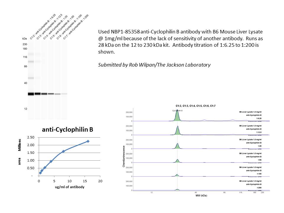
Application: Simple WesternSample Tested: mouse liver lysateSpecies: MouseVerified Customer | Posted 04/21/2017Used NBP1-85358 anti-Cyclophilin B antibody with B6 Mouse Liver Lysate @ 1mg/ml because of the lack of sensitivity of another antibody. Runs as 28 kDa on the 12 to 230 kDa kit. Antibody titration of 1:6.25 to 1:200 is shown.

There are no reviews that match your criteria.
Protocols
Find general support by application which include: protocols, troubleshooting, illustrated assays, videos and webinars.
- Antigen Retrieval Protocol (PIER)
- Antigen Retrieval for Frozen Sections Protocol
- Appropriate Fixation of IHC/ICC Samples
- Cellular Response to Hypoxia Protocols
- Chromogenic IHC Staining of Formalin-Fixed Paraffin-Embedded (FFPE) Tissue Protocol
- Chromogenic Immunohistochemistry Staining of Frozen Tissue
- ClariTSA™ Fluorophore Kits
- Detection & Visualization of Antibody Binding
- Fluorescent IHC Staining of Frozen Tissue Protocol
- Graphic Protocol for Heat-induced Epitope Retrieval
- Graphic Protocol for the Preparation and Fluorescent IHC Staining of Frozen Tissue Sections
- Graphic Protocol for the Preparation and Fluorescent IHC Staining of Paraffin-embedded Tissue Sections
- Graphic Protocol for the Preparation of Gelatin-coated Slides for Histological Tissue Sections
- ICC Cell Smear Protocol for Suspension Cells
- ICC Immunocytochemistry Protocol Videos
- ICC for Adherent Cells
- IHC Sample Preparation (Frozen sections vs Paraffin)
- Immunocytochemistry (ICC) Protocol
- Immunocytochemistry Troubleshooting
- Immunofluorescence of Organoids Embedded in Cultrex Basement Membrane Extract
- Immunofluorescent IHC Staining of Formalin-Fixed Paraffin-Embedded (FFPE) Tissue Protocol
- Immunohistochemistry (IHC) and Immunocytochemistry (ICC) Protocols
- Immunohistochemistry Frozen Troubleshooting
- Immunohistochemistry Paraffin Troubleshooting
- Preparing Samples for IHC/ICC Experiments
- Preventing Non-Specific Staining (Non-Specific Binding)
- Primary Antibody Selection & Optimization
- Protocol for Heat-Induced Epitope Retrieval (HIER)
- Protocol for Making a 4% Formaldehyde Solution in PBS
- Protocol for VisUCyte™ HRP Polymer Detection Reagent
- Protocol for the Fluorescent ICC Staining of Cell Smears - Graphic
- Protocol for the Fluorescent ICC Staining of Cultured Cells on Coverslips - Graphic
- Protocol for the Preparation & Fixation of Cells on Coverslips
- Protocol for the Preparation and Chromogenic IHC Staining of Frozen Tissue Sections
- Protocol for the Preparation and Chromogenic IHC Staining of Frozen Tissue Sections - Graphic
- Protocol for the Preparation and Chromogenic IHC Staining of Paraffin-embedded Tissue Sections
- Protocol for the Preparation and Chromogenic IHC Staining of Paraffin-embedded Tissue Sections - Graphic
- Protocol for the Preparation and Fluorescent ICC Staining of Cells on Coverslips
- Protocol for the Preparation and Fluorescent ICC Staining of Non-adherent Cells
- Protocol for the Preparation and Fluorescent ICC Staining of Stem Cells on Coverslips
- Protocol for the Preparation and Fluorescent IHC Staining of Frozen Tissue Sections
- Protocol for the Preparation and Fluorescent IHC Staining of Paraffin-embedded Tissue Sections
- Protocol for the Preparation of Gelatin-coated Slides for Histological Tissue Sections
- Protocol for the Preparation of a Cell Smear for Non-adherent Cell ICC - Graphic
- R&D Systems Quality Control Western Blot Protocol
- TUNEL and Active Caspase-3 Detection by IHC/ICC Protocol
- The Importance of IHC/ICC Controls
- Troubleshooting Guide: Immunohistochemistry
- Troubleshooting Guide: Western Blot Figures
- Western Blot Conditions
- Western Blot Protocol
- Western Blot Protocol for Cell Lysates
- Western Blot Troubleshooting
- Western Blot Troubleshooting Guide
- View all Protocols, Troubleshooting, Illustrated assays and Webinars
Loading...