Cytokeratin, pan Antibody (AE-1/AE-3)
Novus Biologicals | Catalog # NBP2-29429
Key Product Details
Validated by
Orthogonal Validation
Species Reactivity
Validated:
Human, Mouse, Rat, Bovine, Canine, Chicken, Monkey, Rabbit, Reptile, Zebrafish
Cited:
Human, Mouse, Rat, Avian - Chicken, Bovine, Canine, Fish - Danio rerio (Zebrafish), Lizards
Applications
Validated:
Multiplex Immunofluorescence, Immunohistochemistry, Immunohistochemistry-Paraffin, Immunohistochemistry-Frozen, Western Blot, Flow Cytometry, Flow (Intracellular), Dual RNAscope ISH-IHC, Immunocytochemistry/ Immunofluorescence, COMET, Single Cell Western, CyTOF-reported
Cited:
Immunohistochemistry, Immunohistochemistry-Paraffin, Immunohistochemistry-Frozen, Western Blot, Block/Neutralize, Flow Cytometry, Immunocytochemistry, Immunocytochemistry/ Immunofluorescence, IF/IHC
Label
Unconjugated
Antibody Source
Monoclonal Mouse IgG1 Kappa/IgG1 Kappa Clone # AE-1/AE-3
Loading...
Product Specifications
Immunogen
Human epidermal keratin
Reactivity Notes
Reptile reactivity reported in scientific literature (PMID: 11351328). Zebrafish reactivity reported in scientific literature (PMID: 30970016).
Localization
Cytoplasmic
Marker
Epithelial Marker
Specificity
Twenty human keratins are resolved with two-dimensional gel electrophoresis into acidic (pI 6.0) subfamilies. This antibody cocktail recognizes acidic (Type I or LMW) and basic (Type II or HMW) cytokeratins, which 67kDa (CK1); 64kDa (CK3); 59kDa (CK4); 58kDa (CK5); 56kDa (CK6); 52kDa (CK8); 56.5kDa (CK10); 50kDa (CK14); 50kDa (CK15); 48kDa (CK16); 40kDa (CK19). Many studies have shown the usefulness of keratins as markers in cancer research and tumor diagnosis. AE-1/AE-3 is a broad spectrum anti pan-cytokeratin antibody cocktail, which differentiates epithelial tumors from non-epithelial tumors e.g. squamous vs. adenocarcinoma of the lung, liver carcinoma, breast cancer, and esophageal cancer. It has been used to characterize the source of various neoplasms and to study the distribution of cytokeratin containing cells in epithelia during normal development and during the development of epithelial neoplasms. This antibody stains cytokeratins present in normal and abnormal human tissues and has shown high sensitivity in the recognition of epithelial cells and carcinomas.
Clonality
Monoclonal
Host
Mouse
Isotype
IgG1 Kappa/IgG1 Kappa
Description
200ug/ml of antibody purified from Bioreactor Concentrate by Protein A or G. Prepared in 10 mM PBS with 0.05% BSA & 0.05% azide. Also available WITHOUT BSA & azide at 1.0 mg/ml. (NBP2-33200)
Antibody with azide - store at 2 to 8C. Antibody without azide - store at -20 to -80C.
Antibody with azide - store at 2 to 8C. Antibody without azide - store at -20 to -80C.
Scientific Data Images for Cytokeratin, pan Antibody (AE-1/AE-3)
.png "Detection of pan-Cytokeratin in Human Breast via Multiplex Immunofluorescence staining on COMET™")
Detection of pan-Cytokeratin in Human Breast via Multiplex Immunofluorescence staining on COMET™
Pan-Cytokeratin was detected in immersion fixed paraffin-embedded sections of human Breast Tumor using Mouse Anti-Human Cytokeratin, pan Monoclonal Antibody (Catalog # NBP2-29429) at 1:500 at 37 ° Celsius for 4 minutes. Before incubation with the primary antibody, tissue underwent an all-in-one dewaxing and antigen retrieval preprocessing using PreTreatment Module (PT Module) and Dewax and HIER Buffer H (pH 9; Epredia Catalog # TA-999-DHBH). Tissue was stained using the Alexa Fluor™ 555 Goat anti-Mouse IgG Secondary Antibody at 1:100 at 37 ° Celsius for 2 minutes. (Yellow; Lunaphore Catalog # DR555MS) and counterstained with DAPI (blue; Lunaphore Catalog # DR100). Specific staining was localized to the cytoplasm. Protocol available in COMET™ Panel Builder..png "Detection of Cytokeratin, pan in Mouse Kidney via seqIF™ staining on COMET™")
Detection of Cytokeratin, pan in Mouse Kidney via seqIF™ staining on COMET™
Cytokeratin, pan was detected in immersion fixed paraffin-embedded sections of mouse Kidney using Mouse Anti-Human/Mouse Cyokeratin, pan Monoclonal Antibody (Catalog #NBP2-29429) at 1:200 dilution at 37 ° Celsius for 4 minutes. Before incubation with the primary antibody, tissue underwent an all-in-one dewaxing and antigen retrieval preprocessing using PreTreatment Module (PT Module) and Dewax and HIER Buffer H (pH 9; Epredia Catalog # TA-999-DHBH).Tissue was stained using the Alexa Fluor™ 647 Goat anti-Mouse IgG Secondary Antibody at 1:200 at 37 ° Celsius for 2 minutes. (Yellow; Lunaphore Catalog # DR647MS) and counterstained with DAPI (blue; Lunaphore Catalog # DR100). Specific staining was localized to the membrane. Protocol available in COMET™ Panel Builder.
Detection of Cytokeratin, pan in Human Tissue in Dual RNAscope ISH-IHC
FFPE tissue sections of human metastatic tonsil were probed for Pan Cytokeratin mRNA (ACD RNAScope probe, catalog # 310221; Fast Red chromogen, ACD catalog # 322500). Adjacent tissue section was processes for IHC using mouse monoclonal antibody (Novus catalog # NBP2-29429) at 0.3 ug/mL for 1 hour at room temperature followed by incubation with the anti-mouse IgG VisUCyte HRP Polymer Antibody (Novus Catalog # VC001) and DAB chromogen (yellow-brown). Tissue was counterstained with hematoxylin (blue).
Staining of Cytokeratin, pan in MCF-7 Cells Using Conjugated Cytokeratin, pan Antibody
MCF-7 cells stained with Biotin conjugated version of pan Cytokeratin antibody and SAv-A488. A) SAv-A488 only at a dilution of 1:500, B) pan CK at a dilution of 1:200, C) pan CK at a dilution of 1:400, D) pan CK at a dilution of 1:800. ICC/IF image submitted by a verified customer review.
Immunohistochemical Staining of Cytokeratin, pan in Paraffin Embedded Human Colon Carcinoma
Analysis using Azide and BSA Free version of NBP2-29429. Human Colon Carcinoma stained with pan Cytokeratin Monoclonal Antibody cocktail (AE-1/AE3).
Flow Cytometry of HeLa Cells Stained with Conjugated Cytokeratin, pan Antibody
An intracellular stain was performed on HeLa cells with pan Cytokeratin Antibody (AE1 + AE3) NBP2-33200R (blue) and a matched isotype control (orange). Cells were fixed with 4% PFA and then permeabilized with 0.1% saponin. Cells were incubated in an antibody dilution of 10 ug/mL for 30 minutes at room temperature. Both antibodies were conjugated to Dylight 550.
Flow Cytometry of HeLa Cells Stained with PE Conjugated Cytokeratin, pan Antibody
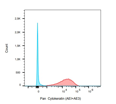
Human Pan-Cytokeratins on HeLa Cells. Black: Cells alone; Green: Isotype Control; Red: PE-labeled Pan-Cytokeratin Monoclonal Antibody (AE-1/AE-3)
CyTOF of Human Fibroblasts Stained for Cytokeratin, pan
Human fibroblasts. Blue: fibroblasts. Red: fibroblast + cytokeratin transfection. CyTOF image submitted by a verified customer review. [NBP2-29429] -")
Flow Cytometry: Cytokeratin, pan Antibody (AE-1/AE-3) [NBP2-29429] -
Flow Cytometry: Cytokeratin, pan Antibody (AE-1/AE-3) [NBP2-29429] - An intracellular stain was performed on HeLa cells with pan Cytokeratin Antibody (AE1 + AE3) NBP2-33200AF647 (blue) and a matched isotype control (orange). Cells were fixed with 4% PFA and then permeabilized with 0.1% saponin. Cells were incubated in an antibody dilution of 5 ug/mL for 30 minutes at room temperature. Both antibodies were conjugated to Alexa Fluor 647. Image from the AF647 version of this antibody. [NBP2-29429] -")
Flow Cytometry: Cytokeratin, pan Antibody (AE-1/AE-3) [NBP2-29429] -
Flow Cytometry: Cytokeratin, pan Antibody (AE-1/AE-3) [NBP2-29429] - An intracellular stain was performed on HeLa cells with pan Cytokeratin [AE1 + AE3] Antibody NBP2-33200AF488 (blue) and a matched isotype control (orange). Cells were fixed with 4% PFA and then permeabilized with 0.1% saponin. Cells were incubated in an antibody dilution of 5 ug/mL for 30 minutes at room temperature. Both antibodies were conjugated to Alexa Fluor 488. Image from the AF488 version of this antibody. [NBP2-29429] -")
Flow Cytometry: Cytokeratin, pan Antibody (AE-1/AE-3) [NBP2-29429] -
Flow Cytometry: Cytokeratin, pan Antibody (AE-1/AE-3) [NBP2-29429] - An intracellular stain was performed on HeLa cells with pan Cytokeratin Antibody (AE1 + AE3) NBP2-33200PE (blue) and a matched isotype control (orange). Cells were fixed with 4% PFA and then permeabilized with 0.1% saponin. Cells were incubated in an antibody dilution of 2.5 ug/mL for 30 minutes at room temperature. Both antibodies were conjugated to phycoerythrin. Image from the PE version of this antibody.Applications for Cytokeratin, pan Antibody (AE-1/AE-3)
Application
Recommended Usage
Flow Cytometry
1-2 ug/million cells
Immunocytochemistry/ Immunofluorescence
1-2 ug/ml
Immunohistochemistry-Frozen
0.5-1.0ug/ml
Immunohistochemistry-Paraffin
0.25-0.5 ug/ml
Single Cell Western
1:10
Western Blot
1-2 ug/ml
Application Notes
Use in ICC/IF reported in scientific literature (PMID:34376789)
Immunohistochemistry (Formalin-fixed): 0.25-0.5ug/ml for 30 min at RT. Staining of formalin-fixed tissues requires heating tissue sections in 10mM Tris with 1mM EDTA, pH 9.0, for 45 min at 95C followed by cooling at RT for 20 minutes.
Optimal dilution for a specific application should be determined.
Western Blot: 1-2ug/ml for 2 hours at RT.
The staining pattern of the pan cytokeratin antibody cocktail may be different than that of either antibody separately.
This antibody cocktail recognizes acidic (Type I or LMW) and basic (Type II or HMW) cytokeratins, which 67 kDa (CK1) ; 64 kDa (CK3) ; 59 kDa (CK4) ; 58 kDa (CK5) ; 56 kDa (CK6) ; 52 kDa (CK8) ; 56.5 kDa (CK10) ; 50k Da (CK14) ; 50 kDa (CK15) ; 48 kDa (CK16) ; 40 kDa (CK19). The pan cytokeratin cocktail does not react with keratin 18, which is also expressed in carcinomas. As such, negative staining with NBP2-29429 in of itself may not be sufficient evidence to rule out the possibility of a carcinoma (Ordonez, 2013).
For example, hepatocellular, adrenal cortical, clear cell renal and chromophobe renal cell carcinomas have been reported to be negative for the pan cytokeratin antibody. In this regard, the pan cytokeratin antibody can be used as part of a screening panel to more extensively define the tumor cell lineages.
The pan cytokeratin antibody may cross-react with GFAP, leading to aberrant positive staining of glial tumors such as ependymoma, glioblastoma, or schwannoma (Ordonez, 2013). Use in Immunohistochemistry reported in scientific literature (PMID: 29169625). This Cytokeratin, pan Antibody (AE-1/AE-3) is validated for CyTOF from a verified customer review.
Reviewed Applications
Read 1 review rated 4 using NBP2-29429 in the following applications:
Flow Cytometry Panel Builder
Bio-Techne Knows Flow Cytometry
Save time and reduce costly mistakes by quickly finding compatible reagents using the Panel Builder Tool.
Advanced Features
- Spectra Viewer - Custom analysis of spectra from multiple fluorochromes
- Spillover Popups - Visualize the spectra of individual fluorochromes
- Antigen Density Selector - Match fluorochrome brightness with antigen density

Formulation, Preparation, and Storage
Purification
Protein A or G purified
Formulation
10 mM PBS with 0.05% BSA
Preservative
0.05% Sodium Azide
Concentration
0.2 mg/ml
Shipping
The product is shipped with polar packs. Upon receipt, store it immediately at the temperature recommended below.
Stability & Storage
Store at 4C.
Background: Cytokeratin, pan
Epithelial cells express multiple subtypes of cytokeratins which can be used to classify epithelial cell type or differentiation status, as well tumor progression or diagnosis (2). Cytokeratins are important for both stability and integrity of epithelial cells and function in intracellular signaling, from wound healing to apoptosis (1). Cytokeratins are useful immunohistochemistry tumor markers and antibodies to cytokeratins are a common pathological tool (1,3,6). Cytokeratin pan antibody is an antibody cocktail mixture that can detect multiple cytokeratins and reacts to multiple epithelial tissues (1,3,6). For example, AE-1/AE-3 is a commonly used specific pan cytokeratin that detects cytokeratins 1-8, 10, 14-16 and 19 (1,3,6).
Given the role of cytokeratins in the structural integrity of epithelial cells, mutations in cytokeratins have been shown to play a role in a variety of human diseases including epidermolysis bullosa simplex (EBS) (4,5). EBS is an autosomal dominant disorder that is caused by missense mutations in either CK5 or CK14 (5). Other known cytokeratin-related disorders include bullous ichthyosis, a skin disorder characterized by redness, blistering, and hyperkeratosis, and epidermolytic palmoplantar keratoderma (EPPK), which results in hyperkeratosis on the palms and soles of the body (7).
References
1. Awasthi, P., Thahriani, A., Bhattacharya, A., Awasthi, P., & Keratins, B. A. (2016). Keratins or cytokeratins: a review article. Journal of Advanced Medical and Dental Sciences Research. https://10.21276/jamdsr.2016.4.4.30
2. Southgate, J., Harnden, P., & Trejdosiewicz, L. K. (1999). Cytokeratin expression patterns in normal and malignant urothelium: a review of the biological and diagnostic implications. Histology and histopathology. https://doi.org/10.14670/HH-14.657
3. Belaldavar, C., Mane, D. R., Hallikerimath, S., Kale, A. D. (2016). Cytokeratins: Its role and expression profile in oral health and disease. Journal of Oral and Maxillofacial Surgery, Medicine, and Pathology. https://doi.org/10.1016/j.ajoms.2015.08.001.
4. Linder S. (2007). Cytokeratin markers come of age. Tumour biology : the journal of the International Society for Oncodevelopmental Biology and Medicine. https://doi.org/10.1159/000107582
5. Jacob, J. T., Coulombe, P. A., Kwan, R., & Omary, M. B. (2018). Types I and II Keratin Intermediate Filaments. Cold Spring Harbor perspectives in biology. https://doi.org/10.1101/cshperspect.a018275
6. Ordonez N. G. (2013). Broad-spectrum immunohistochemical epithelial markers: a review. Human pathology. https://doi.org/10.1016/j.humpath.2012.11.016
7. McLean, W. H., & Moore, C. B. (2011). Keratin disorders: from gene to therapy. Human molecular genetics. https://doi.org/10.1093/hmg/ddr379
Alternate Names
AEI2, CK1, EHK, EPPK, K1, KRT1A, NEPPK
Gene Symbol
KRT1
UniProt
Additional Cytokeratin, pan Products
Product Documents for Cytokeratin, pan Antibody (AE-1/AE-3)
Certificate of Analysis
To download a Certificate of Analysis, please enter a lot or batch number in the search box below.
Product Specific Notices for Cytokeratin, pan Antibody (AE-1/AE-3)
This product is for research use only and is not approved for use in humans or in clinical diagnosis. Primary Antibodies are guaranteed for 1 year from date of receipt.
Citations for Cytokeratin, pan Antibody (AE-1/AE-3)
 Powered by Bioz
Customer Reviews for Cytokeratin, pan Antibody (AE-1/AE-3) (1)
4 out of 5
1 Customer Rating
Have you used Cytokeratin, pan Antibody (AE-1/AE-3)?
Submit a review and receive an Amazon gift card!
$25/€18/£15/$25CAN/¥2500 Yen for a review with an image
$10/€7/£6/$10CAN/¥1110 Yen for a review without an image
Submit a review
Customer Images
Showing
1
-
1 of
1 review
Showing All
Filter By:
-
Application: CyTOFSample Tested: fibroblastsSpecies: HumanVerified Customer | Posted 03/31/2021Blue: fibroblast Red: fibroblast + cytokeratin transfection
 Bio-Techne ResponseThis review was submitted through the legacy Novus Innovators Program, reflecting a new species or application tested on a primary antibody.
There are no reviews that match your criteria.
Protocols
Find general support by application which include: protocols, troubleshooting, illustrated assays, videos and webinars.
- 7-Amino Actinomycin D (7-AAD) Cell Viability Flow Cytometry Protocol
- Antigen Retrieval Protocol (PIER)
- Antigen Retrieval for Frozen Sections Protocol
- Appropriate Fixation of IHC/ICC Samples
- Cellular Response to Hypoxia Protocols
- Chromogenic IHC Staining of Formalin-Fixed Paraffin-Embedded (FFPE) Tissue Protocol
- Chromogenic Immunohistochemistry Staining of Frozen Tissue
- ClariTSA™ Fluorophore Kits
- Detection & Visualization of Antibody Binding
- Extracellular Membrane Flow Cytometry Protocol
- Flow Cytometry Protocol for Cell Surface Markers
- Flow Cytometry Protocol for Staining Membrane Associated Proteins
- Flow Cytometry Staining Protocols
- Flow Cytometry Troubleshooting Guide
- Fluorescent IHC Staining of Frozen Tissue Protocol
- Graphic Protocol for Heat-induced Epitope Retrieval
- Graphic Protocol for the Preparation and Fluorescent IHC Staining of Frozen Tissue Sections
- Graphic Protocol for the Preparation and Fluorescent IHC Staining of Paraffin-embedded Tissue Sections
- Graphic Protocol for the Preparation of Gelatin-coated Slides for Histological Tissue Sections
- ICC Cell Smear Protocol for Suspension Cells
- ICC Immunocytochemistry Protocol Videos
- ICC for Adherent Cells
- IHC Sample Preparation (Frozen sections vs Paraffin)
- ISH-IHC Protocol for Chromogenic Detection on Formalin Fixed Paraffin Embedded (FFPE) Tissue
- Immunocytochemistry (ICC) Protocol
- Immunocytochemistry Troubleshooting
- Immunofluorescence of Organoids Embedded in Cultrex Basement Membrane Extract
- Immunofluorescent IHC Staining of Formalin-Fixed Paraffin-Embedded (FFPE) Tissue Protocol
- Immunohistochemistry (IHC) and Immunocytochemistry (ICC) Protocols
- Immunohistochemistry Frozen Troubleshooting
- Immunohistochemistry Paraffin Troubleshooting
- Intracellular Flow Cytometry Protocol Using Alcohol (Methanol)
- Intracellular Flow Cytometry Protocol Using Detergents
- Intracellular Nuclear Staining Flow Cytometry Protocol Using Detergents
- Intracellular Staining Flow Cytometry Protocol Using Alcohol Permeabilization
- Intracellular Staining Flow Cytometry Protocol Using Detergents to Permeabilize Cells
- Preparing Samples for IHC/ICC Experiments
- Preventing Non-Specific Staining (Non-Specific Binding)
- Primary Antibody Selection & Optimization
- Propidium Iodide Cell Viability Flow Cytometry Protocol
- Protocol for Heat-Induced Epitope Retrieval (HIER)
- Protocol for Liperfluo
- Protocol for Making a 4% Formaldehyde Solution in PBS
- Protocol for VisUCyte™ HRP Polymer Detection Reagent
- Protocol for the Characterization of Human Th22 Cells
- Protocol for the Characterization of Human Th9 Cells
- Protocol for the Fluorescent ICC Staining of Cell Smears - Graphic
- Protocol for the Fluorescent ICC Staining of Cultured Cells on Coverslips - Graphic
- Protocol for the Preparation & Fixation of Cells on Coverslips
- Protocol for the Preparation and Chromogenic IHC Staining of Frozen Tissue Sections
- Protocol for the Preparation and Chromogenic IHC Staining of Frozen Tissue Sections - Graphic
- Protocol for the Preparation and Chromogenic IHC Staining of Paraffin-embedded Tissue Sections
- Protocol for the Preparation and Chromogenic IHC Staining of Paraffin-embedded Tissue Sections - Graphic
- Protocol for the Preparation and Fluorescent ICC Staining of Cells on Coverslips
- Protocol for the Preparation and Fluorescent ICC Staining of Non-adherent Cells
- Protocol for the Preparation and Fluorescent ICC Staining of Stem Cells on Coverslips
- Protocol for the Preparation and Fluorescent IHC Staining of Frozen Tissue Sections
- Protocol for the Preparation and Fluorescent IHC Staining of Paraffin-embedded Tissue Sections
- Protocol for the Preparation of Gelatin-coated Slides for Histological Tissue Sections
- Protocol for the Preparation of a Cell Smear for Non-adherent Cell ICC - Graphic
- Protocol: Annexin V and PI Staining by Flow Cytometry
- Protocol: Annexin V and PI Staining for Apoptosis by Flow Cytometry
- R&D Systems Quality Control Western Blot Protocol
- TUNEL and Active Caspase-3 Detection by IHC/ICC Protocol
- The Importance of IHC/ICC Controls
- Troubleshooting Guide: Fluorokine Flow Cytometry Kits
- Troubleshooting Guide: Immunohistochemistry
- Troubleshooting Guide: Western Blot Figures
- Western Blot Conditions
- Western Blot Protocol
- Western Blot Protocol for Cell Lysates
- Western Blot Troubleshooting
- Western Blot Troubleshooting Guide
- View all Protocols, Troubleshooting, Illustrated assays and Webinars
FAQs for Cytokeratin, pan Antibody (AE-1/AE-3)
Showing
1
-
1 of
1 FAQ
Showing All
-
Q: Could you tell us recommended antigen retrieval method for this antibody?-EndFragment-->
A: Staining of formalin-fixed tissues requires boiling tissue sections in 10mM citrate buffer, pH 6.0, for 10-20 min followed by cooling at RT for 20 minutes. Optimal dilution of antibody for IHC should be determined by the user.
Loading...