DLL4 Antibody - BSA Free
Novus Biologicals | Catalog # NB600-892
![Western Blot: DLL4 Antibody [NB600-892]](https://resources.rndsystems.com/images/products/DLL4-Antibody-Western-Blot-NB600-892-img0006.jpg "Western Blot: DLL4 Antibody [NB600-892]")
Key Product Details
Species Reactivity
Validated:
Cited:
Applications
Validated:
Cited:
Label
Antibody Source
Format
Product Specifications
Immunogen
Reactivity Notes
Mouse reactivity reported in scientific literature (PMID: 27735989).
Rat reactivity reported in scientific literature (PMID: 19828677).
Localization
Clonality
Host
Isotype
Description
Store vial at -20C prior to opening. Aliquot contents and freeze at -20C or below for extended storage. Avoid cycles of freezing and thawing. Centrifuge product if not completely clear after standing at room temperature. This product is stable for several weeks at 4C as an undiluted liquid. Dilute only prior to immediate use.
Scientific Data Images for DLL4 Antibody - BSA Free
Western Blot: DLL4 Antibody [NB600-892]
Western Blot: DLL4 Antibody [NB600-892] - Western Blot of DLL4 antibody. Lane 1: Mouse Pancreas Whole Cell Lysate. Lane 2: HUVEK Whole Cell Lysate.Lane 3: Human rDLL4.Lane 4: HEK293 Whole Cell Lysate + Human rDLL4. Lane 5: HEK293 Whole Cell Lysate.Load: 10 ug per lane for Whole Cell Lysates or 50 ng for recombinant protein.Primary antibody: DLL4 antibody at 1:1,000 overnight at 4C.Secondary antibody: Peroxidase rabbit secondary antibody at 1:40,000 for 30 min at RT.Block: Blocking Buffer for Fluorescent Western Blotting for 30 min at RT.Predicted/Observed size: 74 kDa, 74 kDa for DLL4. Other band(s): Not Identified.![Immunohistochemistry: DLL4 Antibody [NB600-892]](https://resources.rndsystems.com/images/products/DLL4-Antibody-Immunohistochemistry-NB600-892-img0007.jpg "Immunohistochemistry: DLL4 Antibody [NB600-892]")
Immunohistochemistry: DLL4 Antibody [NB600-892]
Immunohistochemistry: DLL4 Antibody [NB600-892] - Affinity Purified DLL4 antibody was used at 20 ug/ml to detect DLL4 in a variety of tissues including colon, liver, skeletal muscle, ovary, pancreas, prostate, testes, thymus, tonsil and uterus. In contrast to reported findings, no staining was observed in vascular tissue. This image shows DLL4 staining of human ovary. Tissue was formalin-fixed and paraffin embedded.![Western Blot: DLL4 Antibody [NB600-892]](https://resources.rndsystems.com/images/products/DLL4-Antibody-Western-Blot-NB600-892-img0005.jpg "Western Blot: DLL4 Antibody [NB600-892]")
Western Blot: DLL4 Antibody [NB600-892]
Western Blot: DLL4 Antibody [NB600-892] - Western Blot of DLL4 antibody. Lane 1: mouse pancreatic tissue. Load: 20 ug per lane.Primary antibody: DLL4 antibody at 1:500 for overnight at 4C.Secondary antibody: HRP-labeled Goat anti-Rabbit IgG secondary antibody at 1:5,000 for 1 hour at RT. A chemiluminescence system was used for signal detection using a 3 min exposure time.Block: 5% Goat Serum overnight at 4C.Predicted/Observed size: 74 kDa, 70 kDa for Delta 4. Other band(s): none.![ELISA: DLL4 Antibody [NB600-892]](https://resources.rndsystems.com/images/products/DLL4-Antibody-ELISA-NB600-892-img0008.jpg "ELISA: DLL4 Antibody [NB600-892]")
ELISA: DLL4 Antibody [NB600-892]
ELISA: DLL4 Antibody [NB600-892] - ELISA results of purified DLL4 antibody tested against BSA-conjugated peptide of immunizing peptide. Each well was coated in duplicate with 0.1ug of conjugate. The starting dilution of antibody was 5ug/ml and the X-axis represents the Log10 of a 3-fold dilution. This titration is a 4-parameter curve fit where the IC50 is defined as the titer of the antibody. Assay performed using 3% fish gel, Goat anti-Rabbit IgG Antibody Peroxidase Conjugated (Min X Bv Ch Gt GP Ham Hs Hu Ms Rt & Sh Serum Proteins) and TMB ELISA Peroxidase Substrate.
DLL4 Antibody
Affinity Purified anti-Delta-4 antibody was used at 20 ug/ml to detect Delta-4 in a variety of tissues including colon, liver, skeletal muscle, ovary, pancreas, prostate, testes, thymus, tonsil and uterus. In contrast to reported findings, no staining was observed in vascular tissue. This image shows Delta-4 staining of human ovary. Tissue was formalin-fixed and paraffin embedded. Personal Communication, Tina Roush, LifeSpanBiosciences, Seattle, WA.
Western Blot: DLL4 Antibody - BSA Free [NB600-892] -
Expression of NICD in human platelets.(A) Immunoblot showing expression of NICD in DLL-4 (15 ug/ml for 10 min)-treated platelets in absence or presence of either DAPT (10 uM) or DBZ (10 uM) or vehicle. (B) Corresponding densitometric analysis of NICD normalised with beta -actin (n=6). (C, D and E) Immunoblot of NICD expression in either stored or A23187 (1 uM)-treated platelets under conditions as indicated. Data are represented as mean +/- SEM of at least three individual experiments and analyzed by RM one-way ANOVA with Dunnett’s multiple comparisons test.Figure 2—source data 1.Excel sheet shows numerical data of Figure 2.Figure 2—source data 2.Unedited and labelled blots of Figure 2.Figure 2—source data 3.Unedited and unlabelled blots of Figure 2.Excel sheet shows numerical data of Figure 2.Unedited and labelled blots of Figure 2.Unedited and unlabelled blots of Figure 2. Image collected and cropped by CiteAb from the following open publication (https://pubmed.ncbi.nlm.nih.gov/36190110), licensed under a CC-BY license. Not internally tested by Novus Biologicals.Applications for DLL4 Antibody - BSA Free
ELISA
Immunocytochemistry/ Immunofluorescence
Immunohistochemistry
Immunohistochemistry-Paraffin
Western Blot
Use in Immunocytochemistry/immunofluorescence reported in scientific literature (PMID 28352669).
Use in FLOW reported in scientific literature (PMID: 19915058).
Reviewed Applications
Read 1 review rated 1 using NB600-892 in the following applications:
Flow Cytometry Panel Builder
Bio-Techne Knows Flow Cytometry
Save time and reduce costly mistakes by quickly finding compatible reagents using the Panel Builder Tool.
Advanced Features
- Spectra Viewer - Custom analysis of spectra from multiple fluorochromes
- Spillover Popups - Visualize the spectra of individual fluorochromes
- Antigen Density Selector - Match fluorochrome brightness with antigen density

Formulation, Preparation, and Storage
Purification
Formulation
Format
Preservative
Concentration
Shipping
Stability & Storage
Background: DLL4
Long Name
Alternate Names
Entrez Gene IDs
Gene Symbol
UniProt
Additional DLL4 Products
Product Documents for DLL4 Antibody - BSA Free
Certificate of Analysis
To download a Certificate of Analysis, please enter a lot or batch number in the search box below.
Product Specific Notices for DLL4 Antibody - BSA Free
This product is for research use only and is not approved for use in humans or in clinical diagnosis. Primary Antibodies are guaranteed for 1 year from date of receipt.
Related Research Areas
Citations for DLL4 Antibody - BSA Free
 Powered by Bioz
Customer Reviews for DLL4 Antibody - BSA Free (1)
Have you used DLL4 Antibody - BSA Free?
Submit a review and receive an Amazon gift card!
$25/€18/£15/$25CAN/¥2500 Yen for a review with an image
$10/€7/£6/$10CAN/¥1110 Yen for a review without an image
Submit a review
Customer Images
-
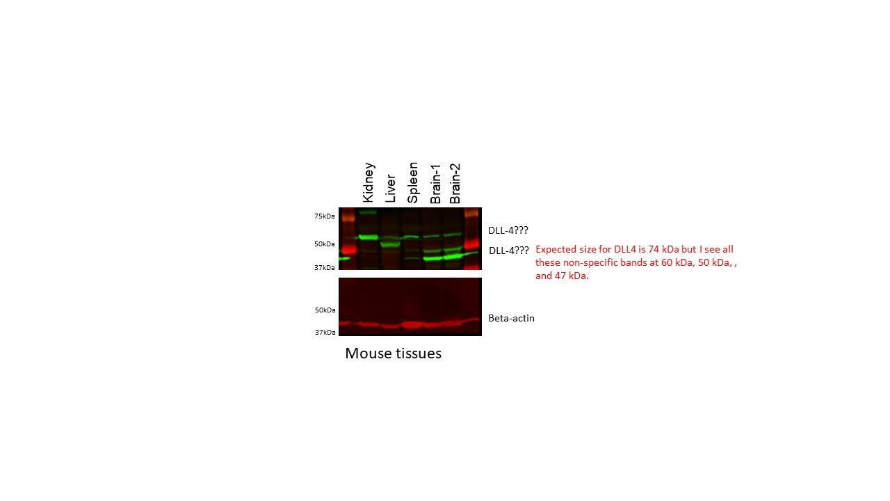
Application: Western BlotSample Tested: Adult kidney, Liver, Mouse Spleen and BrainSpecies: MouseVerified Customer | Posted 02/24/2020Mouse tissue lysate were separated on 12% SDS-PAGE. Western blotting was performed on PVDF membrane, antibody used at 1:200 diltuion and b-actin was used as normalizer. Licor IRDye® 800CW Goat anti-Rabbit IgG was used as a secondary for DLL4.Lot of non-specific bands

There are no reviews that match your criteria.
Protocols
Find general support by application which include: protocols, troubleshooting, illustrated assays, videos and webinars.
- 7-Amino Actinomycin D (7-AAD) Cell Viability Flow Cytometry Protocol
- Antigen Retrieval Protocol (PIER)
- Antigen Retrieval for Frozen Sections Protocol
- Appropriate Fixation of IHC/ICC Samples
- Cellular Response to Hypoxia Protocols
- Chromogenic IHC Staining of Formalin-Fixed Paraffin-Embedded (FFPE) Tissue Protocol
- Chromogenic Immunohistochemistry Staining of Frozen Tissue
- ClariTSA™ Fluorophore Kits
- Detection & Visualization of Antibody Binding
- ELISA Sample Preparation & Collection Guide
- ELISA Troubleshooting Guide
- Extracellular Membrane Flow Cytometry Protocol
- Flow Cytometry Protocol for Cell Surface Markers
- Flow Cytometry Protocol for Staining Membrane Associated Proteins
- Flow Cytometry Staining Protocols
- Flow Cytometry Troubleshooting Guide
- Fluorescent IHC Staining of Frozen Tissue Protocol
- Graphic Protocol for Heat-induced Epitope Retrieval
- Graphic Protocol for the Preparation and Fluorescent IHC Staining of Frozen Tissue Sections
- Graphic Protocol for the Preparation and Fluorescent IHC Staining of Paraffin-embedded Tissue Sections
- Graphic Protocol for the Preparation of Gelatin-coated Slides for Histological Tissue Sections
- How to Run an R&D Systems DuoSet ELISA
- How to Run an R&D Systems Quantikine ELISA
- How to Run an R&D Systems Quantikine™ QuicKit™ ELISA
- ICC Cell Smear Protocol for Suspension Cells
- ICC Immunocytochemistry Protocol Videos
- ICC for Adherent Cells
- IHC Sample Preparation (Frozen sections vs Paraffin)
- Immunocytochemistry (ICC) Protocol
- Immunocytochemistry Troubleshooting
- Immunofluorescence of Organoids Embedded in Cultrex Basement Membrane Extract
- Immunofluorescent IHC Staining of Formalin-Fixed Paraffin-Embedded (FFPE) Tissue Protocol
- Immunohistochemistry (IHC) and Immunocytochemistry (ICC) Protocols
- Immunohistochemistry Frozen Troubleshooting
- Immunohistochemistry Paraffin Troubleshooting
- Intracellular Flow Cytometry Protocol Using Alcohol (Methanol)
- Intracellular Flow Cytometry Protocol Using Detergents
- Intracellular Nuclear Staining Flow Cytometry Protocol Using Detergents
- Intracellular Staining Flow Cytometry Protocol Using Alcohol Permeabilization
- Intracellular Staining Flow Cytometry Protocol Using Detergents to Permeabilize Cells
- Preparing Samples for IHC/ICC Experiments
- Preventing Non-Specific Staining (Non-Specific Binding)
- Primary Antibody Selection & Optimization
- Propidium Iodide Cell Viability Flow Cytometry Protocol
- Protocol for Heat-Induced Epitope Retrieval (HIER)
- Protocol for Liperfluo
- Protocol for Making a 4% Formaldehyde Solution in PBS
- Protocol for VisUCyte™ HRP Polymer Detection Reagent
- Protocol for the Characterization of Human Th22 Cells
- Protocol for the Characterization of Human Th9 Cells
- Protocol for the Fluorescent ICC Staining of Cell Smears - Graphic
- Protocol for the Fluorescent ICC Staining of Cultured Cells on Coverslips - Graphic
- Protocol for the Preparation & Fixation of Cells on Coverslips
- Protocol for the Preparation and Chromogenic IHC Staining of Frozen Tissue Sections
- Protocol for the Preparation and Chromogenic IHC Staining of Frozen Tissue Sections - Graphic
- Protocol for the Preparation and Chromogenic IHC Staining of Paraffin-embedded Tissue Sections
- Protocol for the Preparation and Chromogenic IHC Staining of Paraffin-embedded Tissue Sections - Graphic
- Protocol for the Preparation and Fluorescent ICC Staining of Cells on Coverslips
- Protocol for the Preparation and Fluorescent ICC Staining of Non-adherent Cells
- Protocol for the Preparation and Fluorescent ICC Staining of Stem Cells on Coverslips
- Protocol for the Preparation and Fluorescent IHC Staining of Frozen Tissue Sections
- Protocol for the Preparation and Fluorescent IHC Staining of Paraffin-embedded Tissue Sections
- Protocol for the Preparation of Gelatin-coated Slides for Histological Tissue Sections
- Protocol for the Preparation of a Cell Smear for Non-adherent Cell ICC - Graphic
- Protocol: Annexin V and PI Staining by Flow Cytometry
- Protocol: Annexin V and PI Staining for Apoptosis by Flow Cytometry
- Quantikine HS ELISA Kit Assay Principle, Alkaline Phosphatase
- Quantikine HS ELISA Kit Principle, Streptavidin-HRP Polymer
- R&D Systems Quality Control Western Blot Protocol
- Sandwich ELISA (Colorimetric) – Biotin/Streptavidin Detection Protocol
- Sandwich ELISA (Colorimetric) – Direct Detection Protocol
- TUNEL and Active Caspase-3 Detection by IHC/ICC Protocol
- The Importance of IHC/ICC Controls
- Troubleshooting Guide: ELISA
- Troubleshooting Guide: Fluorokine Flow Cytometry Kits
- Troubleshooting Guide: Immunohistochemistry
- Troubleshooting Guide: Western Blot Figures
- Western Blot Conditions
- Western Blot Protocol
- Western Blot Protocol for Cell Lysates
- Western Blot Troubleshooting
- Western Blot Troubleshooting Guide
- View all Protocols, Troubleshooting, Illustrated assays and Webinars
FAQs for DLL4 Antibody - BSA Free
-
Q: Is the antibody intended to work on paraffin sections only? Did you test it on frozen sections?
A:
In our QC data, I found that we have successfully tested this antibody for Western Blot and IHC-P. We have not confirmed the antibody for the IHC-Fr application; however, I would not discourage you from trying it on your frozen sections. For IHC-P sections this antibody worked well at concentrations from 10 ug/ml - 20 ug/ml and you may try this antibody for IHC on your frozen sections considering similar dilutions as a starting point. Moreover, considering your case, we can recommend our Innovators Reward Program. Our Innovator's Reward is offered to reward researchers for testing new species and applications with our products. If testing on this antibody is performed on an untested species and/or application, and the results are shared, then the antibody is eligible for the Innovator's Reward. Under this program, Novus will provide you a credit of equal or lesser value on a future product of equal value in exchange for your new data in the form of an online review. The great thing is you are eligible even if the antibody doesn't work because the data is still useful to us.
Associated Pathways
