DNA Polymerase beta Antibody - BSA Free
Novus Biologicals | Catalog # NBP2-38600
![Immunohistochemistry-Paraffin: DNA Polymerase beta Antibody [NBP2-38600]](https://resources.rndsystems.com/images/products/DNA-Polymerase-beta-Antibody-Immunohistochemistry-Paraffin-NBP2-38600-img0006.jpg "Immunohistochemistry-Paraffin: DNA Polymerase beta Antibody [NBP2-38600]")
Loading...
Key Product Details
Validated by
Orthogonal Validation
Species Reactivity
Validated:
Human
Predicted:
Mouse (97%), Rat (97%). Backed by our 100% Guarantee.
Applications
Immunohistochemistry, Immunohistochemistry-Paraffin, Western Blot, Immunocytochemistry/ Immunofluorescence
Label
Unconjugated
Antibody Source
Polyclonal Rabbit IgG
Format
BSA Free
Loading...
Product Specifications
Immunogen
This antibody was developed against a recombinant protein corresponding to amino acids: YCGVLYFTGSDIFNKNMRAHALEKGFTINEYTIRPLGVTGVAGEPLPVDSEKDIFDYIQWKYREPKDRSE
Clonality
Polyclonal
Host
Rabbit
Isotype
IgG
Scientific Data Images for DNA Polymerase beta Antibody - BSA Free
![Immunocytochemistry/ Immunofluorescence: DNA Polymerase beta Antibody [NBP2-38600]](https://resources.rndsystems.com/images/products/DNA-Polymerase-beta-Antibody-Immunocytochemistry-Immunofluorescence-NBP2-38600-img0004.jpg "Immunocytochemistry/ Immunofluorescence: DNA Polymerase beta Antibody [NBP2-38600]")
Immunocytochemistry/ Immunofluorescence: DNA Polymerase beta Antibody [NBP2-38600]
Immunocytochemistry/Immunofluorescence: DNA Polymerase beta Antibody [NBP2-38600] - Staining of human cell line A-431 shows localization to vesicles. Antibody staining is shown in green.![Immunohistochemistry-Paraffin: DNA Polymerase beta Antibody [NBP2-38600]](https://resources.rndsystems.com/images/products/DNA-Polymerase-beta-Antibody-Immunohistochemistry-Paraffin-NBP2-38600-img0005.jpg "Immunohistochemistry-Paraffin: DNA Polymerase beta Antibody [NBP2-38600]")
Immunohistochemistry-Paraffin: DNA Polymerase beta Antibody [NBP2-38600]
Immunohistochemistry-Paraffin: DNA Polymerase beta Antibody [NBP2-38600] - Staining of human liver shows low expression as expected.![Immunohistochemistry: DNA Polymerase beta Antibody [NBP2-38600]](https://resources.rndsystems.com/images/products/DNA-Polymerase-beta-Antibody-Immunohistochemistry-NBP2-38600-img0002.jpg "Immunohistochemistry: DNA Polymerase beta Antibody [NBP2-38600]")
Immunohistochemistry: DNA Polymerase beta Antibody [NBP2-38600]
Immunohistochemistry: DNA Polymerase beta Antibody [NBP2-38600] - Staining of human testis shows strong nuclear positivity in cells in seminiferous ducts.
DNA Polymerase beta Antibody [NBP2-38600] -
Analysis in human cell line MOLT-4.
DNA Polymerase beta Antibody [NBP2-38600] -
Staining of human urinary bladder shows moderate nuclear positivity in urothelial cells.
DNA Polymerase beta Antibody [NBP2-38600] -
Staining of human prostate shows moderate nuclear positivity in glandular cells.
DNA Polymerase beta Antibody [NBP2-38600] -
Staining of human colon shows no positivity in glandular cells as expected.Applications for DNA Polymerase beta Antibody - BSA Free
Application
Recommended Usage
Immunocytochemistry/ Immunofluorescence
0.25-2 ug/ml
Immunohistochemistry
1:200 - 1:500
Immunohistochemistry-Paraffin
1:200 - 1:500
Western Blot
0.04-0.4 ug/ml
Application Notes
For IHC-Paraffin, HIER pH 6 retrieval is recommended. ICC/IF Fixation Permeabilization: Use PFA/Triton X-100.
Reviewed Applications
Read 1 review rated 5 using NBP2-38600 in the following applications:
Formulation, Preparation, and Storage
Purification
Affinity purified
Formulation
PBS (pH 7.2) and 40% Glycerol
Format
BSA Free
Preservative
0.02% Sodium Azide
Concentration
Concentrations vary lot to lot. See vial label for concentration. If unlisted please contact technical services.
Shipping
The product is shipped with polar packs. Upon receipt, store it immediately at the temperature recommended below.
Stability & Storage
Store at 4C short term. Aliquot and store at -20C long term. Avoid freeze-thaw cycles.
Background: DNA Polymerase beta
Additional DNA Polymerase beta Products
Product Documents for DNA Polymerase beta Antibody - BSA Free
Certificate of Analysis
To download a Certificate of Analysis, please enter a lot or batch number in the search box below.
Product Specific Notices for DNA Polymerase beta Antibody - BSA Free
This product is for research use only and is not approved for use in humans or in clinical diagnosis. Primary Antibodies are guaranteed for 1 year from date of receipt.
Related Research Areas
Citations for DNA Polymerase beta Antibody - BSA Free
 Powered by Bioz
Customer Reviews for DNA Polymerase beta Antibody - BSA Free (1)
5 out of 5
1 Customer Rating
Have you used DNA Polymerase beta Antibody - BSA Free?
Submit a review and receive an Amazon gift card!
$25/€18/£15/$25CAN/¥2500 Yen for a review with an image
$10/€7/£6/$10CAN/¥1110 Yen for a review without an image
Submit a review
Customer Images
Showing
1
-
1 of
1 review
Showing All
Filter By:
-
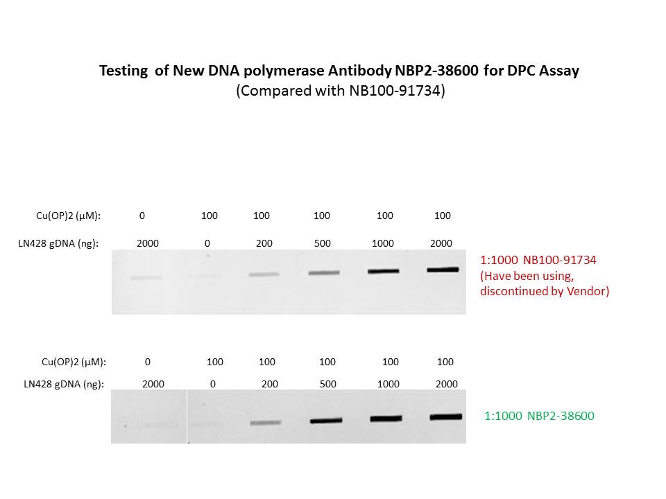
Application: Slot blot DPC assaySample Tested: Genomic DNA samples from LN428 cells -treated by copper complex to induce DNA-protein cross-linksSpecies: HumanVerified Customer | Posted 03/24/2017After the rabbit anti-DNA polymerase beta antibody from Novus Biologicals NB100-91734, which this lab has been using for DPC assay, being discontinued by Novus biologicals, we have been looking for an available antibody for the application of DPC assay. LN428 human cancer cells were treated by 100 uM of 1,10-Phenanthroline-Copper (II) complex to induce genomic DNA damages (DNA-protein cross-links); a serial of amount of gDNA samples (0, 200, 500, 1000 and 2000ng) were prepared and gDNA samples were blotted onto nitrocellulose membrane and probed for DNA polymerase beta which was trapped on the sites of gDNA damages. The NBP2-38600 antibody was compared with the discontinued DNA Polymerase beta antibody NB100-91734, which we have been using and has been working very well for DPC assay, The result showed that NBP2-38600 worked as good as, or evenbetter than, the discontinued NB100-91734. Slot blot DPC assay, a new and widely-used technique to detect DNA-Protein Cross-links (DPC) - induced by oxidative stresses in vitro (in tubes) or in vivo (in cells).

There are no reviews that match your criteria.
Protocols
Find general support by application which include: protocols, troubleshooting, illustrated assays, videos and webinars.
- Antigen Retrieval Protocol (PIER)
- Antigen Retrieval for Frozen Sections Protocol
- Appropriate Fixation of IHC/ICC Samples
- Cellular Response to Hypoxia Protocols
- Chromogenic IHC Staining of Formalin-Fixed Paraffin-Embedded (FFPE) Tissue Protocol
- Chromogenic Immunohistochemistry Staining of Frozen Tissue
- ClariTSA™ Fluorophore Kits
- Detection & Visualization of Antibody Binding
- Fluorescent IHC Staining of Frozen Tissue Protocol
- Graphic Protocol for Heat-induced Epitope Retrieval
- Graphic Protocol for the Preparation and Fluorescent IHC Staining of Frozen Tissue Sections
- Graphic Protocol for the Preparation and Fluorescent IHC Staining of Paraffin-embedded Tissue Sections
- Graphic Protocol for the Preparation of Gelatin-coated Slides for Histological Tissue Sections
- ICC Cell Smear Protocol for Suspension Cells
- ICC Immunocytochemistry Protocol Videos
- ICC for Adherent Cells
- IHC Sample Preparation (Frozen sections vs Paraffin)
- Immunocytochemistry (ICC) Protocol
- Immunocytochemistry Troubleshooting
- Immunofluorescence of Organoids Embedded in Cultrex Basement Membrane Extract
- Immunofluorescent IHC Staining of Formalin-Fixed Paraffin-Embedded (FFPE) Tissue Protocol
- Immunohistochemistry (IHC) and Immunocytochemistry (ICC) Protocols
- Immunohistochemistry Frozen Troubleshooting
- Immunohistochemistry Paraffin Troubleshooting
- Preparing Samples for IHC/ICC Experiments
- Preventing Non-Specific Staining (Non-Specific Binding)
- Primary Antibody Selection & Optimization
- Protocol for Heat-Induced Epitope Retrieval (HIER)
- Protocol for Making a 4% Formaldehyde Solution in PBS
- Protocol for VisUCyte™ HRP Polymer Detection Reagent
- Protocol for the Fluorescent ICC Staining of Cell Smears - Graphic
- Protocol for the Fluorescent ICC Staining of Cultured Cells on Coverslips - Graphic
- Protocol for the Preparation & Fixation of Cells on Coverslips
- Protocol for the Preparation and Chromogenic IHC Staining of Frozen Tissue Sections
- Protocol for the Preparation and Chromogenic IHC Staining of Frozen Tissue Sections - Graphic
- Protocol for the Preparation and Chromogenic IHC Staining of Paraffin-embedded Tissue Sections
- Protocol for the Preparation and Chromogenic IHC Staining of Paraffin-embedded Tissue Sections - Graphic
- Protocol for the Preparation and Fluorescent ICC Staining of Cells on Coverslips
- Protocol for the Preparation and Fluorescent ICC Staining of Non-adherent Cells
- Protocol for the Preparation and Fluorescent ICC Staining of Stem Cells on Coverslips
- Protocol for the Preparation and Fluorescent IHC Staining of Frozen Tissue Sections
- Protocol for the Preparation and Fluorescent IHC Staining of Paraffin-embedded Tissue Sections
- Protocol for the Preparation of Gelatin-coated Slides for Histological Tissue Sections
- Protocol for the Preparation of a Cell Smear for Non-adherent Cell ICC - Graphic
- R&D Systems Quality Control Western Blot Protocol
- TUNEL and Active Caspase-3 Detection by IHC/ICC Protocol
- The Importance of IHC/ICC Controls
- Troubleshooting Guide: Immunohistochemistry
- Troubleshooting Guide: Western Blot Figures
- Western Blot Conditions
- Western Blot Protocol
- Western Blot Protocol for Cell Lysates
- Western Blot Troubleshooting
- Western Blot Troubleshooting Guide
- View all Protocols, Troubleshooting, Illustrated assays and Webinars
Loading...