Dopamine beta-Hydroxylase Antibody
Novus Biologicals | Catalog # NBP1-31386
![Immunohistochemistry-Paraffin: Dopamine beta-Hydroxylase Antibody [NBP1-31386]](https://resources.rndsystems.com/images/products/Dopamine-beta-Hydroxylase-Antibody-Immunohistochemistry-Paraffin-NBP1-31386-img0013.jpg "Immunohistochemistry-Paraffin: Dopamine beta-Hydroxylase Antibody [NBP1-31386]")
Loading...
Key Product Details
Validated by
Orthogonal Validation
Species Reactivity
Validated:
Human, Mouse, Rat
Cited:
Human, Mouse
Applications
Validated:
Immunohistochemistry, Immunohistochemistry-Paraffin, Western Blot
Cited:
Western Blot
Label
Unconjugated
Antibody Source
Polyclonal Rabbit IgG
Loading...
Product Specifications
Immunogen
Recombinant protein encompassing a sequence within the center region of human Dopamine beta-Hydroxylase. The exact sequence is proprietary.
Reactivity Notes
Immunogen displays the following percentage of sequence identity for non-tested species: Rhesus Macaque (86%), Bovine (80%).
Localization
Soluble dopamine beta-hydroxylase: Cytoplasmic vesicle, secretory vesicle lumen, Cytoplasmic vesicle, secretory vesicle, chromaffin granule lumen, secretory vesicle membrane, chromaffin granule membrane
Clonality
Polyclonal
Host
Rabbit
Isotype
IgG
Theoretical MW
69 kDa.
Disclaimer note: The observed molecular weight of the protein may vary from the listed predicted molecular weight due to post translational modifications, post translation cleavages, relative charges, and other experimental factors.
Disclaimer note: The observed molecular weight of the protein may vary from the listed predicted molecular weight due to post translational modifications, post translation cleavages, relative charges, and other experimental factors.
Scientific Data Images for Dopamine beta-Hydroxylase Antibody
Immunohistochemistry-Paraffin: Dopamine beta-Hydroxylase Antibody [NBP1-31386]
Immunohistochemistry-Paraffin: Dopamine beta-Hydroxylase Antibody [NBP1-31386] - RT2 xenograft. Dopamine beta hydroxylase antibody dilution: 1:500. Antigen Retrieval: Trilogy™ (EDTA based, pH 8.0) buffer, 15min.![Immunohistochemistry-Paraffin: Dopamine beta-Hydroxylase Antibody [NBP1-31386]](https://resources.rndsystems.com/images/products/Dopamine-beta-Hydroxylase-Antibody-Immunohistochemistry-Paraffin-NBP1-31386-img0011.jpg "Immunohistochemistry-Paraffin: Dopamine beta-Hydroxylase Antibody [NBP1-31386]")
Immunohistochemistry-Paraffin: Dopamine beta-Hydroxylase Antibody [NBP1-31386]
Immunohistochemistry-Paraffin: Dopamine beta-Hydroxylase Antibody [NBP1-31386] - U87 xenograft. Dopamine beta hydroxylase antibody dilution: 1:500. Antigen Retrieval: Trilogy™ (EDTA based, pH 8.0) buffer, 15min.![Immunohistochemistry-Paraffin: Dopamine beta-Hydroxylase Antibody [NBP1-31386]](https://resources.rndsystems.com/images/products/Dopamine-beta-Hydroxylase-Antibody-Immunohistochemistry-Paraffin-NBP1-31386-img0012.jpg "Immunohistochemistry-Paraffin: Dopamine beta-Hydroxylase Antibody [NBP1-31386]")
Immunohistochemistry-Paraffin: Dopamine beta-Hydroxylase Antibody [NBP1-31386]
Immunohistochemistry-Paraffin: Dopamine beta-Hydroxylase Antibody [NBP1-31386] - Mouse brain. Dopamine beta hydroxylase antibody dilution: 1:500. Antigen Retrieval: Trilogy™ (EDTA based, pH 8.0) buffer, 15min.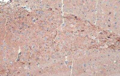
Dopamine beta-Hydroxylase Antibody [NBP1-31386] - Mouse brain. Dopamine beta Hydroxylase stained by Dopamine beta Hydroxylase antibody diluted at 1:500. Antigen Retrieval: Citrate buffer, pH 6.0, 15 min.

Western Blot: Dopamine beta-Hydroxylase Antibody [NBP1-31386] -
Western Blot: Dopamine beta-Hydroxylase Antibody [NBP1-31386] - Various whole cell extracts (30 ug) were separated by 7.5% SDS-PAGE, and the membrane was blotted with Dopamine beta Hydroxylase antibody diluted at 1:2000. The HRP-conjugated anti-rabbit IgG antibody was used to detect the primary antibody. Corresponding RNA expression data for the same cell lines are based on Human Protein Atlas program.
Western Blot: Dopamine beta-Hydroxylase Antibody [NBP1-31386] -
Expression levels of monoamine-regulating enzymes in the brain of SMN delta 7 mice.a, h, o Representative autoradiograms of brain lysates immunoblots of SMN delta 7 and wild-type (WT) mice at a post-natal day 3, h day 6, and o day 11. b–g Protein levels quantification of b Tyrosine hydroxylase (TH), c phospho-Tyrosine hydroxylase at Ser-40 (P-Ser40-TH), d Dopamine beta hydroxylase (D beta H), e Tryptophan hydroxylase 2 (TPH2), f Aromatic amino acid decarboxylase (AADC) and g Monoamine Oxidase A (MAO-A) in SMN delta 7 (n = 4) and WT (n = 5) mice (except for TPH2: n = 4 mice/genotype) at post-natal day 3. i–n Protein levels quantification of i TH (n = 4 WT, n = 5 SMN delta 7), j P-Ser40-TH (n = 4 WT, n = 5 SMN delta 7), k D beta H (n = 4 WT, n = 4 SMN delta 7), l TPH2 (n = 4 WT, n = 3 SMN delta 7), m AADC (n = 4 WT, n = 5 SMN delta 7) and n MAO-A (n = 4 WT, n = 5 SMN delta 7) at post-natal day 6. p–u Protein levels quantification of p TH (n = 13 WT, n = 14 SMN delta 7), q P-Ser40-TH (n = 4 WT, n = 5 SMN delta 7), r D beta H (n = 4 WT, n = 5 SMN delta 7), s TPH2 (n = 4 WT, n = 5 SMN delta 7), t AADC (n = 13 WT, n = 14 SMN delta 7) and u MAO-A (n = 8 WT, n = 10 SMN delta 7) at post-natal day 11. Data are normalized to beta -Actin levels and shown as box and whisker plots representing the median with interquartile range (IQR). Dots represent individual mice values. *p < 0.05, **p < 0.01, compared with age-matched WT mice (unpaired t-test). Image collected and cropped by CiteAb from the following open publication (https://pubmed.ncbi.nlm.nih.gov/37957344), licensed under a CC-BY license. Not internally tested by Novus Biologicals.
Western Blot: Dopamine beta-Hydroxylase Antibody [NBP1-31386] -
Expression levels of monoamine-regulating enzymes in the brain of SMN delta 7 mice.a, h, o Representative autoradiograms of brain lysates immunoblots of SMN delta 7 and wild-type (WT) mice at a post-natal day 3, h day 6, and o day 11. b–g Protein levels quantification of b Tyrosine hydroxylase (TH), c phospho-Tyrosine hydroxylase at Ser-40 (P-Ser40-TH), d Dopamine beta hydroxylase (D beta H), e Tryptophan hydroxylase 2 (TPH2), f Aromatic amino acid decarboxylase (AADC) and g Monoamine Oxidase A (MAO-A) in SMN delta 7 (n = 4) and WT (n = 5) mice (except for TPH2: n = 4 mice/genotype) at post-natal day 3. i–n Protein levels quantification of i TH (n = 4 WT, n = 5 SMN delta 7), j P-Ser40-TH (n = 4 WT, n = 5 SMN delta 7), k D beta H (n = 4 WT, n = 4 SMN delta 7), l TPH2 (n = 4 WT, n = 3 SMN delta 7), m AADC (n = 4 WT, n = 5 SMN delta 7) and n MAO-A (n = 4 WT, n = 5 SMN delta 7) at post-natal day 6. p–u Protein levels quantification of p TH (n = 13 WT, n = 14 SMN delta 7), q P-Ser40-TH (n = 4 WT, n = 5 SMN delta 7), r D beta H (n = 4 WT, n = 5 SMN delta 7), s TPH2 (n = 4 WT, n = 5 SMN delta 7), t AADC (n = 13 WT, n = 14 SMN delta 7) and u MAO-A (n = 8 WT, n = 10 SMN delta 7) at post-natal day 11. Data are normalized to beta -Actin levels and shown as box and whisker plots representing the median with interquartile range (IQR). Dots represent individual mice values. *p < 0.05, **p < 0.01, compared with age-matched WT mice (unpaired t-test). Image collected and cropped by CiteAb from the following open publication (https://pubmed.ncbi.nlm.nih.gov/37957344), licensed under a CC-BY license. Not internally tested by Novus Biologicals.Applications for Dopamine beta-Hydroxylase Antibody
Application
Recommended Usage
Immunohistochemistry
1:100-1:1000
Immunohistochemistry-Paraffin
1:100-1:1000
Western Blot
1:500-1:3000
Formulation, Preparation, and Storage
Purification
Antigen Affinity-purified
Formulation
PBS, 1% BSA, 20% Glycerol
Preservative
0.025% Proclin 300
Concentration
Concentrations vary lot to lot. See vial label for concentration. If unlisted please contact technical services.
Shipping
The product is shipped with polar packs. Upon receipt, store it immediately at the temperature recommended below.
Stability & Storage
Aliquot and store at -20C or -80C. Avoid freeze-thaw cycles.
Background: Dopamine beta-Hydroxylase
Alternate Names
DBH, DBM, Dopamine betaHydroxylase, DOPBHY
Entrez Gene IDs
1621 (Human)
Gene Symbol
DBH
UniProt
Additional Dopamine beta-Hydroxylase Products
Product Documents for Dopamine beta-Hydroxylase Antibody
Certificate of Analysis
To download a Certificate of Analysis, please enter a lot or batch number in the search box below.
Product Specific Notices for Dopamine beta-Hydroxylase Antibody
This product is for research use only and is not approved for use in humans or in clinical diagnosis. Primary Antibodies are guaranteed for 1 year from date of receipt.
Citations for Dopamine beta-Hydroxylase Antibody
 Powered by Bioz
Customer Reviews for Dopamine beta-Hydroxylase Antibody
There are currently no reviews for this product. Be the first to review Dopamine beta-Hydroxylase Antibody and earn rewards!
Have you used Dopamine beta-Hydroxylase Antibody?
Submit a review and receive an Amazon gift card!
$25/€18/£15/$25CAN/¥2500 Yen for a review with an image
$10/€7/£6/$10CAN/¥1110 Yen for a review without an image
Submit a review
Protocols
Find general support by application which include: protocols, troubleshooting, illustrated assays, videos and webinars.
- Antigen Retrieval Protocol (PIER)
- Antigen Retrieval for Frozen Sections Protocol
- Appropriate Fixation of IHC/ICC Samples
- Cellular Response to Hypoxia Protocols
- Chromogenic IHC Staining of Formalin-Fixed Paraffin-Embedded (FFPE) Tissue Protocol
- Chromogenic Immunohistochemistry Staining of Frozen Tissue
- ClariTSA™ Fluorophore Kits
- Detection & Visualization of Antibody Binding
- Fluorescent IHC Staining of Frozen Tissue Protocol
- Graphic Protocol for Heat-induced Epitope Retrieval
- Graphic Protocol for the Preparation and Fluorescent IHC Staining of Frozen Tissue Sections
- Graphic Protocol for the Preparation and Fluorescent IHC Staining of Paraffin-embedded Tissue Sections
- Graphic Protocol for the Preparation of Gelatin-coated Slides for Histological Tissue Sections
- IHC Sample Preparation (Frozen sections vs Paraffin)
- Immunofluorescent IHC Staining of Formalin-Fixed Paraffin-Embedded (FFPE) Tissue Protocol
- Immunohistochemistry (IHC) and Immunocytochemistry (ICC) Protocols
- Immunohistochemistry Frozen Troubleshooting
- Immunohistochemistry Paraffin Troubleshooting
- Preparing Samples for IHC/ICC Experiments
- Preventing Non-Specific Staining (Non-Specific Binding)
- Primary Antibody Selection & Optimization
- Protocol for Heat-Induced Epitope Retrieval (HIER)
- Protocol for Making a 4% Formaldehyde Solution in PBS
- Protocol for VisUCyte™ HRP Polymer Detection Reagent
- Protocol for the Preparation & Fixation of Cells on Coverslips
- Protocol for the Preparation and Chromogenic IHC Staining of Frozen Tissue Sections
- Protocol for the Preparation and Chromogenic IHC Staining of Frozen Tissue Sections - Graphic
- Protocol for the Preparation and Chromogenic IHC Staining of Paraffin-embedded Tissue Sections
- Protocol for the Preparation and Chromogenic IHC Staining of Paraffin-embedded Tissue Sections - Graphic
- Protocol for the Preparation and Fluorescent IHC Staining of Frozen Tissue Sections
- Protocol for the Preparation and Fluorescent IHC Staining of Paraffin-embedded Tissue Sections
- Protocol for the Preparation of Gelatin-coated Slides for Histological Tissue Sections
- R&D Systems Quality Control Western Blot Protocol
- TUNEL and Active Caspase-3 Detection by IHC/ICC Protocol
- The Importance of IHC/ICC Controls
- Troubleshooting Guide: Immunohistochemistry
- Troubleshooting Guide: Western Blot Figures
- Western Blot Conditions
- Western Blot Protocol
- Western Blot Protocol for Cell Lysates
- Western Blot Troubleshooting
- Western Blot Troubleshooting Guide
- View all Protocols, Troubleshooting, Illustrated assays and Webinars
Loading...