![Western Blot: Drosha Antibody [NBP1-03349]](https://resources.rndsystems.com/images/products/Drosha-Antibody-Western-Blot-NBP1-03349-img0006.jpg "Western Blot: Drosha Antibody [NBP1-03349]")
Loading...
Key Product Details
Validated by
Independent Antibodies, Biological Validation
Species Reactivity
Validated:
Human, Mouse
Cited:
Human, Mouse
Applications
Validated:
Immunohistochemistry, Immunohistochemistry-Frozen, Western Blot, Immunocytochemistry/ Immunofluorescence, Immunoprecipitation
Cited:
Immunohistochemistry-Frozen, Western Blot, Immunocytochemistry/ Immunofluorescence, Immunoprecipitation, IF/IHC
Label
Unconjugated
Antibody Source
Polyclonal Rabbit IgG
Loading...
Product Specifications
Immunogen
The immunogen recognized by this antibody maps to a region between residue 1324 and 1374 of human double-stranded RNA-specific endoribonuclease using the numbering given in entry NP_037367.3 (GeneID 29102).
Reactivity Notes
Mouse reactivity reported in scientific literature (PMID: 25351346).
Clonality
Polyclonal
Host
Rabbit
Isotype
IgG
Scientific Data Images for Drosha Antibody
Western Blot: Drosha Antibody [NBP1-03349]
Western Blot: Drosha Antibody [NBP1-03349] - Detection of Human Drosha by Western Blot. Samples: Whole cell lysate (50 ug) from HeLa and 293T cells prepared using NETN lysis buffer. Antibody: Affinity purified rabbit anti-Drosha antibody NBP1-03349 used for WB at 0.1 ug/ml. Detection: Chemiluminescence with an exposure time of 10 seconds.![Immunoprecipitation: Drosha Antibody [NBP1-03349]](https://resources.rndsystems.com/images/products/Drosha-Antibody-Immunoprecipitation-NBP1-03349-img0007.jpg "Immunoprecipitation: Drosha Antibody [NBP1-03349]")
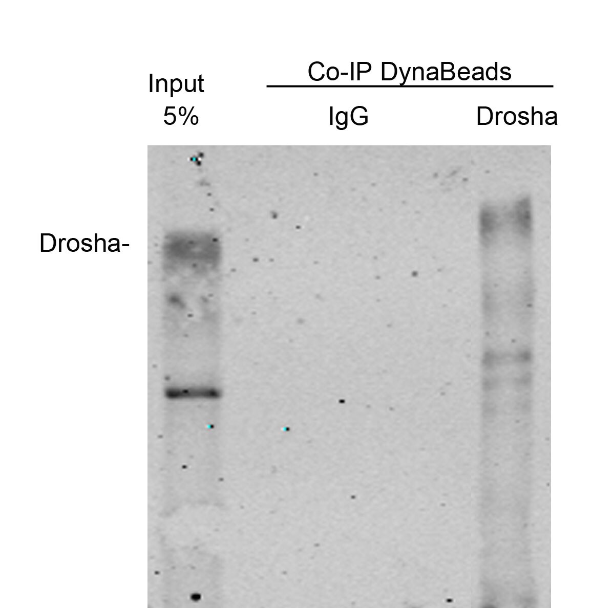
Immunoprecipitation: Drosha Antibody [NBP1-03349]
Immunoprecipitation: Drosha Antibody [NBP1-03349] - Detection of human Drosha by western blot of immunoprecipitates. Samples: Whole cell lysate (0.5 or 1.0 mg per IP reaction; 20% of IP loaded) from HeLa cells prepared using NETN lysis buffer. Antibodies: Affinity purified rabbit anti-Drosha antibody NBP1-03349 used for IP at 6 ug per reaction. Drosha was also immunoprecipitated by another rabbit anti-Drosha antibody. For blotting immunoprecipitated Drosha, NBP1-03349 was used at 1 ug/ml. Detection: Chemiluminescence with an exposure time of 10 seconds.Applications for Drosha Antibody
Application
Recommended Usage
Immunocytochemistry/ Immunofluorescence
Reported in scientific literature (PMID 25351346)
Immunohistochemistry
1:250
Immunohistochemistry-Frozen
Reported in scientific literature (PMID 25351346)
Immunoprecipitation
2 - 10 ug/mg lysate
Western Blot
1:2000 - 1:10000
Application Notes
Centrifuge tube to remove product from lid. Optimal working dilutions should be determined experimentally by the investigator. Prepare working dilution immediately before use.
Reviewed Applications
Read 3 reviews rated 4 using NBP1-03349 in the following applications:
Formulation, Preparation, and Storage
Purification
Immunogen affinity purified
Formulation
TBS and 0.1% BSA
Preservative
0.09% Sodium Azide
Concentration
0.2 mg/ml
Shipping
The product is shipped with polar packs. Upon receipt, store it immediately at the temperature recommended below.
Stability & Storage
Store at 4C. Do not freeze.
Background: Drosha
Alternate Names
drosha, double-stranded RNA-specific endoribonuclease, drosha, ribonuclease type III, EC 3.1.26.3, ETOHI2, nuclear RNase III Drosha, p241, Protein Drosha, putative protein p241 which interacts with transcription factor Sp1, putative ribonuclease III, RANSE3L, ribonuclease 3, Ribonuclease III, ribonuclease III, nuclear, RN3nuclear, RNase III
Entrez Gene IDs
29102 (Human)
Gene Symbol
DROSHA
UniProt
Additional Drosha Products
Product Documents for Drosha Antibody
Certificate of Analysis
To download a Certificate of Analysis, please enter a lot or batch number in the search box below.
Product Specific Notices for Drosha Antibody
This product is for research use only and is not approved for use in humans or in clinical diagnosis. Primary Antibodies are guaranteed for 1 year from date of receipt.
Citations for Drosha Antibody
 Powered by Bioz
Customer Reviews for Drosha Antibody (3)
4 out of 5
3 Customer Ratings
Have you used Drosha Antibody?
Submit a review and receive an Amazon gift card!
$25/€18/£15/$25CAN/¥2500 Yen for a review with an image
$10/€7/£6/$10CAN/¥1110 Yen for a review without an image
Submit a review
Customer Images
Showing
1
-
3 of
3 reviews
Showing All
Filter By:
-
Application: ImmunoprecipitationSample Tested: A549 cellsSpecies: HumanVerified Customer | Posted 07/06/2015A549 cells

-
Application: Western BlotSample Tested: A549 cellsSpecies: HumanVerified Customer | Posted 07/06/2015A549 cells
-
Application: Western BlotSample Tested: Human ovarian cancer cell linesSpecies: HumanVerified Customer | Posted 10/06/2014
There are no reviews that match your criteria.
Protocols
Find general support by application which include: protocols, troubleshooting, illustrated assays, videos and webinars.
- Antigen Retrieval Protocol (PIER)
- Antigen Retrieval for Frozen Sections Protocol
- Appropriate Fixation of IHC/ICC Samples
- Cellular Response to Hypoxia Protocols
- Chromogenic IHC Staining of Formalin-Fixed Paraffin-Embedded (FFPE) Tissue Protocol
- Chromogenic Immunohistochemistry Staining of Frozen Tissue
- ClariTSA™ Fluorophore Kits
- Detection & Visualization of Antibody Binding
- Fluorescent IHC Staining of Frozen Tissue Protocol
- Graphic Protocol for Heat-induced Epitope Retrieval
- Graphic Protocol for the Preparation and Fluorescent IHC Staining of Frozen Tissue Sections
- Graphic Protocol for the Preparation and Fluorescent IHC Staining of Paraffin-embedded Tissue Sections
- Graphic Protocol for the Preparation of Gelatin-coated Slides for Histological Tissue Sections
- ICC Cell Smear Protocol for Suspension Cells
- ICC Immunocytochemistry Protocol Videos
- ICC for Adherent Cells
- IHC Sample Preparation (Frozen sections vs Paraffin)
- Immunocytochemistry (ICC) Protocol
- Immunocytochemistry Troubleshooting
- Immunofluorescence of Organoids Embedded in Cultrex Basement Membrane Extract
- Immunofluorescent IHC Staining of Formalin-Fixed Paraffin-Embedded (FFPE) Tissue Protocol
- Immunohistochemistry (IHC) and Immunocytochemistry (ICC) Protocols
- Immunohistochemistry Frozen Troubleshooting
- Immunohistochemistry Paraffin Troubleshooting
- Immunoprecipitation Protocol
- Preparing Samples for IHC/ICC Experiments
- Preventing Non-Specific Staining (Non-Specific Binding)
- Primary Antibody Selection & Optimization
- Protocol for Heat-Induced Epitope Retrieval (HIER)
- Protocol for Making a 4% Formaldehyde Solution in PBS
- Protocol for VisUCyte™ HRP Polymer Detection Reagent
- Protocol for the Fluorescent ICC Staining of Cell Smears - Graphic
- Protocol for the Fluorescent ICC Staining of Cultured Cells on Coverslips - Graphic
- Protocol for the Preparation & Fixation of Cells on Coverslips
- Protocol for the Preparation and Chromogenic IHC Staining of Frozen Tissue Sections
- Protocol for the Preparation and Chromogenic IHC Staining of Frozen Tissue Sections - Graphic
- Protocol for the Preparation and Chromogenic IHC Staining of Paraffin-embedded Tissue Sections
- Protocol for the Preparation and Chromogenic IHC Staining of Paraffin-embedded Tissue Sections - Graphic
- Protocol for the Preparation and Fluorescent ICC Staining of Cells on Coverslips
- Protocol for the Preparation and Fluorescent ICC Staining of Non-adherent Cells
- Protocol for the Preparation and Fluorescent ICC Staining of Stem Cells on Coverslips
- Protocol for the Preparation and Fluorescent IHC Staining of Frozen Tissue Sections
- Protocol for the Preparation and Fluorescent IHC Staining of Paraffin-embedded Tissue Sections
- Protocol for the Preparation of Gelatin-coated Slides for Histological Tissue Sections
- Protocol for the Preparation of a Cell Smear for Non-adherent Cell ICC - Graphic
- R&D Systems Quality Control Western Blot Protocol
- TUNEL and Active Caspase-3 Detection by IHC/ICC Protocol
- The Importance of IHC/ICC Controls
- Troubleshooting Guide: Immunohistochemistry
- Troubleshooting Guide: Western Blot Figures
- Western Blot Conditions
- Western Blot Protocol
- Western Blot Protocol for Cell Lysates
- Western Blot Troubleshooting
- Western Blot Troubleshooting Guide
- View all Protocols, Troubleshooting, Illustrated assays and Webinars
Loading...