DUOX2 Antibody - BSA Free
Novus Biologicals | Catalog # NB110-61576
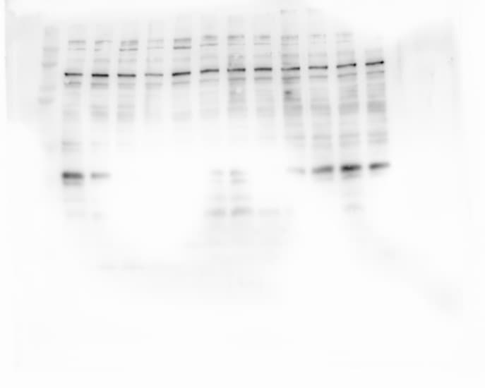
![Knockdown Validated: DUOX2 Antibody - BSA Free [NB110-61576]](https://resources.rndsystems.com/images/products/DUOX2-Antibody-Knockdown-Validated-NB110-61576-img0006.jpg "Western Blot: DUOX2 Antibody - BSA Free [NB110-61576]")
Key Product Details
Validated by
Species Reactivity
Validated:
Cited:
Applications
Validated:
Cited:
Label
Antibody Source
Format
Product Specifications
Immunogen
Localization
Clonality
Host
Isotype
Scientific Data Images for DUOX2 Antibody - BSA Free
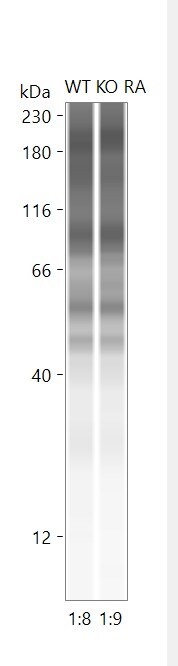
![Western Blot: DUOX2 AntibodyBSA Free [NB110-61576]](https://resources.rndsystems.com/images/products/DUOX2-Antibody-Western-Blot-NB110-61576-img0002.jpg "Western Blot: DUOX2 AntibodyBSA Free [NB110-61576]")
Western Blot: DUOX2 AntibodyBSA Free [NB110-61576]
Western Blot: DUOX2 Antibody [NB110-61576] - Detection of DUOX2 in A549 in cell lysate.![Immunocytochemistry/ Immunofluorescence: DUOX2 Antibody - BSA Free [NB110-61576]](https://resources.rndsystems.com/images/products/DUOX2-Antibody-Immunocytochemistry-Immunofluorescence-NB110-61576-img0004.jpg "Immunocytochemistry/ Immunofluorescence: DUOX2 Antibody - BSA Free [NB110-61576]")
Immunocytochemistry/ Immunofluorescence: DUOX2 Antibody - BSA Free [NB110-61576]
Immunocytochemistry/Immunofluorescence: DUOX2 Antibody [NB110-61576] - A431 cells were fixed for 10 minutes using 10% formalin and then permeabilized for 5 minutes using 1X PBS + 0.05% Triton-X100. The cells were incubated with anti-DUOX at 5 ug/ml overnight at 4C and detected with an anti-rabbit Dylight 488 (Green) at a 1:500 dilution. Nuclei were counterstained with DAPI (Blue). Cells were imaged using a 40X objective.![Western Blot: DUOX2 AntibodyBSA Free [NB110-61576]](https://resources.rndsystems.com/images/products/DUOX2-Antibody-Western-Blot-NB110-61576-img0005.jpg "Western Blot: DUOX2 AntibodyBSA Free [NB110-61576]")
![Immunocytochemistry/ Immunofluorescence: DUOX2 Antibody - BSA Free [NB110-61576]](https://resources.rndsystems.com/images/products/DUOX2-Antibody-Immunocytochemistry-Immunofluorescence-NB110-61576-img0003.jpg "Immunocytochemistry/ Immunofluorescence: DUOX2 Antibody - BSA Free [NB110-61576]")
Immunocytochemistry/ Immunofluorescence: DUOX2 Antibody - BSA Free [NB110-61576]
Immunocytochemistry/Immunofluorescence: DUOX2 Antibody [NB110-61576] - DUOX2 antibody was tested in A431 cells with Dylight 488 (green). Nuclei and alpha-tubulin were counterstained with DAPI (blue) and Dylight 550 (red).Applications for DUOX2 Antibody - BSA Free
Immunocytochemistry/ Immunofluorescence
Immunohistochemistry-Paraffin
Western Blot
Reviewed Applications
Read 2 reviews rated 2 using NB110-61576 in the following applications:
Formulation, Preparation, and Storage
Purification
Formulation
Format
Preservative
Concentration
Shipping
Stability & Storage
Background: DUOX2
Alternate Names
Gene Symbol
Additional DUOX2 Products
Product Documents for DUOX2 Antibody - BSA Free
Certificate of Analysis
To download a Certificate of Analysis, please enter a lot or batch number in the search box below.
Product Specific Notices for DUOX2 Antibody - BSA Free
This product is for research use only and is not approved for use in humans or in clinical diagnosis. Primary Antibodies are guaranteed for 1 year from date of receipt.
Citations for DUOX2 Antibody - BSA Free
 Powered by Bioz
Customer Reviews for DUOX2 Antibody - BSA Free (2)
Have you used DUOX2 Antibody - BSA Free?
Submit a review and receive an Amazon gift card!
$25/€18/£15/$25CAN/¥2500 Yen for a review with an image
$10/€7/£6/$10CAN/¥1110 Yen for a review without an image
Submit a review
Customer Images
-
Application: Western BlotSample Tested: Lung tissueSpecies: MouseVerified Customer | Posted 09/08/2025Protein expression of duox2 in mouse lung homogenatesI tested the protein expression of duox2 in mouse lung homogenates treated with smoke and room air

-
Application: Simple WesternSample Tested: Lung tissueSpecies: MouseVerified Customer | Posted 09/08/2025duox2 protein expression using simple western JESSI checked for the protein expression of duox2 in mouse lung homogenate
Bio-Techne ResponseThis review reflects a new species or application tested on a primary antibody.
There are no reviews that match your criteria.
Protocols
View specific protocols for DUOX2 Antibody - BSA Free (NB110-61576):
Immunocytochemistry Protocol
Culture cells to appropriate density in 35 mm culture dishes or 6-well plates.
1. Remove culture medium and add 10% formalin to the dish. Fix at room temperature for 30 minutes.
2. Remove the formalin and add ice cold methanol. Incubate for 5-10 minutes.
3. Remove methanol and add washing solution (i.e. PBS). Be sure to not let the specimen dry out. Wash three times for 10 minutes.
4. To block nonspecific antibody binding incubate in 10% normal goat serum from 1 hour to overnight at room temperature.
5. Add primary antibody at appropriate dilution and incubate at room temperature from 2 hours to overnight at room temperature.
6. Remove primary antibody and replace with washing solution. Wash three times for 10 minutes.
7. Add secondary antibody at appropriate dilution. Incubate for 1 hour at room temperature.
8. Remove antibody and replace with wash solution, then wash for 10 minutes. Add Hoechst 33258 to wash solution at 1:25,0000 and incubate for 10 minutes. Wash a third time for 10 minutes.
9. Cells can be viewed directly after washing. The plates can also be stored in PBS containing Azide covered in Parafilm (TM). Cells can also be cover-slipped using Fluoromount, with appropriate sealing.
*The above information is only intended as a guide. The researcher should determine what protocol best meets their needs. Please follow safe laboratory procedures.
Western Blot Protocol
1. Perform SDS-PAGE (4-12% MOPS) on samples to be analyzed, loading 38 ug of total protein per lane.
2. Transfer proteins to Nitrocellulose according to the instructions provided by the manufacturer of the transfer
apparatus.
3. Rinse membrane with dH2O and then stain the blot using Ponceau S for 1-2 minutes to access the transfer of proteins onto the nitrocellulose membrane. Rinse the blot in water to remove excess stain and mark the lane locations and locations of molecular weight markers using a pencil.
4. Rinse the blot in TBS for approximately 5 minutes.
5. Block the membrane using 5% NFDM + 1% BSA in TBS + Tween, 1 hour at RT.
6. Rinse the membrane in dH2O and then wash the membrane in wash buffer [TBS + 0.1% Tween] 3 times for 10 minutes each.
7. Dilute the rabbit anti-DUOX2 primary antibody (NB 110-61576) in blocking buffer and incubate 1 hour at room temperature.
8. Rinse the membrane in dH2O and then wash the membrane in wash buffer [TBS + 0.1% Tween] 3 times for 10 minutes each.
9. Apply the diluted rabbit-IgG HRP-conjugated secondary antibody in blocking buffer (as per manufacturers
instructions) and incubate 1 hour at room temperature.
10. Wash the blot in wash buffer [TBS + 0.1% Tween] 3 times for 10 minutes each (this step can be repeated as required to reduce background).
11. Apply the detection reagent of choice in accordance with the manufacturers instructions (Pierce ECL).
Note: Tween-20 can be added to the blocking or antibody dilution buffer at a final concentration of 0.05-0.2%, provided it does not interfere with antibody-antigen binding.
Find general support by application which include: protocols, troubleshooting, illustrated assays, videos and webinars.
- Antigen Retrieval Protocol (PIER)
- Antigen Retrieval for Frozen Sections Protocol
- Appropriate Fixation of IHC/ICC Samples
- Cellular Response to Hypoxia Protocols
- Chromogenic IHC Staining of Formalin-Fixed Paraffin-Embedded (FFPE) Tissue Protocol
- Chromogenic Immunohistochemistry Staining of Frozen Tissue
- ClariTSA™ Fluorophore Kits
- Detection & Visualization of Antibody Binding
- Fluorescent IHC Staining of Frozen Tissue Protocol
- Graphic Protocol for Heat-induced Epitope Retrieval
- Graphic Protocol for the Preparation and Fluorescent IHC Staining of Frozen Tissue Sections
- Graphic Protocol for the Preparation and Fluorescent IHC Staining of Paraffin-embedded Tissue Sections
- Graphic Protocol for the Preparation of Gelatin-coated Slides for Histological Tissue Sections
- ICC Cell Smear Protocol for Suspension Cells
- ICC Immunocytochemistry Protocol Videos
- ICC for Adherent Cells
- IHC Sample Preparation (Frozen sections vs Paraffin)
- Immunocytochemistry (ICC) Protocol
- Immunocytochemistry Troubleshooting
- Immunofluorescence of Organoids Embedded in Cultrex Basement Membrane Extract
- Immunofluorescent IHC Staining of Formalin-Fixed Paraffin-Embedded (FFPE) Tissue Protocol
- Immunohistochemistry (IHC) and Immunocytochemistry (ICC) Protocols
- Immunohistochemistry Frozen Troubleshooting
- Immunohistochemistry Paraffin Troubleshooting
- Preparing Samples for IHC/ICC Experiments
- Preventing Non-Specific Staining (Non-Specific Binding)
- Primary Antibody Selection & Optimization
- Protocol for Heat-Induced Epitope Retrieval (HIER)
- Protocol for Making a 4% Formaldehyde Solution in PBS
- Protocol for VisUCyte™ HRP Polymer Detection Reagent
- Protocol for the Fluorescent ICC Staining of Cell Smears - Graphic
- Protocol for the Fluorescent ICC Staining of Cultured Cells on Coverslips - Graphic
- Protocol for the Preparation & Fixation of Cells on Coverslips
- Protocol for the Preparation and Chromogenic IHC Staining of Frozen Tissue Sections
- Protocol for the Preparation and Chromogenic IHC Staining of Frozen Tissue Sections - Graphic
- Protocol for the Preparation and Chromogenic IHC Staining of Paraffin-embedded Tissue Sections
- Protocol for the Preparation and Chromogenic IHC Staining of Paraffin-embedded Tissue Sections - Graphic
- Protocol for the Preparation and Fluorescent ICC Staining of Cells on Coverslips
- Protocol for the Preparation and Fluorescent ICC Staining of Non-adherent Cells
- Protocol for the Preparation and Fluorescent ICC Staining of Stem Cells on Coverslips
- Protocol for the Preparation and Fluorescent IHC Staining of Frozen Tissue Sections
- Protocol for the Preparation and Fluorescent IHC Staining of Paraffin-embedded Tissue Sections
- Protocol for the Preparation of Gelatin-coated Slides for Histological Tissue Sections
- Protocol for the Preparation of a Cell Smear for Non-adherent Cell ICC - Graphic
- R&D Systems Quality Control Western Blot Protocol
- TUNEL and Active Caspase-3 Detection by IHC/ICC Protocol
- The Importance of IHC/ICC Controls
- Troubleshooting Guide: Immunohistochemistry
- Troubleshooting Guide: Western Blot Figures
- Western Blot Conditions
- Western Blot Protocol
- Western Blot Protocol for Cell Lysates
- Western Blot Troubleshooting
- Western Blot Troubleshooting Guide
- View all Protocols, Troubleshooting, Illustrated assays and Webinars
FAQs for DUOX2 Antibody - BSA Free
-
Q:
Can you please let me know the homology of the immunogen peptide with mouse, for NB110-61576? Here is a link to the mouse DUOX2 protein information: ncbi
A: The homology is 84.6%.
-
Q: Hi, I am looking for an antibody to human DUOX2 that will work on human formalin fixed paraffin embedded sections. I notice from your website that you have several recommended for WB and wondered if you have any experience of their use in other areas?
A: At this time we do not have an antibody for DUOX2 that works in IHC paraffin embedded sections. If you choose to try one in this application we have our Innovator's Reward program where we reward our customers for trying antibodies in previously untested applications and species.
-
Q:
Can you please let me know the homology of the immunogen peptide with mouse, for NB110-61576? Here is a link to the mouse DUOX2 protein information: ncbi
A: The homology is 84.6%.
-
Q: Hi, I am looking for an antibody to human DUOX2 that will work on human formalin fixed paraffin embedded sections. I notice from your website that you have several recommended for WB and wondered if you have any experience of their use in other areas?
A: At this time we do not have an antibody for DUOX2 that works in IHC paraffin embedded sections. If you choose to try one in this application we have our Innovator's Reward program where we reward our customers for trying antibodies in previously untested applications and species.