E-Cadherin Antibody (DECMA-1)
Novus Biologicals | Catalog # NB120-11512
![Western Blot: E-Cadherin Antibody (DECMA-1) [NB120-11512]](https://resources.rndsystems.com/images/products/E-Cadherin-Antibody-DECMA-1-Western-Blot-NB120-11512-img0006.jpg "Western Blot: E-Cadherin Antibody (DECMA-1) [NB120-11512]")
Loading...
Key Product Details
Species Reactivity
Validated:
Human, Mouse, Bovine, Canine, Feline
Cited:
Mouse, Bovine
Applications
Validated:
Immunohistochemistry, Immunohistochemistry-Frozen, Western Blot, Immunocytochemistry/ Immunofluorescence, Immunoprecipitation, Microarray
Cited:
Immunohistochemistry-Frozen, Immunocytochemistry/ Immunofluorescence
Label
Unconjugated
Antibody Source
Monoclonal Rat IgG1 Clone # DECMA-1
Loading...
Product Specifications
Immunogen
Mouse embryonal carcinoma cell line PCC4 Aza RI.
Localization
Cell membrane; cell-cell junction; single-pass type I membrane protein
Marker
Epithelial Cell Marker, Adherens Junctions Marker
Specificity
Antibody localizes the cell surface glycoprotein uvomorulin/E-cadherin
Clonality
Monoclonal
Host
Rat
Isotype
IgG1
Scientific Data Images for E-Cadherin Antibody (DECMA-1)
Western Blot: E-Cadherin Antibody (DECMA-1) [NB120-11512]
Western Blot: E-Cadherin Antibody (DECMA-1) [NB120-11512] - MDCK cell extract separated on SDS-PAGE and probed with Rat Anti-Uvomorulin/E-Cadherin Clone: DECMA-1. Detection by Rabbit Anti-Rat IgG-Peroxidase and a chemiluminescent substrate. Antibody dilution 1:250.![Immunocytochemistry/ Immunofluorescence: E-Cadherin Antibody (DECMA-1) [NB120-11512]](https://resources.rndsystems.com/images/products/E-Cadherin-Antibody-DECMA-1-Immunocytochemistry-Immunofluorescence-NB120-11512-img0009.jpg "Immunocytochemistry/ Immunofluorescence: E-Cadherin Antibody (DECMA-1) [NB120-11512]")
Immunocytochemistry/ Immunofluorescence: E-Cadherin Antibody (DECMA-1) [NB120-11512]
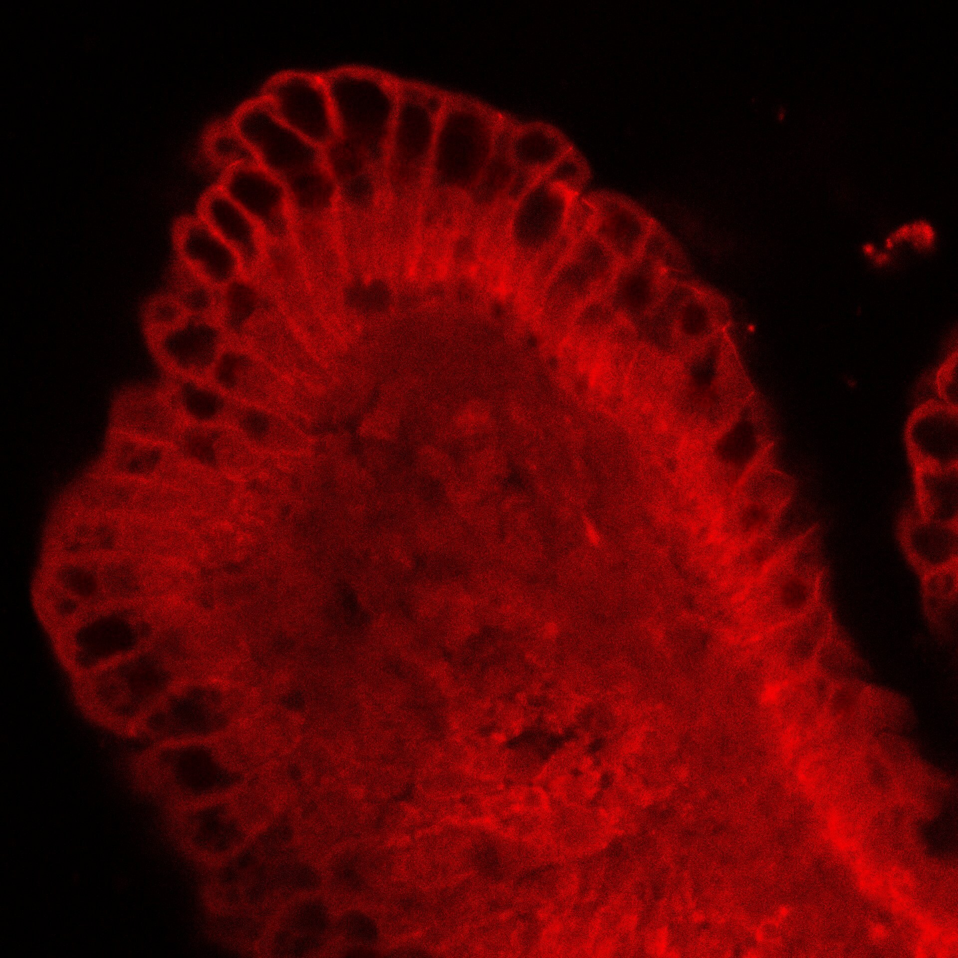
Immunocytochemistry/Immunofluorescence: E-Cadherin Antibody (DECMA-1) [NB120-11512] - Adult mouse small intestine stained with NB12-11512. Image from verified customer review.![Western Blot: E-Cadherin Antibody (DECMA-1) [NB120-11512]](https://resources.rndsystems.com/images/products/E-Cadherin-Antibody-DECMA-1-Western-Blot-NB120-11512-img0005.jpg "Western Blot: E-Cadherin Antibody (DECMA-1) [NB120-11512]")
Western Blot: E-Cadherin Antibody (DECMA-1) [NB120-11512]
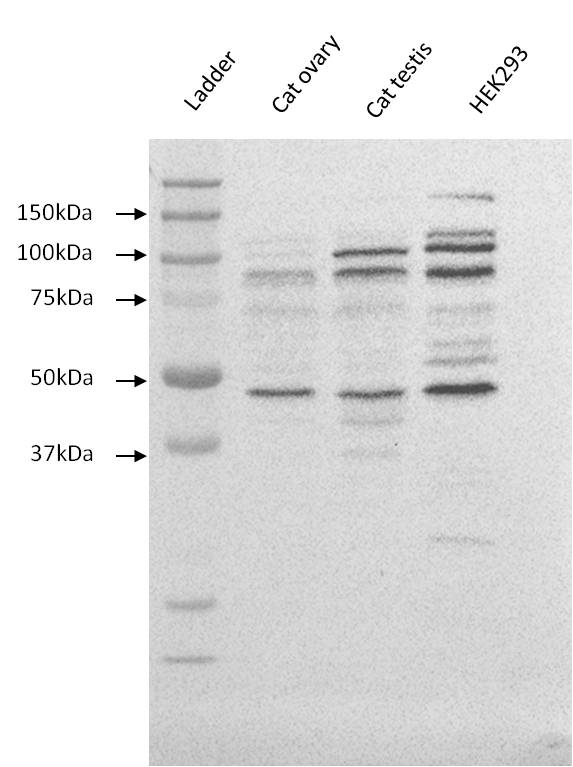
Western Blot: E-Cadherin Antibody (DECMA-1) [NB120-11512] - Cat testis, cat ovary and HEK293 whole cell lysates. Image from verified customer review.![Immunocytochemistry/ Immunofluorescence: E-Cadherin Antibody (DECMA-1) [NB120-11512]](https://resources.rndsystems.com/images/products/E-Cadherin-Antibody-DECMA-1-Immunocytochemistry-Immunofluorescence-NB120-11512-img0007.jpg "Immunocytochemistry/ Immunofluorescence: E-Cadherin Antibody (DECMA-1) [NB120-11512]")
Immunocytochemistry/ Immunofluorescence: E-Cadherin Antibody (DECMA-1) [NB120-11512]
Immunocytochemistry/Immunofluorescence: E-Cadherin Antibody (DECMA-1) [NB120-11512] - MDCK cells were fixed and permeabilized with methanol followed by acetone. Fixed cells were stained with 1:3200 Rat Monoclonal Anti-Uvomorulin/E-Cadherin, Clone: DECMA-1. Detection using Goat Anti-Rat IgG, FITC conjugate.![Immunocytochemistry/ Immunofluorescence: E-Cadherin Antibody (DECMA-1) [NB120-11512]](https://resources.rndsystems.com/images/products/E-Cadherin-Antibody-DECMA-1-Immunocytochemistry-Immunofluorescence-NB120-11512-img0008.jpg "Immunocytochemistry/ Immunofluorescence: E-Cadherin Antibody (DECMA-1) [NB120-11512]")
Immunocytochemistry/ Immunofluorescence: E-Cadherin Antibody (DECMA-1) [NB120-11512]
Immunocytochemistry/Immunofluorescence: E-Cadherin Antibody (DECMA-1) [NB120-11512] - MDCK cells were fixed and permeabilized with cold methanol followed by aceton. Fixed cells were stained with Rat Anti-Uvomorulin/E-Cadherin Clone: DECMA-1 diluted to 1:3200. The antibody was developed using Rabbit Anti-Rat IgG, FITC-conjugate.Applications for E-Cadherin Antibody (DECMA-1)
Application
Recommended Usage
Immunocytochemistry/ Immunofluorescence
1:1600
Immunohistochemistry
1:10-1:500
Immunohistochemistry-Frozen
1:10-1:500
Immunoprecipitation
1:10-1:500
Western Blot
1:3000
Application Notes
The antibody may be used for studies of embryonal development, cell-cell interactions of cultured cells, and localization of uvomorulin/E-cadherin in westernblot or immunohistochemical assays.
Reviewed Applications
Read 2 reviews rated 4 using NB120-11512 in the following applications:
Formulation, Preparation, and Storage
Purification
Unpurified
Formulation
Raw ascites
Preservative
0.09% Sodium Azide
Concentration
This product is unpurified. The exact concentration of antibody is not quantifiable.
Shipping
The product is shipped with polar packs. Upon receipt, store it immediately at the temperature recommended below.
Stability & Storage
Store at -20C. Avoid freeze-thaw cycles.
Background: E-Cadherin
Alternate Names
Arc-1, CAD1, Cadherin-1, CD324, CDH1, Cell-CAM120/80, ECAD, ECadherin, L-CAM, Uvomorulin
Gene Symbol
CDH1
UniProt
Additional E-Cadherin Products
Product Documents for E-Cadherin Antibody (DECMA-1)
Certificate of Analysis
To download a Certificate of Analysis, please enter a lot or batch number in the search box below.
Product Specific Notices for E-Cadherin Antibody (DECMA-1)
This product is for research use only and is not approved for use in humans or in clinical diagnosis. Primary Antibodies are guaranteed for 1 year from date of receipt.
Citations for E-Cadherin Antibody (DECMA-1)
 Powered by Bioz
Customer Reviews for E-Cadherin Antibody (DECMA-1) (2)
4 out of 5
2 Customer Ratings
Have you used E-Cadherin Antibody (DECMA-1)?
Submit a review and receive an Amazon gift card!
$25/€18/£15/$25CAN/¥2500 Yen for a review with an image
$10/€7/£6/$10CAN/¥1110 Yen for a review without an image
Submit a review
Customer Images
Showing
1
-
2 of
2 reviews
Showing All
Filter By:
-
Application: ImmunocytochemistrySample Tested: Adult small intestineSpecies: MouseVerified Customer | Posted 06/06/2019small intestine stained with NB120-11512

-
Application: Western BlotSample Tested: cat testis, cat ovary, HEK293 whole cell lysateSpecies: OtherVerified Customer | Posted 11/18/2013
There are no reviews that match your criteria.
Protocols
Find general support by application which include: protocols, troubleshooting, illustrated assays, videos and webinars.
- Antigen Retrieval Protocol (PIER)
- Antigen Retrieval for Frozen Sections Protocol
- Appropriate Fixation of IHC/ICC Samples
- Cellular Response to Hypoxia Protocols
- Chromogenic IHC Staining of Formalin-Fixed Paraffin-Embedded (FFPE) Tissue Protocol
- Chromogenic Immunohistochemistry Staining of Frozen Tissue
- ClariTSA™ Fluorophore Kits
- Detection & Visualization of Antibody Binding
- Fluorescent IHC Staining of Frozen Tissue Protocol
- Graphic Protocol for Heat-induced Epitope Retrieval
- Graphic Protocol for the Preparation and Fluorescent IHC Staining of Frozen Tissue Sections
- Graphic Protocol for the Preparation and Fluorescent IHC Staining of Paraffin-embedded Tissue Sections
- Graphic Protocol for the Preparation of Gelatin-coated Slides for Histological Tissue Sections
- ICC Cell Smear Protocol for Suspension Cells
- ICC Immunocytochemistry Protocol Videos
- ICC for Adherent Cells
- IHC Sample Preparation (Frozen sections vs Paraffin)
- Immunocytochemistry (ICC) Protocol
- Immunocytochemistry Troubleshooting
- Immunofluorescence of Organoids Embedded in Cultrex Basement Membrane Extract
- Immunofluorescent IHC Staining of Formalin-Fixed Paraffin-Embedded (FFPE) Tissue Protocol
- Immunohistochemistry (IHC) and Immunocytochemistry (ICC) Protocols
- Immunohistochemistry Frozen Troubleshooting
- Immunohistochemistry Paraffin Troubleshooting
- Immunoprecipitation Protocol
- Preparing Samples for IHC/ICC Experiments
- Preventing Non-Specific Staining (Non-Specific Binding)
- Primary Antibody Selection & Optimization
- Protocol for Heat-Induced Epitope Retrieval (HIER)
- Protocol for Making a 4% Formaldehyde Solution in PBS
- Protocol for VisUCyte™ HRP Polymer Detection Reagent
- Protocol for the Fluorescent ICC Staining of Cell Smears - Graphic
- Protocol for the Fluorescent ICC Staining of Cultured Cells on Coverslips - Graphic
- Protocol for the Preparation & Fixation of Cells on Coverslips
- Protocol for the Preparation and Chromogenic IHC Staining of Frozen Tissue Sections
- Protocol for the Preparation and Chromogenic IHC Staining of Frozen Tissue Sections - Graphic
- Protocol for the Preparation and Chromogenic IHC Staining of Paraffin-embedded Tissue Sections
- Protocol for the Preparation and Chromogenic IHC Staining of Paraffin-embedded Tissue Sections - Graphic
- Protocol for the Preparation and Fluorescent ICC Staining of Cells on Coverslips
- Protocol for the Preparation and Fluorescent ICC Staining of Non-adherent Cells
- Protocol for the Preparation and Fluorescent ICC Staining of Stem Cells on Coverslips
- Protocol for the Preparation and Fluorescent IHC Staining of Frozen Tissue Sections
- Protocol for the Preparation and Fluorescent IHC Staining of Paraffin-embedded Tissue Sections
- Protocol for the Preparation of Gelatin-coated Slides for Histological Tissue Sections
- Protocol for the Preparation of a Cell Smear for Non-adherent Cell ICC - Graphic
- R&D Systems Quality Control Western Blot Protocol
- TUNEL and Active Caspase-3 Detection by IHC/ICC Protocol
- The Importance of IHC/ICC Controls
- Troubleshooting Guide: Immunohistochemistry
- Troubleshooting Guide: Western Blot Figures
- Western Blot Conditions
- Western Blot Protocol
- Western Blot Protocol for Cell Lysates
- Western Blot Troubleshooting
- Western Blot Troubleshooting Guide
- View all Protocols, Troubleshooting, Illustrated assays and Webinars
Loading...
Associated Pathways
