Endorepellin/Perlecan/Heparan Sulfate Proteoglycan Antibody (A76) - BSA Free
Novus Biologicals | Catalog # NBP1-05170
![Immunocytochemistry/ Immunofluorescence: Endorepellin/Perlecan/Heparan Sulfate Proteoglycan Antibody (A76) - BSA Free [NBP1-05170]](https://resources.rndsystems.com/images/products/Endorepellin-Perlecan-Heparan-Sulfate-Proteoglycan-Antibody-A76-BSA-Free-Immunocytochemistry-Immunofluorescence-NBP1-05170-img0001.jpg "Immunocytochemistry/ Immunofluorescence: Endorepellin/Perlecan/Heparan Sulfate Proteoglycan Antibody (A76) - BSA Free [NBP1-05170]")
Key Product Details
Species Reactivity
Validated:
Human, Bovine
Cited:
Human
Applications
Validated:
Immunohistochemistry, Immunohistochemistry-Paraffin, Immunohistochemistry-Frozen, Western Blot, ELISA, Immunocytochemistry/ Immunofluorescence, Immunoprecipitation, Immunoaffinity Purification
Cited:
Immunohistochemistry-Paraffin, Immunocytochemistry/ Immunofluorescence
Label
Unconjugated
Antibody Source
Monoclonal Mouse IgG1 Clone # A76
Format
BSA Free
Loading...
Product Specifications
Immunogen
Lysed bovine corneal endothelial cells and extracellular matrix
Specificity
Highly specific for perlecan. There is no evidence for cross-reactivity with other connective tissue proteins (vitronectin, fibronectin, elastin, collagen, laminin).
Clonality
Monoclonal
Host
Mouse
Isotype
IgG1
Description
Concentration for any given batch will be between 0.85 - 1.15 mg/mL.
Scientific Data Images
Immunocytochemistry/ Immunofluorescence: Endorepellin/Perlecan/Heparan Sulfate Proteoglycan Antibody (A76) - BSA Free [NBP1-05170]
Endorepellin-Perlecan-Heparan-Sulfate-Proteoglycan-Antibody-A76-BSA-Free-Immunocytochemistry-Immunofluorescence-NBP1-05170-img0001.jpg - BSA Free [NBP1-05170] -")
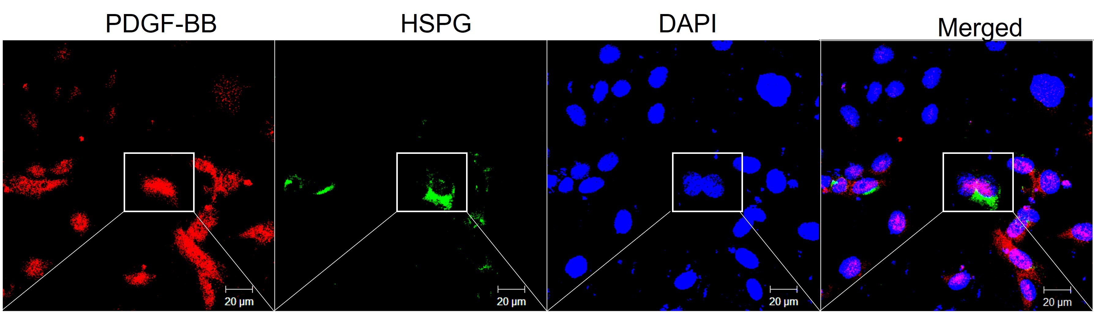
Immunocytochemistry/Immunofluorescence: Mouse Monoclonal Endorepellin/Perlecan/Heparan Sulfate Proteoglycan Antibody (A76) - BSA Free [NBP1-05170] -
Immunocytochemistry/Immunofluorescence: Mouse Monoclonal Endorepellin/Perlecan/Heparan Sulfate Proteoglycan Antibody (A76) - BSA Free [NBP1-05170] - Human primary brain microvascular endothelial cells stained for PDGF-BB (NBP1-58279), red and HSPG (NBP1-05170), green and nuclear DAPI blue. Image from a verified customer review. - BSA Free [NBP1-05170] -")
Immunocytochemistry/ Immunofluorescence: Endorepellin/Perlecan/Heparan Sulfate Proteoglycan Antibody (A76) - BSA Free [NBP1-05170] -
hS/PCs sequentially secrete basement membrane proteins during microstructure reorganization and function. Encapsulated hS/PCs in HA-PEGDA hydrogels initially produce laminin (red) (A–C) and collagen IV (green) (D–F) even at the single-cell state (A,D). Perlecan (green) (G–I) secretion follows to stabilize the laminin and collagen IV networks. Scalebar = 20 μm in all confocal micrographs. Image collected and cropped by CiteAb from the following open publication (https://pubmed.ncbi.nlm.nih.gov/31750298), licensed under a CC-BY license. Not internally tested by Novus Biologicals.Applications
Application
Recommended Usage
ELISA
1:8000
Immunohistochemistry
1:10-1:500
Immunohistochemistry-Frozen
1:10-1:500
Immunohistochemistry-Paraffin
1:10-1:500
Immunoprecipitation
1:10-1:500
Western Blot
1:100-1:2000
Application Notes
Use in ICC/IF was reported in scientific literature (PMID: 31750298). IHC: NBP1-05170 can be used in immunostaining of frozen PLP-fixed sections of bovine and human tissues. AP: NBP1-05170 is more sensitive than NBP2-23522 or NBP2-23523 in ELISA. (2) IP: NBP1-05170 can be used in immunoprecipitation.
Reviewed Applications
Read 1 review rated 5 using NBP1-05170 in the following applications:
Formulation, Preparation, and Storage
Purification
Protein A or G purified
Formulation
10mM Phosphate (pH 7.4) and 0.5M NaCl
Format
BSA Free
Preservative
0.09% Sodium Azide
Concentration
Please see the vial label for concentration. If unlisted please contact technical services.
Shipping
The product is shipped with polar packs. Upon receipt, store it immediately at the temperature recommended below.
Stability & Storage
Store at 4C in the dark.
Background: Endorepellin/Perlecan
Long Name
Heparan Sulfate Proteoglycan
Alternate Names
HSPG2, Perlecan
Gene Symbol
HSPG2
Additional Endorepellin/Perlecan Products
Product Documents
Certificate of Analysis
To download a Certificate of Analysis, please enter a lot or batch number in the search box below.
Product Specific Notices
This product is for research use only and is not approved for use in humans or in clinical diagnosis. Primary Antibodies are guaranteed for 1 year from date of receipt.
Related Research Areas
Citations for Endorepellin/Perlecan/Heparan Sulfate Proteoglycan Antibody (A76) - BSA Free
 Powered by Bioz
Customer Reviews (1)
5 out of 5
1 Customer Rating
Have you used Endorepellin/Perlecan/Heparan Sulfate Proteoglycan Antibody (A76) - BSA Free?
Submit a review and receive an Amazon gift card!
$25/€18/£15/$25CAN/¥2500 Yen for a review with an image
$10/€7/£6/$10CAN/¥1110 Yen for a review without an image
Submit a review
Customer Images
Showing
1
-
1 of
1 review
Showing All
Filter By:
-
Application: ImmunocytochemistrySample Tested: Brain microvascular endothelial cellsSpecies: HumanVerified Customer | Posted 01/23/2024Human brain microvascular endothelial cells stained for PDGF-BB (Red) and HSPG (Green) and nuclear DAPI (Blue) show the endothelial deposition of basement membrane molecule, HSPG.

There are no reviews that match your criteria.
Protocols
Find general support by application which include: protocols, troubleshooting, illustrated assays, videos and webinars.
- Antigen Retrieval Protocol (PIER)
- Antigen Retrieval for Frozen Sections Protocol
- Appropriate Fixation of IHC/ICC Samples
- Cellular Response to Hypoxia Protocols
- Chromogenic IHC Staining of Formalin-Fixed Paraffin-Embedded (FFPE) Tissue Protocol
- Chromogenic Immunohistochemistry Staining of Frozen Tissue
- ClariTSA™ Fluorophore Kits
- Detection & Visualization of Antibody Binding
- ELISA Sample Preparation & Collection Guide
- ELISA Troubleshooting Guide
- Fluorescent IHC Staining of Frozen Tissue Protocol
- Graphic Protocol for Heat-induced Epitope Retrieval
- Graphic Protocol for the Preparation and Fluorescent IHC Staining of Frozen Tissue Sections
- Graphic Protocol for the Preparation and Fluorescent IHC Staining of Paraffin-embedded Tissue Sections
- Graphic Protocol for the Preparation of Gelatin-coated Slides for Histological Tissue Sections
- How to Run an R&D Systems DuoSet ELISA
- How to Run an R&D Systems Quantikine ELISA
- How to Run an R&D Systems Quantikine™ QuicKit™ ELISA
- ICC Cell Smear Protocol for Suspension Cells
- ICC Immunocytochemistry Protocol Videos
- ICC for Adherent Cells
- IHC Sample Preparation (Frozen sections vs Paraffin)
- Immunocytochemistry (ICC) Protocol
- Immunocytochemistry Troubleshooting
- Immunofluorescence of Organoids Embedded in Cultrex Basement Membrane Extract
- Immunofluorescent IHC Staining of Formalin-Fixed Paraffin-Embedded (FFPE) Tissue Protocol
- Immunohistochemistry (IHC) and Immunocytochemistry (ICC) Protocols
- Immunohistochemistry Frozen Troubleshooting
- Immunohistochemistry Paraffin Troubleshooting
- Immunoprecipitation Protocol
- Preparing Samples for IHC/ICC Experiments
- Preventing Non-Specific Staining (Non-Specific Binding)
- Primary Antibody Selection & Optimization
- Protocol for Heat-Induced Epitope Retrieval (HIER)
- Protocol for Making a 4% Formaldehyde Solution in PBS
- Protocol for VisUCyte™ HRP Polymer Detection Reagent
- Protocol for the Fluorescent ICC Staining of Cell Smears - Graphic
- Protocol for the Fluorescent ICC Staining of Cultured Cells on Coverslips - Graphic
- Protocol for the Preparation & Fixation of Cells on Coverslips
- Protocol for the Preparation and Chromogenic IHC Staining of Frozen Tissue Sections
- Protocol for the Preparation and Chromogenic IHC Staining of Frozen Tissue Sections - Graphic
- Protocol for the Preparation and Chromogenic IHC Staining of Paraffin-embedded Tissue Sections
- Protocol for the Preparation and Chromogenic IHC Staining of Paraffin-embedded Tissue Sections - Graphic
- Protocol for the Preparation and Fluorescent ICC Staining of Cells on Coverslips
- Protocol for the Preparation and Fluorescent ICC Staining of Non-adherent Cells
- Protocol for the Preparation and Fluorescent ICC Staining of Stem Cells on Coverslips
- Protocol for the Preparation and Fluorescent IHC Staining of Frozen Tissue Sections
- Protocol for the Preparation and Fluorescent IHC Staining of Paraffin-embedded Tissue Sections
- Protocol for the Preparation of Gelatin-coated Slides for Histological Tissue Sections
- Protocol for the Preparation of a Cell Smear for Non-adherent Cell ICC - Graphic
- Quantikine HS ELISA Kit Assay Principle, Alkaline Phosphatase
- Quantikine HS ELISA Kit Principle, Streptavidin-HRP Polymer
- R&D Systems Quality Control Western Blot Protocol
- Sandwich ELISA (Colorimetric) – Biotin/Streptavidin Detection Protocol
- Sandwich ELISA (Colorimetric) – Direct Detection Protocol
- TUNEL and Active Caspase-3 Detection by IHC/ICC Protocol
- The Importance of IHC/ICC Controls
- Troubleshooting Guide: ELISA
- Troubleshooting Guide: Immunohistochemistry
- Troubleshooting Guide: Western Blot Figures
- Western Blot Conditions
- Western Blot Protocol
- Western Blot Protocol for Cell Lysates
- Western Blot Troubleshooting
- Western Blot Troubleshooting Guide
- View all Protocols, Troubleshooting, Illustrated assays and Webinars
Loading...