Endothelin-1 Antibody (TR.ET.48.5) - BSA Free
Novus Biologicals | Catalog # NB300-526
![Western Blot: Endothelin-1 Antibody (TR.ET.48.5)BSA Free [NB300-526]](https://resources.rndsystems.com/images/products/Endothelin-1-Antibody-TR-ET-48-5-Western-Blot-NB300-526-img0018.jpg "Western Blot: Endothelin-1 Antibody (TR.ET.48.5)BSA Free [NB300-526]")
Key Product Details
Species Reactivity
Validated:
Human, Mouse, Rat, Porcine, Canine, Rabbit, Sheep
Cited:
Human, Mouse, Rat
Applications
Validated:
Immunohistochemistry, Immunohistochemistry-Paraffin, Immunohistochemistry-Frozen, Western Blot, Flow Cytometry, Immunocytochemistry/ Immunofluorescence, Radioimmunoassay
Cited:
Immunohistochemistry, Immunohistochemistry-Paraffin, Western Blot, IF/IHC
Label
Unconjugated
Antibody Source
Monoclonal Mouse IgG1 Clone # TR.ET.48.5
Format
BSA Free
Loading...
Product Specifications
Immunogen
Pure endothelin-1 conjugated to KLH.
Epitope
Studies suggest that this antibody binds to an epitope in the region of ET-1 represented by amino acids 8-16.
Reactivity Notes
Canine reactivity reported in scientific literature (PMID: 11673223). Rabbit reactivity reported in scientific literature (PMID: 12181591). Porcine reactivity reported in scientific literature (PMID: 21587116). Please note that this antibody is reactive to Mouse and derived from the same host, Mouse. Additional Mouse on Mouse blocking steps may be required for IHC and ICC experiments. Please contact Technical Support for more information.
Localization
Secreted
Clonality
Monoclonal
Host
Mouse
Isotype
IgG1
Scientific Data Images for Endothelin-1 Antibody (TR.ET.48.5) - BSA Free
Western Blot: Endothelin-1 Antibody (TR.ET.48.5)BSA Free [NB300-526]
Western Blot: Endothelin-1 Antibody (TR.ET.48.5) [NB300-526] - Analysis of 25 ug of PC12 cell lysates.![Immunocytochemistry/ Immunofluorescence: Endothelin-1 Antibody (TR.ET.48.5) - BSA Free [NB300-526]](https://resources.rndsystems.com/images/products/Endothelin-1-Antibody-TR-ET-48-5-Immunocytochemistry-Immunofluorescence-NB300-526-img0015.jpg "Immunocytochemistry/ Immunofluorescence: Endothelin-1 Antibody (TR.ET.48.5) - BSA Free [NB300-526]")
Immunocytochemistry/ Immunofluorescence: Endothelin-1 Antibody (TR.ET.48.5) - BSA Free [NB300-526]
Immunocytochemistry/Immunofluorescence: Endothelin-1 Antibody (TR.ET.48.5) [NB300-526] - Analysis of Endothelin 1 (green) showing positive staining in the secretion of Hela cells (right) compared with a negative control in the absence of primary antibody (left).![Immunohistochemistry: Endothelin-1 Antibody (TR.ET.48.5) - BSA Free [NB300-526]](https://resources.rndsystems.com/images/products/Endothelin-1-Antibody-TR-ET-48-5-Immunohistochemistry-NB300-526-img0017.jpg "Immunohistochemistry: Endothelin-1 Antibody (TR.ET.48.5) - BSA Free [NB300-526]")
Immunohistochemistry: Endothelin-1 Antibody (TR.ET.48.5) - BSA Free [NB300-526]
Immunohistochemistry: Endothelin-1 Antibody (TR.ET.48.5) [NB300-526] - Immunolocalization of ET-1 in human bowel.![Flow Cytometry: Endothelin-1 Antibody (TR.ET.48.5) - BSA Free [NB300-526]](https://resources.rndsystems.com/images/products/Endothelin-1-Antibody-TR-ET-48-5-Flow-Cytometry-NB300-526-img0012.jpg "Flow Cytometry: Endothelin-1 Antibody (TR.ET.48.5) - BSA Free [NB300-526]")
Flow Cytometry: Endothelin-1 Antibody (TR.ET.48.5) - BSA Free [NB300-526]
Flow Cytometry: Endothelin-1 Antibody (TR.ET.48.5) [NB300-526] - Analysis of Endothelin 1 in 3T3 cells compared to an isotype control (blue).![Immunocytochemistry/ Immunofluorescence: Endothelin-1 Antibody (TR.ET.48.5) - BSA Free [NB300-526]](https://resources.rndsystems.com/images/products/Endothelin-1-Antibody-TR-ET-48-5-Immunocytochemistry-Immunofluorescence-NB300-526-img0013.jpg "Immunocytochemistry/ Immunofluorescence: Endothelin-1 Antibody (TR.ET.48.5) - BSA Free [NB300-526]")
Immunocytochemistry/ Immunofluorescence: Endothelin-1 Antibody (TR.ET.48.5) - BSA Free [NB300-526]
Immunocytochemistry/Immunofluorescence: Endothelin-1 Antibody (TR.ET.48.5) [NB300-526] - Analysis of Endothelin 1 (green) showing positive staining in the secretion of PC12 cells (right) compared with a negative control in the absence of primary antibody (left).![Immunocytochemistry/ Immunofluorescence: Endothelin-1 Antibody (TR.ET.48.5) - BSA Free [NB300-526]](https://resources.rndsystems.com/images/products/Endothelin-1-Antibody-TR-ET-48-5-Immunocytochemistry-Immunofluorescence-NB300-526-img0014.jpg "Immunocytochemistry/ Immunofluorescence: Endothelin-1 Antibody (TR.ET.48.5) - BSA Free [NB300-526]")
Immunocytochemistry/ Immunofluorescence: Endothelin-1 Antibody (TR.ET.48.5) - BSA Free [NB300-526]
Immunocytochemistry/Immunofluorescence: Endothelin-1 Antibody (TR.ET.48.5) [NB300-526] - Analysis of Endothelin 1 (green) showing positive staining in the secretion of HUVEC cells (right) compared with a negative control in the absence of primary antibody (left).![Immunohistochemistry: Endothelin-1 Antibody (TR.ET.48.5) - BSA Free [NB300-526]](https://resources.rndsystems.com/images/products/Endothelin-1-Antibody-TR-ET-48-5-Immunohistochemistry-NB300-526-img0016.jpg "Immunohistochemistry: Endothelin-1 Antibody (TR.ET.48.5) - BSA Free [NB300-526]")
Immunohistochemistry: Endothelin-1 Antibody (TR.ET.48.5) - BSA Free [NB300-526]
Immunohistochemistry: Endothelin-1 Antibody (TR.ET.48.5) [NB300-526] - Immunolocalization of ET-1 in human bowel.![Flow Cytometry: Endothelin-1 Antibody (TR.ET.48.5) - BSA Free [NB300-526]](https://resources.rndsystems.com/images/products/Endothelin-1-Antibody-TR-ET-48-5-Flow-Cytometry-NB300-526-img0006.jpg "Flow Cytometry: Endothelin-1 Antibody (TR.ET.48.5) - BSA Free [NB300-526]")
Flow Cytometry: Endothelin-1 Antibody (TR.ET.48.5) - BSA Free [NB300-526]
Flow Cytometry: Endothelin-1 Antibody (TR.ET.48.5) [NB300-526] - Analysis of Endothelin 1 in HepG2 cells compared to an isotype control (blue).![Flow Cytometry: Endothelin-1 Antibody (TR.ET.48.5) - BSA Free [NB300-526]](https://resources.rndsystems.com/images/products/Endothelin-1-Antibody-TR-ET-48-5-Flow-Cytometry-NB300-526-img0007.jpg "Flow Cytometry: Endothelin-1 Antibody (TR.ET.48.5) - BSA Free [NB300-526]")
Flow Cytometry: Endothelin-1 Antibody (TR.ET.48.5) - BSA Free [NB300-526]
Flow Cytometry: Endothelin-1 Antibody (TR.ET.48.5) [NB300-526] - Analysis of Endothelin 1 in 293T cells compared to an isotype control (blue).![Flow Cytometry: Endothelin-1 Antibody (TR.ET.48.5) - BSA Free [NB300-526]](https://resources.rndsystems.com/images/products/Endothelin-1-Antibody-TR-ET-48-5-Flow-Cytometry-NB300-526-img0011.jpg "Flow Cytometry: Endothelin-1 Antibody (TR.ET.48.5) - BSA Free [NB300-526]")
Flow Cytometry: Endothelin-1 Antibody (TR.ET.48.5) - BSA Free [NB300-526]
Flow Cytometry: Endothelin-1 Antibody (TR.ET.48.5) [NB300-526] - Analysis of Endothelin 1 in HepG2 cells compared to an isotype control (blue).Applications for Endothelin-1 Antibody (TR.ET.48.5) - BSA Free
Application
Recommended Usage
Flow Cytometry
0.5 ug
Immunocytochemistry/ Immunofluorescence
1:200 - 1:1000
Immunohistochemistry
1:250
Immunohistochemistry-Frozen
1:250
Immunohistochemistry-Paraffin
1:250
Radioimmunoassay
1:25000
Western Blot
1:100 - 1:1000
Application Notes
IHC: Staining of ET-1 in human corpus cavernosum tissue with this antibody results in staining of endothelial cells. Radioimmune assays can be used to concentrate ET-1 in solution (e.g. serum/plasma, milk, urine).
Reviewed Applications
Read 1 review rated 4 using NB300-526 in the following applications:
Flow Cytometry Panel Builder
Bio-Techne Knows Flow Cytometry
Save time and reduce costly mistakes by quickly finding compatible reagents using the Panel Builder Tool.
Advanced Features
- Spectra Viewer - Custom analysis of spectra from multiple fluorochromes
- Spillover Popups - Visualize the spectra of individual fluorochromes
- Antigen Density Selector - Match fluorochrome brightness with antigen density

Formulation, Preparation, and Storage
Purification
Immunogen affinity purified
Formulation
PBS
Format
BSA Free
Preservative
0.05% Sodium Azide
Concentration
2.5 mg/ml
Shipping
The product is shipped with polar packs. Upon receipt, store it immediately at the temperature recommended below.
Stability & Storage
Store at -20C. Avoid freeze-thaw cycles.
Background: Endothelin-1
Alternate Names
ARCND3, EDN1, ET1, HDLCQ7, PPET1, QME
Entrez Gene IDs
1906 (Human)
Gene Symbol
EDN1
UniProt
Additional Endothelin-1 Products
Product Documents for Endothelin-1 Antibody (TR.ET.48.5) - BSA Free
Certificate of Analysis
To download a Certificate of Analysis, please enter a lot or batch number in the search box below.
Product Specific Notices for Endothelin-1 Antibody (TR.ET.48.5) - BSA Free
This product is for research use only and is not approved for use in humans or in clinical diagnosis. Primary Antibodies are guaranteed for 1 year from date of receipt.
Related Research Areas
Citations for Endothelin-1 Antibody (TR.ET.48.5) - BSA Free
 Powered by Bioz
Customer Reviews for Endothelin-1 Antibody (TR.ET.48.5) - BSA Free (1)
4 out of 5
1 Customer Rating
Have you used Endothelin-1 Antibody (TR.ET.48.5) - BSA Free?
Submit a review and receive an Amazon gift card!
$25/€18/£15/$25CAN/¥2500 Yen for a review with an image
$10/€7/£6/$10CAN/¥1110 Yen for a review without an image
Submit a review
Customer Images
Showing
1
-
1 of
1 review
Showing All
Filter By:
-
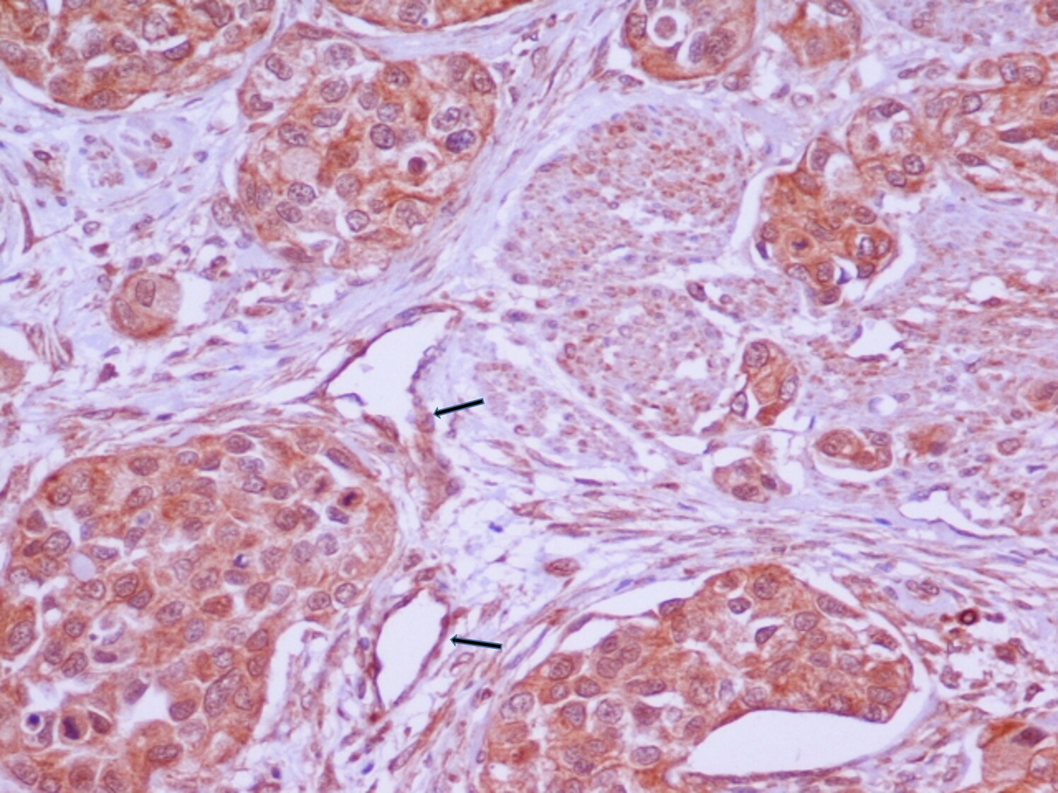
Application: ImmunocytochemistrySample Tested:Species: HumanVerified Customer | Posted 06/24/2015Muscle-invasive,high-grade,stage pT2 bladder TCC.Strong cytoplasmic immunostaining.Positive marker(arrows):capillary endothelium

There are no reviews that match your criteria.
Protocols
Find general support by application which include: protocols, troubleshooting, illustrated assays, videos and webinars.
- 7-Amino Actinomycin D (7-AAD) Cell Viability Flow Cytometry Protocol
- Antigen Retrieval Protocol (PIER)
- Antigen Retrieval for Frozen Sections Protocol
- Appropriate Fixation of IHC/ICC Samples
- Cellular Response to Hypoxia Protocols
- Chromogenic IHC Staining of Formalin-Fixed Paraffin-Embedded (FFPE) Tissue Protocol
- Chromogenic Immunohistochemistry Staining of Frozen Tissue
- ClariTSA™ Fluorophore Kits
- Detection & Visualization of Antibody Binding
- Extracellular Membrane Flow Cytometry Protocol
- Flow Cytometry Protocol for Cell Surface Markers
- Flow Cytometry Protocol for Staining Membrane Associated Proteins
- Flow Cytometry Staining Protocols
- Flow Cytometry Troubleshooting Guide
- Fluorescent IHC Staining of Frozen Tissue Protocol
- Graphic Protocol for Heat-induced Epitope Retrieval
- Graphic Protocol for the Preparation and Fluorescent IHC Staining of Frozen Tissue Sections
- Graphic Protocol for the Preparation and Fluorescent IHC Staining of Paraffin-embedded Tissue Sections
- Graphic Protocol for the Preparation of Gelatin-coated Slides for Histological Tissue Sections
- ICC Cell Smear Protocol for Suspension Cells
- ICC Immunocytochemistry Protocol Videos
- ICC for Adherent Cells
- IHC Sample Preparation (Frozen sections vs Paraffin)
- Immunocytochemistry (ICC) Protocol
- Immunocytochemistry Troubleshooting
- Immunofluorescence of Organoids Embedded in Cultrex Basement Membrane Extract
- Immunofluorescent IHC Staining of Formalin-Fixed Paraffin-Embedded (FFPE) Tissue Protocol
- Immunohistochemistry (IHC) and Immunocytochemistry (ICC) Protocols
- Immunohistochemistry Frozen Troubleshooting
- Immunohistochemistry Paraffin Troubleshooting
- Intracellular Flow Cytometry Protocol Using Alcohol (Methanol)
- Intracellular Flow Cytometry Protocol Using Detergents
- Intracellular Nuclear Staining Flow Cytometry Protocol Using Detergents
- Intracellular Staining Flow Cytometry Protocol Using Alcohol Permeabilization
- Intracellular Staining Flow Cytometry Protocol Using Detergents to Permeabilize Cells
- Preparing Samples for IHC/ICC Experiments
- Preventing Non-Specific Staining (Non-Specific Binding)
- Primary Antibody Selection & Optimization
- Propidium Iodide Cell Viability Flow Cytometry Protocol
- Protocol for Heat-Induced Epitope Retrieval (HIER)
- Protocol for Liperfluo
- Protocol for Making a 4% Formaldehyde Solution in PBS
- Protocol for VisUCyte™ HRP Polymer Detection Reagent
- Protocol for the Characterization of Human Th22 Cells
- Protocol for the Characterization of Human Th9 Cells
- Protocol for the Fluorescent ICC Staining of Cell Smears - Graphic
- Protocol for the Fluorescent ICC Staining of Cultured Cells on Coverslips - Graphic
- Protocol for the Preparation & Fixation of Cells on Coverslips
- Protocol for the Preparation and Chromogenic IHC Staining of Frozen Tissue Sections
- Protocol for the Preparation and Chromogenic IHC Staining of Frozen Tissue Sections - Graphic
- Protocol for the Preparation and Chromogenic IHC Staining of Paraffin-embedded Tissue Sections
- Protocol for the Preparation and Chromogenic IHC Staining of Paraffin-embedded Tissue Sections - Graphic
- Protocol for the Preparation and Fluorescent ICC Staining of Cells on Coverslips
- Protocol for the Preparation and Fluorescent ICC Staining of Non-adherent Cells
- Protocol for the Preparation and Fluorescent ICC Staining of Stem Cells on Coverslips
- Protocol for the Preparation and Fluorescent IHC Staining of Frozen Tissue Sections
- Protocol for the Preparation and Fluorescent IHC Staining of Paraffin-embedded Tissue Sections
- Protocol for the Preparation of Gelatin-coated Slides for Histological Tissue Sections
- Protocol for the Preparation of a Cell Smear for Non-adherent Cell ICC - Graphic
- Protocol: Annexin V and PI Staining by Flow Cytometry
- Protocol: Annexin V and PI Staining for Apoptosis by Flow Cytometry
- R&D Systems Quality Control Western Blot Protocol
- TUNEL and Active Caspase-3 Detection by IHC/ICC Protocol
- The Importance of IHC/ICC Controls
- Troubleshooting Guide: Fluorokine Flow Cytometry Kits
- Troubleshooting Guide: Immunohistochemistry
- Troubleshooting Guide: Western Blot Figures
- Western Blot Conditions
- Western Blot Protocol
- Western Blot Protocol for Cell Lysates
- Western Blot Troubleshooting
- Western Blot Troubleshooting Guide
- View all Protocols, Troubleshooting, Illustrated assays and Webinars
Loading...