eNOS Antibody - BSA Free
Novus Biologicals | Catalog # NB300-500
Key Product Details
Validated by
Biological Validation
Species Reactivity
Validated:
Human, Mouse, Rat, Porcine, Amphibian, Bovine, Canine, Drosophila, Sheep
Cited:
Human, Mouse, Rat, Porcine, Ovine
Applications
Validated:
Immunohistochemistry, Immunohistochemistry-Paraffin, Immunohistochemistry-Frozen, Western Blot
Cited:
Immunohistochemistry, Western Blot, Flow Cytometry, IF/IHC, Electron Microscopy
Label
Unconjugated
Antibody Source
Polyclonal Rabbit IgG
Format
BSA Free
Loading...
Product Specifications
Immunogen
Synthetic peptide corresponding to residues S(1179) LQERQLRGAVPWAFD(1194) of human eNOS.
Epitope
SLQERQLRGAVPWAFD
Reactivity Notes
Sheep reactivity reported in scientific literature (PMID: 31352175).
Specificity
This does not detect human inducible NOS or rat brain NOS.
Clonality
Polyclonal
Host
Rabbit
Isotype
IgG
Theoretical MW
133 kDa.
Disclaimer note: The observed molecular weight of the protein may vary from the listed predicted molecular weight due to post translational modifications, post translation cleavages, relative charges, and other experimental factors.
Disclaimer note: The observed molecular weight of the protein may vary from the listed predicted molecular weight due to post translational modifications, post translation cleavages, relative charges, and other experimental factors.
Scientific Data Images for eNOS Antibody - BSA Free
![Immunohistochemistry-Paraffin: eNOS Antibody - BSA Free [NB300-500]](https://resources.rndsystems.com/images/products/eNOS-Antibody---BSA-Free-Immunohistochemistry-Paraffin-NB300-500-img0006.jpg "Immunohistochemistry-Paraffin: eNOS Antibody - BSA Free [NB300-500]")
Immunohistochemistry-Paraffin: eNOS Antibody - BSA Free [NB300-500]
eNOS-Antibody---BSA-Free-Immunohistochemistry-Paraffin-NB300-500-img0006.jpg![Immunohistochemistry-Paraffin: eNOS Antibody - BSA Free [NB300-500]](https://resources.rndsystems.com/images/products/eNOS-Antibody---BSA-Free-Immunohistochemistry-Paraffin-NB300-500-img0003.jpg "Immunohistochemistry-Paraffin: eNOS Antibody - BSA Free [NB300-500]")
Immunohistochemistry-Paraffin: eNOS Antibody - BSA Free [NB300-500]
Immunohistochemistry-Paraffin: eNOS Antibody - BSA Free [NB300-500] - Staining of eNOS in human lung tissue.![Immunohistochemistry-Paraffin: eNOS Antibody - BSA Free [NB300-500]](https://resources.rndsystems.com/images/products/eNOS-Antibody---BSA-Free-Immunohistochemistry-Paraffin-NB300-500-img0004.jpg "Immunohistochemistry-Paraffin: eNOS Antibody - BSA Free [NB300-500]")
Immunohistochemistry-Paraffin: eNOS Antibody - BSA Free [NB300-500]
Immunohistochemistry-Paraffin: eNOS Antibody - BSA Free [NB300-500] - Mouse Carotid Artery stained with eNOS antibody. Image from verified customer review.![Immunohistochemistry: eNOS Antibody - BSA Free [NB300-500]](https://resources.rndsystems.com/images/products/eNOS-Antibody---BSA-Free-Immunohistochemistry-NB300-500-img0005.jpg "Immunohistochemistry: eNOS Antibody - BSA Free [NB300-500]")
Immunohistochemistry: eNOS Antibody - BSA Free [NB300-500]
Immunohistochemistry: eNOS Antibody - BSA Free [NB300-500] - Staining of representative pulmonary artery endothelial cells from rats subjected to CON=Control Group, ALI=Acute lung injury Group, LV=Low tidal volume Group, VLV=Very low tidal volume Group, MV=Large tidal volume Group. Dark brown staining of indicates eNOS-positive cells. Magnification x400.
Immunohistochemistry: eNOS Antibody - BSA Free [NB300-500] -
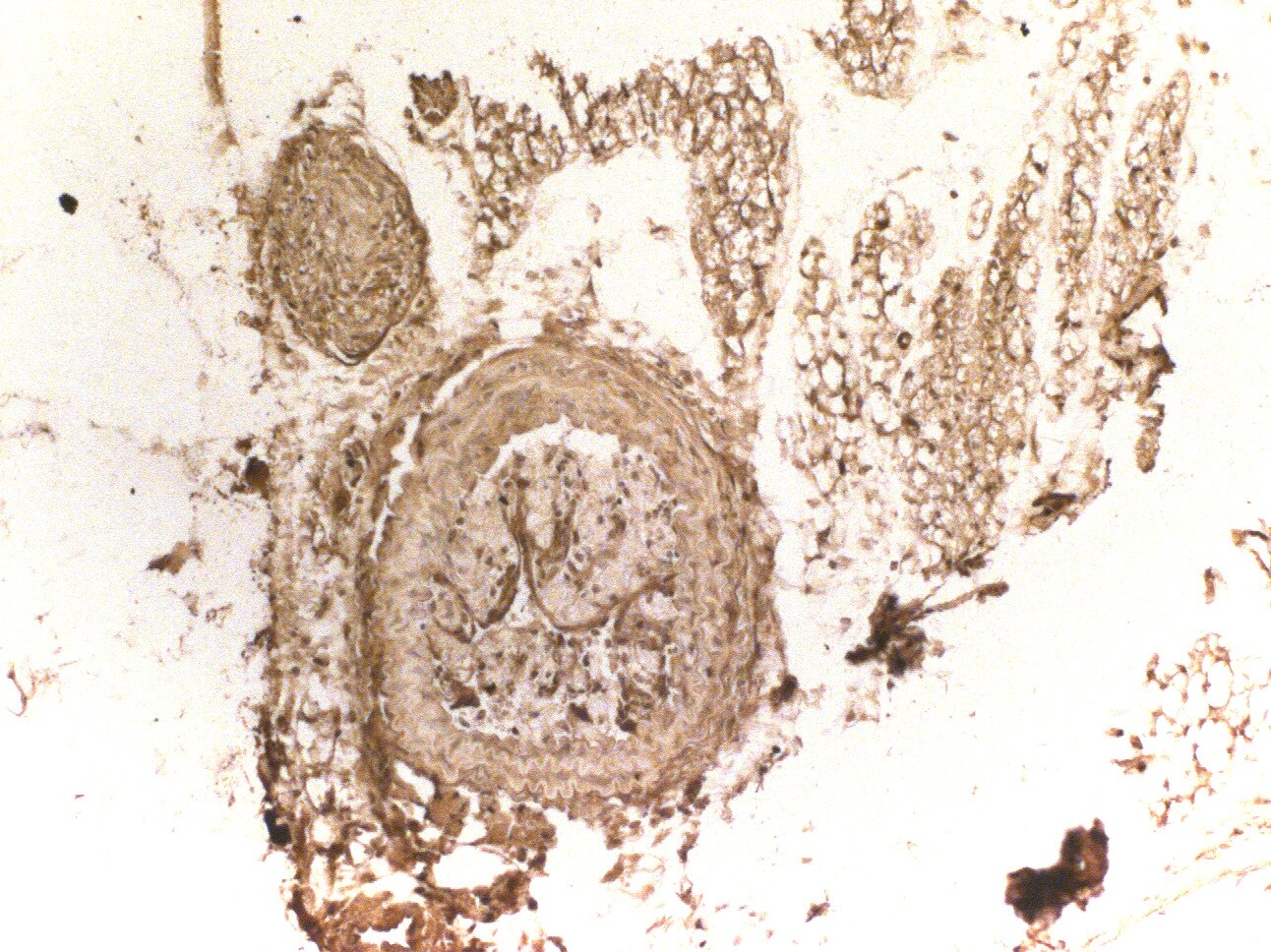
Expression of endothelial markers indicate functional endothelium in TEVG.A. Whole mount staining of native aorta and TEVG. VE-cadherin is a marker of cellular borders of endothelial cells (green). Endothelial nitric oxide synthase (eNOS) is a marker of a functional endothelium (red). DAPI is a nuclear stain (blue). B. Real-time PCR analysis of ephrinB2 and eNOS. Ephrin-B2, a marker of arterial vessels. (n = 5–10 in each group)
Western Blot: eNOS Antibody - BSA Free [NB300-500] -
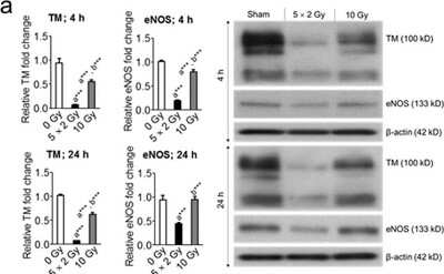
Western Blot: eNOS Antibody - BSA Free [NB300-500] - Mevalonate pathway inhibitors reversed fractionated-radiation–induced suppression of KLF2 & its downstream target molecules. Representative Western blot analysis & quantification of KLF2, TM, & eNOS 4 h after exposure to five fractions of 2 Gy (a) in presence or absence of atorvastatin (1 μM) or GT3 (5 μM) & (b) in presence or absence of GGTi (10 μM) (n = 3). beta -actin served as a loading control. (n, number of independent experiments performed; a, significant statistical difference between nonirradiated & irradiated groups; b, significant statistical difference between fractionated irradiation & single exposure; *, p < 0.05; **, p < 0.01; ***, p < 0.001). Image collected & cropped by CiteAb from the following publication (https://pubmed.ncbi.nlm.nih.gov/32382091), licensed under a CC-BY license. Not internally tested by Novus Biologicals.
Western Blot: eNOS Antibody - BSA Free [NB300-500] -
Western Blot: eNOS Antibody - BSA Free [NB300-500] - Upregulation of HMG-CoA reductase in shPLD2 cells decreases eNOS expression. (A) Representative western blot of HMG-CoA reductase in Scramble & shPLD2 cells. The regions of the Western blots containing the HMG-CoA reductase & actin immunoreactive bands were scanned & the relative abundance of the individual samples quantified using an Odyssey CLx imaging system (B) Quantification of western blots with HMG-CoA reductase normalized to actin (n = 4 experiments). (C) N-SIM microscopy of HMG-CoA reductase; Bar, 7.5 μM. (D) RT-PCR of HMG-CoA reductase in Scramble & shPLD2 cells (n = 3). (E) Western blot of eNOS after treatment with NFOT & Simvastatin. Representative image of 3 experiments. The regions of the Western blots (Suppl. Figure 2) containing the eNOS & actin immunoreactive bands were scanned & the relative abundance of the individual samples quantified using an Odyssey CLx imaging system. (F) Quantification of western blots with eNOS levels normalized to actin (n = 3). Comparisons without bars made to untreated Scramble values. (G) eNOS activity after treatment with Simvastatin (n = 7). Mean ± SEM; *p < 0.05; **p < 0.01; ***p ≤ 0.001; One-way ANOVA with Bonferroni’s Multiple Comparison Test. Image collected & cropped by CiteAb from the following publication (https://pubmed.ncbi.nlm.nih.gov/28831159), licensed under a CC-BY license. Not internally tested by Novus Biologicals.
Western Blot: eNOS Antibody - BSA Free [NB300-500] -
Western Blot: eNOS Antibody - BSA Free [NB300-500] - The human PLD2 polymorphism R172C does not alter eNOS signaling but does decrease caveolin-1 protein levels. Overexpression of HA-tagged hPLD2 (A) & HA-R172C-PLD2 (B) in HeLa cells, visualized using anti-HA immunofluorescent staining (green). Arrowhead, PLD2 localization in filopodia; chevron, in peripheral actin ruffles; *, in dorsal actin ruffles; arrow, in subcortical actin network. Bar, 7.5 μM. Representative image of multiple experiments. (C) Representative western blot of eNOS after transfection of HA-hPLD2 or HA-R172C-PLD2 into shPLD2 cells. The regions of the Western blots containing the eNOS & actin immunoreactive bands were scanned & the relative abundance of the individual samples quantified using an Odyssey CLx imaging system. (D) Quantification of western blotting with eNOS levels normalized to actin (n = 3). (E) eNOS activity as measured by nitrate formation (n = 7). Lane 1 vs 2, P = 0.0021; lane 2 vs 3, P = 0.00035; lane 2 vs 4, P = 0.00012. (F–I) N-SIM microscopy of plasma membrane eNOS & caveolin-1. Bar, 7.5 μM. Representative image of multiple Scramble or shPLD2 cells imaged. Cells in H & I were selected for imaging based on expression of HA-hPLD2 or HA-R172C-PLD2 as visualized by anti-HA immunofluorescence in a separate channel (not shown). Mean ± SEM; *p < 0.05; **p < 0.01; ***p ≤ 0.001; One-way ANOVA with Bonferroni’s Multiple Comparison Test. Image collected & cropped by CiteAb from the following publication (https://pubmed.ncbi.nlm.nih.gov/28831159), licensed under a CC-BY license. Not internally tested by Novus Biologicals.
Immunocytochemistry/ Immunofluorescence: eNOS Antibody - BSA Free [NB300-500] -
Expression of endothelial markers indicate functional endothelium in TEVG.A. Whole mount staining of native aorta and TEVG. VE-cadherin is a marker of cellular borders of endothelial cells (green). Endothelial nitric oxide synthase (eNOS) is a marker of a functional endothelium (red). DAPI is a nuclear stain (blue). B. Real-time PCR analysis of ephrinB2 and eNOS. Ephrin-B2, a marker of arterial vessels. (n = 5–10 in each group) Image collected and cropped by CiteAb from the following open publication (https://pubmed.ncbi.nlm.nih.gov/25830942), licensed under a CC-BY license. Not internally tested by Novus Biologicals.Applications for eNOS Antibody - BSA Free
Application
Recommended Usage
Immunohistochemistry
1:10 - 1:500
Immunohistochemistry-Frozen
1:100
Immunohistochemistry-Paraffin
1:100
Western Blot
1:50-1:1000
Application Notes
In IHC, antigen retrieval is not essential but may optimize staining. By Western blot, a 140kDa band is seen in human endothelial cells. Optimal dilutions should be determined by the end user.
Reviewed Applications
Read 3 reviews rated 4.7 using NB300-500 in the following applications:
Formulation, Preparation, and Storage
Purification
Immunogen affinity purified
Formulation
PBS
Format
BSA Free
Preservative
0.02% Sodium Azide
Concentration
1.0 mg/ml
Shipping
The product is shipped with polar packs. Upon receipt, store it immediately at the temperature recommended below.
Stability & Storage
Store at -20C. Avoid freeze-thaw cycles.
Background: eNOS
Long Name
Endothelial Nitric Oxide Synthase
Alternate Names
NOS3
Gene Symbol
NOS3
UniProt
Additional eNOS Products
Product Documents for eNOS Antibody - BSA Free
Certificate of Analysis
To download a Certificate of Analysis, please enter a lot or batch number in the search box below.
Product Specific Notices for eNOS Antibody - BSA Free
This product is for research use only and is not approved for use in humans or in clinical diagnosis. Primary Antibodies are guaranteed for 1 year from date of receipt.
Related Research Areas
Citations for eNOS Antibody - BSA Free
 Powered by Bioz
Customer Reviews for eNOS Antibody - BSA Free (3)
4.7 out of 5
3 Customer Ratings
Have you used eNOS Antibody - BSA Free?
Submit a review and receive an Amazon gift card!
$25/€18/£15/$25CAN/¥2500 Yen for a review with an image
$10/€7/£6/$10CAN/¥1110 Yen for a review without an image
Submit a review
Customer Images
Showing
1
-
3 of
3 reviews
Showing All
Filter By:
-
Application: Western BlotSample Tested: Mouse brainSpecies: MouseVerified Customer | Posted 05/23/2017
-
Application: ImmunohistochemistrySample Tested: Mouse brainSpecies: MouseVerified Customer | Posted 05/23/2017
-
Application: Immunohistochemistry-ParaffinSample Tested: Mouse Carotid ArterySpecies: MouseVerified Customer | Posted 02/22/2016eNOS antibody tested on mouse carotid artery

There are no reviews that match your criteria.
Protocols
Find general support by application which include: protocols, troubleshooting, illustrated assays, videos and webinars.
- Antigen Retrieval Protocol (PIER)
- Antigen Retrieval for Frozen Sections Protocol
- Appropriate Fixation of IHC/ICC Samples
- Cellular Response to Hypoxia Protocols
- Chromogenic IHC Staining of Formalin-Fixed Paraffin-Embedded (FFPE) Tissue Protocol
- Chromogenic Immunohistochemistry Staining of Frozen Tissue
- ClariTSA™ Fluorophore Kits
- Detection & Visualization of Antibody Binding
- Fluorescent IHC Staining of Frozen Tissue Protocol
- Graphic Protocol for Heat-induced Epitope Retrieval
- Graphic Protocol for the Preparation and Fluorescent IHC Staining of Frozen Tissue Sections
- Graphic Protocol for the Preparation and Fluorescent IHC Staining of Paraffin-embedded Tissue Sections
- Graphic Protocol for the Preparation of Gelatin-coated Slides for Histological Tissue Sections
- IHC Sample Preparation (Frozen sections vs Paraffin)
- Immunofluorescent IHC Staining of Formalin-Fixed Paraffin-Embedded (FFPE) Tissue Protocol
- Immunohistochemistry (IHC) and Immunocytochemistry (ICC) Protocols
- Immunohistochemistry Frozen Troubleshooting
- Immunohistochemistry Paraffin Troubleshooting
- Preparing Samples for IHC/ICC Experiments
- Preventing Non-Specific Staining (Non-Specific Binding)
- Primary Antibody Selection & Optimization
- Protocol for Heat-Induced Epitope Retrieval (HIER)
- Protocol for Making a 4% Formaldehyde Solution in PBS
- Protocol for VisUCyte™ HRP Polymer Detection Reagent
- Protocol for the Preparation & Fixation of Cells on Coverslips
- Protocol for the Preparation and Chromogenic IHC Staining of Frozen Tissue Sections
- Protocol for the Preparation and Chromogenic IHC Staining of Frozen Tissue Sections - Graphic
- Protocol for the Preparation and Chromogenic IHC Staining of Paraffin-embedded Tissue Sections
- Protocol for the Preparation and Chromogenic IHC Staining of Paraffin-embedded Tissue Sections - Graphic
- Protocol for the Preparation and Fluorescent IHC Staining of Frozen Tissue Sections
- Protocol for the Preparation and Fluorescent IHC Staining of Paraffin-embedded Tissue Sections
- Protocol for the Preparation of Gelatin-coated Slides for Histological Tissue Sections
- R&D Systems Quality Control Western Blot Protocol
- TUNEL and Active Caspase-3 Detection by IHC/ICC Protocol
- The Importance of IHC/ICC Controls
- Troubleshooting Guide: Immunohistochemistry
- Troubleshooting Guide: Western Blot Figures
- Western Blot Conditions
- Western Blot Protocol
- Western Blot Protocol for Cell Lysates
- Western Blot Troubleshooting
- Western Blot Troubleshooting Guide
- View all Protocols, Troubleshooting, Illustrated assays and Webinars


